

Diagnostic criteria for spontaneous spinal CSF leaks and intracranial hypotension
- PMID: 18258706
- PMCID: PMC8128584
- DOI: 10.3174/ajnr.A0956
Diagnostic criteria for spontaneous spinal CSF leaks and intracranial hypotension
Abstract
Background and purpose: Comprehensive diagnostic criteria encompassing the varied clinical and radiographic manifestations of spontaneous intracranial hypotension are not available. Therefore, we propose a new set of diagnostic criteria.
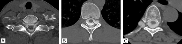
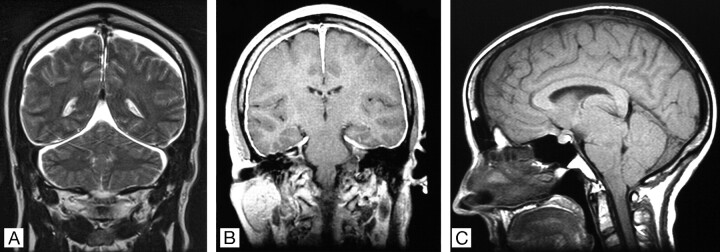
Materials and methods: The diagnostic criteria are based on results of brain and spine imaging, clinical manifestations, results of lumbar puncture, and response to epidural blood patching. The diagnostic criteria include criterion A, the demonstration of extrathecal CSF on spinal imaging. If criterion A is not met, criterion B, which is cranial MR imaging findings of spontaneous intracranial hypotension, follows, with at least one of the following: 1) low opening pressure, 2) spinal meningeal diverticulum, or 3) improvement of symptoms after epidural blood patch. If criteria A and B are not met, there is criterion C, the presence of all of the following or at least 2 of the following if typical orthostatic headaches are present: 1) low opening pressure, 2) spinal meningeal diverticulum, and 3) improvement of symptoms after epidural blood patch. These criteria were applied to a group of 107 consecutive patients evaluated for spontaneous spinal CSF leaks and intracranial hypotension.
Results: The diagnosis was confirmed in 94 patients, with use of criterion A in 78 patients, criterion B in 11 patients, and criterion C in 5 patients.
Conclusions: A new diagnostic scheme is presented reflecting the wide spectrum of clinical and radiographic manifestations of spontaneous spinal CSF leaks and intracranial hypotension.
Figures
Comment in
-
Diagnostic criteria for spontaneous spinal CSF leaks and intracranial hypotension.AJNR Am J Neuroradiol. 2008 Nov;29(10):e94; author reply e85. doi: 10.3174/ajnr.A1156. Epub 2008 Sep 17. AJNR Am J Neuroradiol. 2008. PMID: 18799593 Free PMC article. No abstract available.
References
-
- Schievink WI. Spontaneous spinal cerebrospinal fluid leaks and intracranial hypotension. JAMA 2006;295:2284–96 - PubMed
-
- Schievink WI, Gordon OK, Tourje J. Connective tissue disorders with spontaneous spinal cerebrospinal fluid leaks and intracranial hypotension: a prospective study. Neurosurgery 2004;54:65–71 - PubMed
-
- Schievink WI, Louy C. Precipitating factors of spontaneous spinal CSF leaks and intracranial hypotension. Neurology 2007;69:700–02 - PubMed
-
- Headache Classification Subcommittee of the International Headache Society. The International Classification of Headache Disorders, 2nd ed. Cephalalgia 2004;24:1–160
-
- Mokri B, Posner JB. Spontaneous intracranial hypotension: the broadening clinical and imaging spectrum of CSF leaks. Neurology 2000;55:1771–72 - PubMed
MeSH terms
LinkOut - more resources
Full Text Sources
Medical
Miscellaneous