. 2008 Feb;23(1):26-36.
doi: 10.1177/0748730407311254.

The circadian clock protein BMAL1 is necessary for fertility and proper testosterone production in mice

Affiliations

The circadian clock protein BMAL1 is necessary for fertility and proper testosterone production in mice

J D Alvarez et al. J Biol Rhythms. 2008 Feb.

Abstract

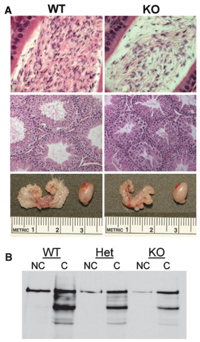
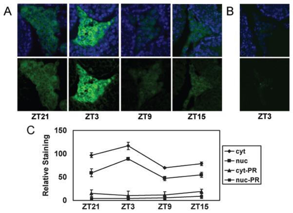
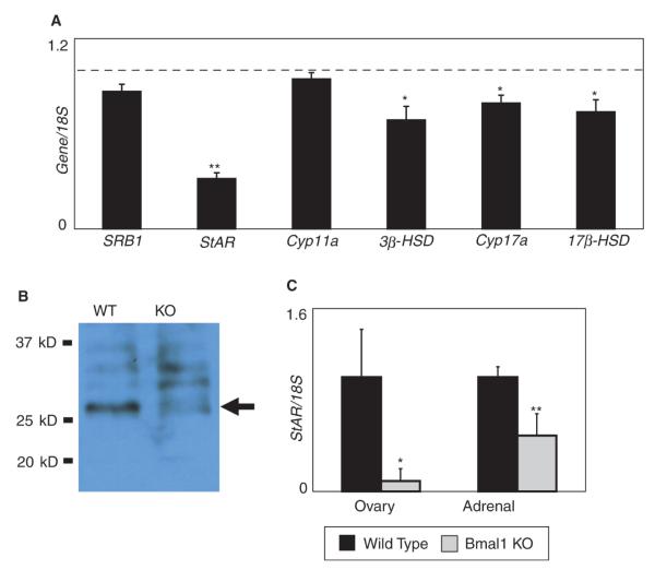
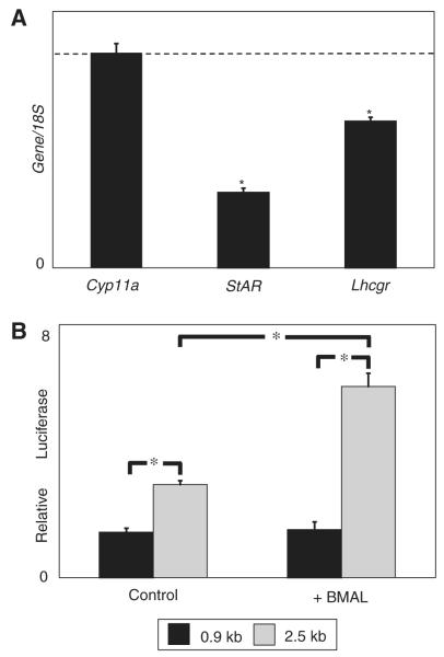
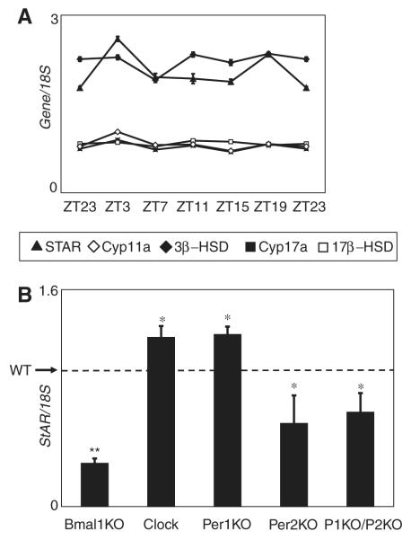
Although it is well established that the circadian clock regulates mammalian reproductive physiology, the molecular mechanisms by which this regulation occurs are not clear. The authors investigated the reproductive capacity of mice lacking Bmal1 (Arntl, Mop3), one of the central circadian clock genes. They found that both male and female Bmal1 knockout (KO) mice are infertile. Gross and microscopic inspection of the reproductive anatomy of both sexes suggested deficiencies in steroidogenesis. Male Bmal1 KO mice had low testosterone and high luteinizing hormone serum concentrations, suggesting a defect in testicular Leydig cells. Importantly, Leydig cells rhythmically express BMAL1 protein, suggesting peripheral control of testosterone production by this clock protein. Expression of steroidogenic genes was reduced in testes and other steroidogenic tissues of Bmal1 KO mice. In particular, expression of the steroidogenic acute regulatory protein (StAR) gene and protein, which regulates the rate-limiting step of steroidogenesis, was decreased in testes from Bmal1 KO mice. A direct effect of BMAL1 on StAR expression in Leydig cells was indicated by in vitro experiments showing enhancement of StAR transcription by BMAL1. Other hormonal defects in male Bmal1 KO mice suggest that BMAL1 also has functions in reproductive physiology outside of the testis. These results enhance understanding of how the circadian clock regulates reproduction.


Figures

Figure 1
Figure 1
Bmal1 knockout (KO) male mice have reproductive deficiencies but functional sperm. (A) Photos are of organs collected from 12-week-old littermates. Genotypes are indicated. Top panel: Sections of caput epididymi showing fewer sperm in the Bmal1 KO animal in comparison to its wild-type littermate. Exact counts are provided in Table 1. Magnification is ×40. Middle panel: Sections of testes showing smaller seminiferous tubules in the Bmal1 KO mice in comparison to the wild-type littermate. Magnification is ×20. Bottom panel: Photos comparing the sizes of the seminal vesicles and a testis from wild-type and Bmal1 KO littermates. (B) There is no difference in capacitation of sperm from Bmal1 KO, heterozygous, and wild-type mice. Sperm were collected from the caudal epididymi of 12-week-old littermates and subjected to capacitation analysis by detection of proteins containing phosphotyrosine. Fewer sperm were collected from Het and KO mice (due to the lower sperm counts in these animals), resulting in lighter bands. However, the pattern of phosphorylated proteins is the same.
Figure 2
Figure 2
BMAL1 expression oscillates in Leydig cells. (A) Immunohistochemical analysis of sectioned testes detected BMAL1 almost exclusively in the Leydig cells. Top panel: Merged images of BMAL1 (green) and nuclear dye Hoechst 33258 (blue). Bottom panel: Staining for BMAL1 only. BMAL1 staining intensity oscillates over the course of day, with a peak at ZT3 and a trough at ZT9. Images shown are representative of 5 testes processed at each time point. (B) Preadsorption of antibody with BMAL1 peptide dramatically reduces staining. Magnification in all pictures is ×40. (C) BMAL1 oscillates in both cytoplasm (cyt) and nuclei (nuc), as measured by densitometry. Data represent the average (± SEM) level of staining in 20 sections derived from 5 animals per time point. The vertical axis represents relative staining intensity, where 0 represents no staining (black) and 255 represents maximum saturated staining (green) assigned by ImageJ software. Significant differences (p < 0.05) in BMAL1 signal in both cytoplasmic and nuclear staining were observed between all time points, except between ZT21 and ZT15 (paired Student t test). Minimal staining using antibody preadsorbed with peptide was observed in cytoplasm (cyt-PR) and nuclei (nuc-PR).
Figure 3
Figure 3
Steroidogenic acute regulatory protein (StAR) expression is reduced in the testis of Bmal1 knockout (KO) mice. (A) Steroidogenic gene expression is reduced in the testis of Bmal1 KO mice. Testis RNA from Bmal1 KO mice and wild-type littermates (8 pairs) was analyzed by quantitative polymerase chain reaction (qPCR). Expression is shown relative to wild-type expression (dashed line). Error bars represent SEM. A single asterisk represents p < 0.05, and a double asterisk represents p < 0.005 (paired Student t test). (B) StAR protein expression is reduced in the testes of Bmal1 KO mice. The arrow indicates the StAR protein. Upper bands represent preprocessed forms of the protein. Data shown are representative of experiments repeated on a total of 3 littermate pairs. (C) StAR expression is reduced in both adrenal glands and in ovaries of Bmal1 KO mice. Adrenal glands were collected from male Bmal1 KO mice and wild-type littermates (2 pairs), and adrenal glands and ovaries were collected from female Bmal1 KO mice and wild-type littermates (3 pairs). Error bars represent standard deviation. Data were normalized such that wild-type expression was set at 1. Data are significant to p = 0.02 (ovary) and <0.001 (adrenal).
Figure 4
Figure 4
BMAL1 directly enhances steroidogenic acute regulatory protein (StAR) expression. (A) Reduced expression of the luteinizing hormone (LH) receptor does not account for the effect on StAR expression observed in Bmal1 knockout (KO) mice. Testis RNA was collected from 5 pairs of Bmal1 KO mice and wild-type littermates and analyzed by quantitative polymerase chain reaction (qPCR). Expression is shown relative to wild-type expression level (dashed line). Error bars represent SEM. Asterisk represents a significant reduction (p < 0.05; paired Student t test). (B) BMAL1 enhances StAR expression in vitro. Expression constructs containing either 966 bp or 2.5 kb of the StAR promoter were transfected into the MA-10 Leydig tumor cell line with either a BMAL1 expression construct or an empty expression construct (pcDNA3.1). After 48 h, the cells were collected and luciferase activity was measured. Luciferase activity is higher with the 2.5-kb promoter sequence and is increased in the presence of BMAL1. Activity from the 966-bp promoter is unaffected by BMAL1. Shown is a single representative experiment with data points collected in triplicate. Differences are significant with p < 0.001 (unpaired Student t test). This experiment has been repeated once with similar results.
Figure 5
Figure 5
Expression of steroidogenic genes in testes is not circadian. Expression of the steroidogenic genes does not oscillate over the course of a day. (A) Testis RNA collected from 12-week-old C57BL6/J individually housed male mice over a continuous 24-h period was examined by quantitative polymerase chain reaction (qPCR). Data shown are from 1 representative experiment. This was repeated 3 times with similar results. (B) Expression of steroidogenic acute regulatory protein (StAR) is affected by mutations of the circadian clock genes. StAR expression in testes from the indicated mutant mice and wild-type mice was measured by qPCR. Expression of the genes is shown relative to wild-type expression level (dashed line). Error bars represent SEM. Clock mutant mice (9 pairs) and Bmal1 knockout (KO) mice (8 pairs) are compared with wild-type littermates. Per1 KO (3 mice), Per2 KO (3 mice), and Per1 KO/Per2 KO mice (4 mice) are compared with age-matched C57BL6/J male mice (4 mice). All data are significant to p < 0.05 (single asterisk), except for the Bmal1 KO mice, which are significant to p = 0.002 (unpaired Student t test).

References

    1. Alvarez JD, Chen D, Storer E, Sehgal A. Non-cyclic and developmental stage-specific expression of circadian clock proteins during murine spermatogenesis. Biol Reprod. 2003;69:81–91. - PubMed
    1. Alvarez JD, Sehgal A. The thymus is similar to the testis in its pattern of circadian clock gene expression. J Biol Rhythms. 2005;20:111–121. - PubMed
    1. Andrews WW, Ojeda SR. A detailed analysis of the serum luteinizing hormone secretory profile in conscious, free-moving female rats during the time of puberty. Endocrinology. 1981;109:2032–2039. - PubMed
    1. Apter D, Butzow T, Laughlin G, Yen S. Gonadotropin-releasing hormone pulse generator activity during pubertal transition in girls: Pulsatile and diurnal patterns of circulating gonadotropins. J Clin Endocrinol Metab. 1993;76:940–949. - PubMed
    1. Ascoli M. Characterization of several clonal lines of cultured Leydig tumor cells: Gonadotropin receptors and steroidogenic responses. Endocrinology. 1981;108:88–95. - PubMed

Publication types

MeSH terms