. 2008 Mar 28;102(6):677-85.
doi: 10.1161/CIRCRESAHA.107.161737. Epub 2008 Feb 7.

c-kit is required for cardiomyocyte terminal differentiation

Affiliations

c-kit is required for cardiomyocyte terminal differentiation

Ming Li et al. Circ Res. .

Abstract

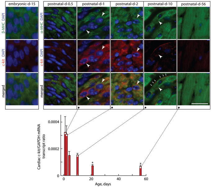
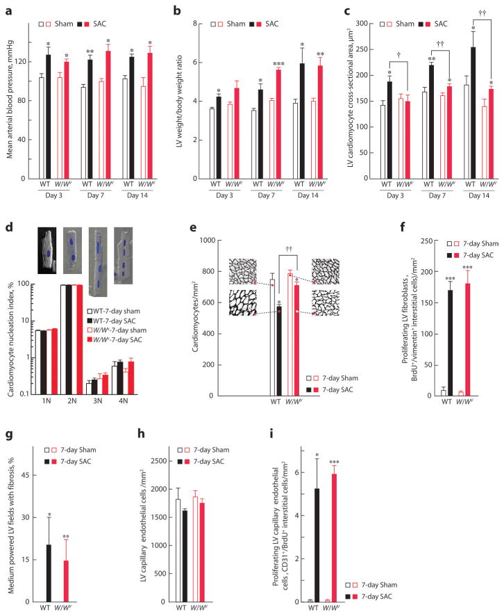
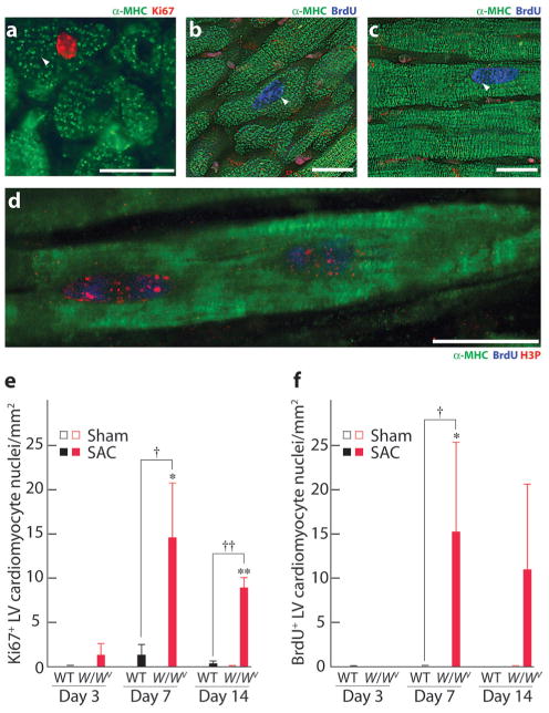
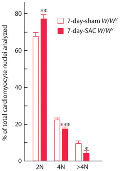
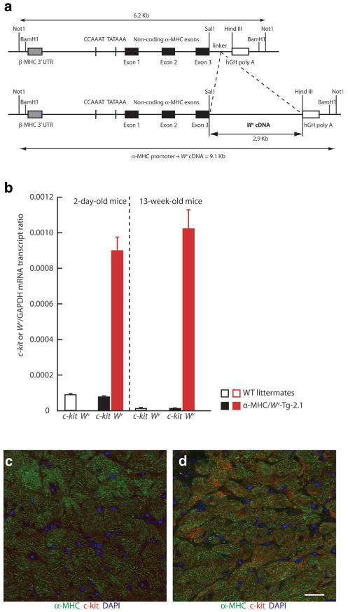
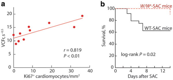
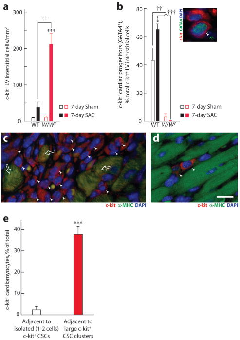
c-kit, the transmembrane tyrosine kinase receptor for stem cell factor, is required for melanocyte and mast cell development, hematopoiesis, and differentiation of spermatogonial stem cells. We show here that in the heart, c-kit is expressed not only by cardiac stem cells but also by cardiomyocytes, commencing immediately after birth and terminating a few days later, coincident with the onset of cardiomyocyte terminal differentiation. To examine the function of c-kit in cardiomyocyte terminal differentiation, we used compound heterozygous mice carrying the W (null) and W(v) (dominant negative) mutations of c-kit. In vivo, adult W/W(v) cardiomyocytes are phenotypically indistinguishable from their wild-type counterparts. After acute pressure overload adult W/W(v) cardiomyocytes reenter the cell cycle and proliferate, leading to left ventricular growth; furthermore in transgenic mice with cardiomyocyte-restricted overexpression of the dominant negative W(v) mutant, pressure overload causes cardiomyocytes to reenter the cell cycle. In contrast, in wild-type mice left ventricular growth after pressure overload results mainly from cardiomyocyte hypertrophy. Importantly, W/W(v) mice with pressure overload-induced cardiomyocyte hyperplasia had improved left ventricular function and survival. In W/W(v) mice, c-kit dysfunction also resulted in an approximately 14-fold decrease (P<0.01) in the number of c-kit(+)/GATA4(+) cardiac progenitors. These findings identify novel functions for c-kit: promotion of cardiac stem cell differentiation and regulation of cardiomyocyte terminal differentiation.


Figures

Figure 1
Figure 1
Expression of c-kit in mouse heart. Cardiac c-kit mRNA levels by quantitative RT-PCR are normalized to GAPDH mRNA levels in postnatal (1- to 10-day-old) and adult (21- and 56-day-old) mice. Levels of cardiac c-kit expression are high 1 day after birth, fall by postnatal day 4, and are barely evident by day 10. Values are means±SEM, and comparisons were made by ANOVA, followed by Tukey's test. *P<0.05 relative to day 1. Insets shows photomicrographs of LV sections from embryonic day 15 and postnatal day 0.5, 1, 2, 10, and 56 mice stained for α-MHC or β-MHC to identify cardiomyocytes and 4′,6′-diamidino-2-phenylindole (DAPI) to detect nuclei and c-kit, with dual staining or merged images as indicated. Arrowheads show some c-kit+ LV cardiomyocytes in postnatal hearts. Arrows show cell surface c-kit+. Scale bar=20 μm.
Figure 2
Figure 2
Hemodynamic and cardiac morphological changes after SAC in WT and W/Wv mice. Three to 7 days of SAC increased mean arterial blood pressure (a) and LV-to-body weight ratio (b) to a similar level in WT and W/Wv mice, compared with sham controls, but SAC-induced LV cardiomyocyte hypertrophy was markedly less in W/Wv mice (c). d, Percentage of cardiomyocytes from WT or W/Wv left ventricles after 7 days of SAC or sham with 1 (1N), 2 (2N), 3 (3N), or 4 (4N) nuclei per cells. Insets are examples of 1N to 4N cardiomyocytes in phase contrast with DAPI-stained nuclei. e, Cardiomyocyte density in 7-day SAC or sham-operated WT or W/Wv left ventricles. Insets are representative LV sections from each group, at the same magnification, stained with laminin to outline cardiomyocytes. Seven days of SAC produced genotype-independent changes in LV fibroblast proliferation (f), fibrosis (g), capillary EC density (h), and EC proliferation (i). Values are means±SEM (n=5 per group), and comparisons within each time-after-operation interval were made by ANOVA, followed by Tukey's test. *P<0.05, **P<0.01, ***P<0.001 for intragenotype comparisons; †P<0.05, ††P<0.01 for intergenotype comparisons.
Figure 3
Figure 3
Cardiomyocytes proliferate in W/Wv mice in response to PO. a, Photomicrograph of a 7-day SAC W/Wv LV section stained for α-MHC to identify cardiomyocytes and Ki67 (arrow) to identify G1/M phase nuclei. b through d, Fourteen-day SAC W/Wv LV sections stained for α-MHC and BrdUrd to identify S phase nuclei (arrows). d, Confocal image of a cardiomyocyte from a 7-day SAC W/Wv LV stained for α-MHC, BrdUrd, and H3P (marker of mitosis). Scale bars=20 μm. SAC-induced changes in Ki67+ (e) and BrdUrd+ (f) cardiomyocytes in WT and W/Wv left ventricles. Values are means±SEM (n=5 per group), and comparisons within each time-after-operation interval were made using ANOVA followed by Tukey's test. *P<0.05, **P<0.01 for intragenotype comparisons; †P<0.05, ††P<0.01 for intergenotype comparisons.
Figure 4
Figure 4
Effect of PO on DNA content (ploidy) in W/Wv cardiomyocyte nuclei. Ventricular cardiomyocytes from each heart were dispersed by enzymatic digestion. The nuclei were isolated and stained with propidium iodide for determination of DNA content by flow cytometric analyses. The results, expressed as 2N, 4N, and >4N, are means±SEM (n=6 to 7 per group). Comparisons were made by t tests. *P<0.05, **P<0.01, ***P<0.001.
Figure 5
Figure 5
Karyokinesis in a 7-day SAC W/Wv LV cardiomyocyte. Photomicrograph (0.5 μm thick; using digital deconvolution) of a cardiomyocyte from a W/Wv SAC LV section stained with α-MHC (to identify the cardiomyocyte), DAPI (to identify nuclei), and aurora B. The arrows show the localization of aurora B between 2 nuclei to illustrate in vivo karyokinesis. Scale bar=20 μm.
Figure 6
Figure 6
c-kit and Wv expression in α-MHC/Wv-Tg-2.1 transgenic mice. a, Structure of the α-MHC/Wv transgene. b, Relative expression of c-kit and the Wv transgene in the LV of 2-day postnatal and adult α-MHC/Wv-Tg-2.1 transgenic mice and their age-matched WT littermates. c-kit-like immunofluorescence in cardiomyocytes from 13-week-old WT (c) and α-MHC/Wv-Tg-2.1 transgenic (d) mice.
Figure 7
Figure 7
Effects of c-kit dysfunction on PO-induced changes in LV function and survival. a, In W/Wv mice subjected to 7 or 14 days of SAC, LV Ki67+ LV cardiomyocyte numbers were positively correlated with ejection-phase LV systolic function (VCFr). b, Actuarial survival in WT SAC and W/Wv SAC mice (n=24 for WT mice and n=21 for W/Wv mice).
Figure 8
Figure 8
c-kit+ LV interstitial cells and cardiac progenitors (GATA4+) in WT and W/Wv left ventricles. Effect of 7 days of SAC on total c-kit+ interstitial cells (a) and the percentage of these cells that were also GATA4+ in WT and W/Wv left ventricles (b). Inset shows photomicrograph showing 2 adjacent cells in a WT LV, stained with c-kit and GATA4 to identify c-kit+ cardiac progenitors (arrowhead). Nuclei were stained using DAPI. Comparisons were made using ANOVA, followed by Tukey's test (n=5 hearts per group). c, Photomicrograph showing a large cluster of c-kit+ interstitial cells (arrowheads) and some c-kit+/α-MHC+ cardiomyocytes (arrows) in the LV. d, Photomicrograph showing isolated c-kit+ LV interstitial cells in the LV of a 7-day sham WT mouse. Magnification was the same in c and d. Scale bar=20 μm. e, Number of c-kit+ cardiomyocytes adjacent to large clusters of c-kit+ LV interstitial cells (filled bar) vs isolated (1 to 2) c-kit+ cells (open bar). Twenty-six clusters and 17 isolated c-kit+ LV interstitial cells from multiple 7-day SAC left ventricles were compared using a t test. Values are means±SEM. *P<0.05, ***P<0.001 for intragenotype comparisons; ††P<0.01, †††P<0.01 for intergenotype comparisons.

Similar articles

Cited by

References

    1. Rubart M, Field LJ. Cardiac regeneration: repopulating the heart. Annu Rev Physiol. 2006;68:29–49. - PubMed
    1. MacLellan WR, Schneider MD. Genetic dissection of cardiac growth control pathways. Annu Rev Physiol. 2000;62:289–313. - PubMed
    1. Jessup M, Brozena S. Heart failure. N Engl J Med. 2003;348:2007–2018. - PubMed
    1. Beltrami AP, Barlucchi L, Torella D, Baker M, Limana F, Chimenti S, Kasahara H, Rota M, Musso E, Urbanek K, Leri A, Kajstura J, Nadal-Ginard B, Anversa P. Adult cardiac stem cells are multipotent and support myocardial regeneration. Cell. 2003;114:763–776. - PubMed
    1. Hsieh PC, Segers VF, Davis ME, MacGillivray C, Gannon J, Molkentin JD, Robbins J, Lee RT. Evidence from a genetic fate-mapping study that stem cells refresh adult mammalian cardiomyocytes after injury. Nat Med. 2007;13:970–974. - PMC - PubMed

Publication types

MeSH terms

Substances