. 2008 Mar;118(3):913-23.
doi: 10.1172/JCI33673.

VEGF-B inhibits apoptosis via VEGFR-1-mediated suppression of the expression of BH3-only protein genes in mice and rats

Affiliations

VEGF-B inhibits apoptosis via VEGFR-1-mediated suppression of the expression of BH3-only protein genes in mice and rats

Yang Li et al. J Clin Invest. 2008 Mar.

Abstract

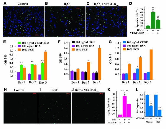
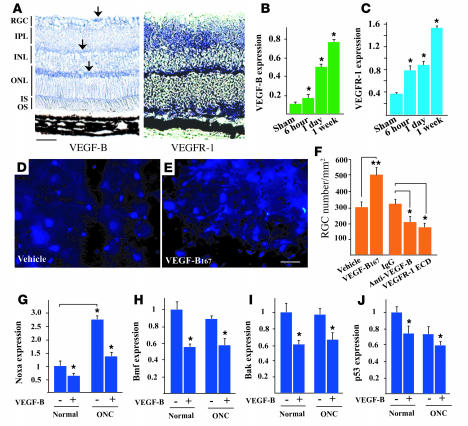
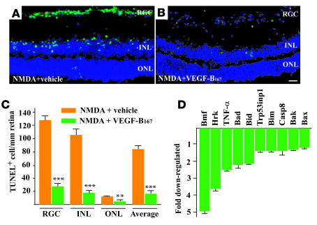
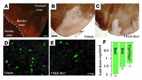
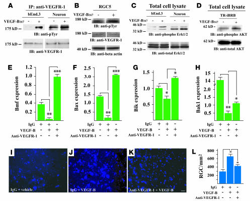
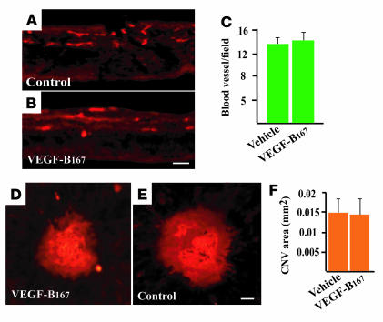
Despite its early discovery and high sequence homology to the other VEGF family members, the biological functions of VEGF-B remain poorly understood. We revealed here a novel function for VEGF-B as a potent inhibitor of apoptosis. Using gene expression profiling of mouse primary aortic smooth muscle cells, and confirming the results by real-time PCR using mouse and rat cell lines, we showed that VEGF-B inhibited the expression of genes encoding the proapoptotic BH3-only proteins and other apoptosis- and cell death-related proteins, including p53 and members of the caspase family, via activation of VEGFR-1. Consistent with this, VEGF-B treatment rescued neurons from apoptosis in the retina and brain in mouse models of ocular neurodegenerative disorders and stroke, respectively. Interestingly, VEGF-B treatment at the dose effective for neuronal survival did not cause retinal neovascularization, suggesting that VEGF-B is the first member of the VEGF family that has a potent antiapoptotic effect while lacking a general angiogenic activity. These findings indicate that VEGF-B may potentially offer a new therapeutic option for the treatment of neurodegenerative diseases.


Figures

Figure 1
Figure 1. VEGF-B167 inhibits the expression of the BH3-only protein and other apoptotic/cell death–related genes in multiple cell lines.
(A) VEGF-B167 treatment inhibited the expression of the BH3-only protein (Bmf, Hrk, Puma, Bid, Bik, Noxa) and other apoptotic/cell death–related genes in the rat RGC-derived cell line RGC5 according to real-time PCR assay. (B) VEGF-B167 treatment inhibited the expression of the BH3-only protein (Hrk, Bik, Bid, Bmf, Noxa) and other apoptotic/cell death–related genes in the immortalized rat retinal pericyte cell line TR-rPCT. (C) VEGF-B167 treatment inhibited the expression of the BH3-only protein (Bmf, Bik, Noxa, Bid, Hrk) and other apoptotic/cell death–related genes in the immortalized rat retinal Müller cell line TR-MUL. (D) VEGF-B167 treatment inhibited the expression of the BH3-only protein (Bik, Bid, Hrk, Noxa, Puma) and other apoptotic/cell death–related genes in the immortalized rat retinal endothelial cell line TR-iBRB. (E) VEGF-B167 treatment inhibited the expression of the BH3-only protein (Bid, Bik, Bbc3, Bad, Hrk, Noxa, Bmf) and other apoptotic/cell death–related genes in mouse primary aortic smooth muscle cells (mSMC).
Figure 2
Figure 2. Survival effect of VEGF-B on the RGC5 cells.
(A and B) H2O2 treatment led to oxidative stress–induced apoptosis in RGC5 cells according to the TUNEL assay. (C and D) VEGF-B167 treatment inhibited the H2O2-induced apoptosis. Scale bar: 50 μm. (E) VEGF-B rescued serum deprivation–induced cell death in RGC5 cells at different time points. (F) PlGF did not rescue serum deprivation–induced cell death in RGC5 cells at different time points. (G) VEGF had a weak survival effect on RGC5 cells. (H and I) Bmf overexpression in the RGC5 cells led to apoptosis according to the TUNEL assay. (J and K) VEGF-B167 treatment inhibited Bmf-induced cellular apoptosis in RGC5 cells. Scale bar: 100 μm. (L) VEGF-B167 treatment inhibited both the endogenous (rat) and exogenous (mouse) Bmf expression in RGC5 cells. *P < 0.05, **P < 0.01.
Figure 3
Figure 3. VEGF-B inhibits axotomy-induced apoptosis in the retina.
(A) VEGF-B is highly expressed in the retina as shown by the in situ hybridization assay. A high level of VEGF-B expression was found primarily in the RGCs and the inner and outer nuclear layers (INL and ONL, arrows). VEGFR-1 expression was mainly found in the inner plexiform layer, part of the inner nuclear layer, and the inner and outer segment layers (IS, OS). Scale bar: 50 μm. (B and C) Real-time PCR assay showed that VEGF-B (B) and VEGFR-1 (C) expression were upregulated in the retinae after ONC injury. The upregulation was seen as early as 6 hours after ONC and reached a high level after 1 week. (DF) A single dose of VEGF-B167 intravitreal treatment increased the number of viable RGCs by about 1.7-fold. VEGF-B neutralizing antibody intravitreal treatment decreased the number of viable RGCs by about 33% (F). VEGFR-1 ECD treatment decreased the number of viable RGCs by about 42% (F). Scale bar: 10 μm. (GJ) Real-time PCR analysis revealed that VEGF-B167 treatment inhibited the expression of the BH3-only protein genes Noxa (G) and Bmf (H), as well as Bak (I) and p53 (J) expression in both normal and ONC-injured retinae. *P < 0.05, **P < 0.01.
Figure 4
Figure 4. VEGF-B inhibits excitotoxin-induced apoptosis in the retina.
(A) NMDA intravitreous treatment led to massive apoptosis in the retina as shown by TUNEL staining. (B and C) VEGF-B167 treatment significantly reduced the number of apoptotic cells in all the 3 layers of the retina. Scale bar: 20 μm. (D) Real-time PCR assay revealed that VEGF-B167 treatment inhibited the expression of the BH3-only protein (Bmf, Hrk, Bad, Bid, Bim) and other apoptotic/cell death–related genes in the NMDA-injured retina. **P < 0.01, ***P < 0.001.
Figure 5
Figure 5. VEGF-B inhibits ischemia-induced neuronal apoptosis and apoptotic gene expression in the brain.
(A) VEGF-B expression is upregulated in the border zone of the brain after MCA occlusion as shown by immunohistochemical staining. Scale bar: 50 μm. (B and C) Recombinant human VEGF-B167 protein treatment decreased brain damage volume by about 32% in the wild-type mice as shown by the MAP-2 staining. Scale bar: 100 μm. (D and E) VEGF-B167 treatment decreased the number of the apoptotic cells in the border zone of the stroke as shown by TUNEL staining. Scale bar: 10 μm. (F) VEGF-B167 treatment inhibited the expression of the BH3-only protein genes Bmf and Hrk and the apoptotic gene Trp53inp1 in the brain with stroke.
Figure 6
Figure 6. VEGFR-1 mediates the effect of VEGF-B.
(A) VEGF-B167 stimulation resulted in VEGFR-1 activation in the bEnd.3 cells and the cortex neurons using the immunoprecipitation assay and anti–VEGFR-1 antibody, followed by the Western blot assay using anti-phosphotyrosine antibody (anti-pTyr). VEGFR-1 was detected in bEnd.3 cells and cortex neurons using Western blot assay. (B) VEGF-B167 induced VEGFR-1 activation in RGC5 cells using Western blot assay and antibodies against phosphotyrosine and VEGFR-1. (C) VEGF-B167 treatment led to ERK1/2 activation in both bEnd.3 cells and cortex neurons using Western blot assay and antibodies against phosphorylated and total ERK1/2. (D) VEGF-B treatment induced Akt phosphorylation in TR-iBRB cells using Western blot assay and antibodies against phosphorylated and total Akt. (EH) VEGFR-1 neutralizing antibody treatment abolished to various degrees the inhibitory effect of VEGF-B on the expression of Bmf (E), Bax (F), Bik (G), and Bak1 (H) in RGC5 cells. (I, J, and L) VEGF-B protein treatment increased RGC survival in the ONC-injured retina. Scale bar: 20 μm. (I, K, and L) VEGFR-1 neutralizing antibody treatment largely abolished the VEGF-B–induced RGC survival in the ONC-injured retina. *P < 0.05, **P < 0.01, ***P < 0.001.
Figure 7
Figure 7. VEGF-B does not affect retinal angiogenesis after intravitreous injection.
(AC) VEGF-B167 treatment did not affect blood vessel density and morphology in the retina using IB4 staining. Scale bar: 50 μm. (DF) VEGF-B167 intravitreous administration after laser treatment did not change the CNV area as shown by IB4 staining. Scale bar: 50 μm.

References

    1. Folkman J. Angiogenesis: an organizing principle for drug discovery? Nat. Rev. Drug Discov. 2007;6:273–286. - PubMed
    1. Ferrara N., Kerbel R.S. Angiogenesis as a therapeutic target. Nature. 2005;438:967–974. - PubMed
    1. Carmeliet P., Jain R.K. Angiogenesis in cancer and other diseases. Nature. 2000;407:249–257. - PubMed
    1. Luttun A., et al. Revascularization of ischemic tissues by PlGF treatment, and inhibition of tumor angiogenesis, arthritis and atherosclerosis by anti-Flt1. Nat. Med. 2002;8:831–840. - PubMed
    1. Alitalo K., Tammela T., Petrova T.V. Lymphangiogenesis in development and human disease. Nature. 2005;438:946–953. - PubMed

Publication types

Substances