

Review
doi: 10.1080/03602530701852917.
Structure-function analyses of single nucleotide polymorphisms in human N-acetyltransferase 1
Affiliations
- PMID: 18259988
- PMCID: PMC2265210
- DOI: 10.1080/03602530701852917
Item in Clipboard
Review
Structure-function analyses of single nucleotide polymorphisms in human N-acetyltransferase 1
Drug Metab Rev.
2008.
Abstract
Human N-acetyltransferase 1 (NAT1) alleles are characterized by one or more single nucleotide polymorphisms (SNPs) associated with rapid and slow acetylation phenotypes. NAT1 both activates and deactivates arylamine drugs and carcinogens, and NAT1 polymorphisms are associated with increased frequencies of many cancers and birth defects. The recently resolved human NAT1 crystal structure was used to evaluate SNPs resulting in the protein substitutions R64W, V149I, R187Q, M205V, S214A, D251V, E261K, and I263V. The analysis enhances knowledge of NAT1 structure-function relationships, important for understanding associations of NAT1 SNPs with genetic predisposition to cancer, birth defects, and other diseases.
Figures
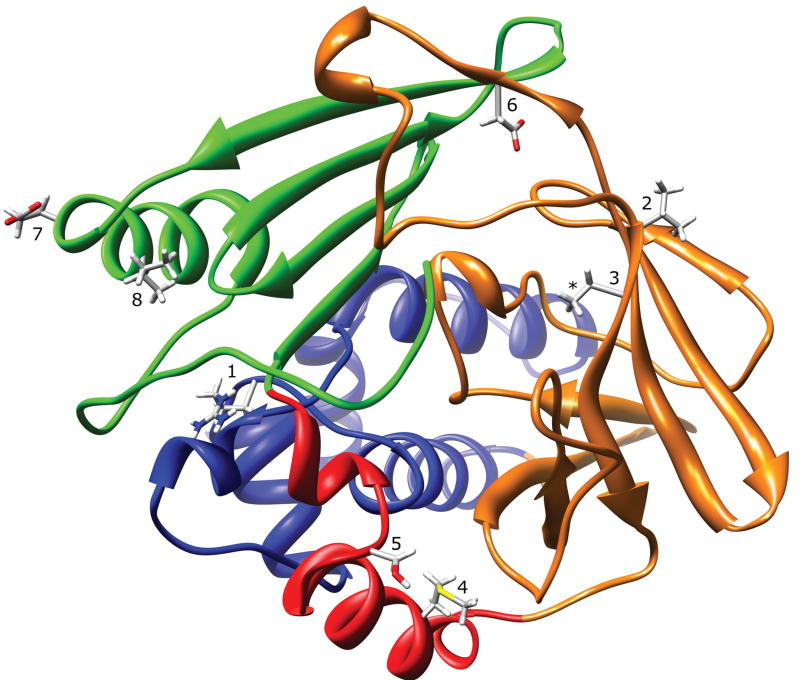
NAT1 Crystal Structure (2PQT) Ribbon Diagram. The ribbon is colored to indicate N-acetyltransferase protein domain I (blue), the interdomain region (red), domain II (orange), and domain III (green). The location of residues R64 (1), V149 (2), R187 (3), M205 (4), S214 (5), D251 (6), E261 (7), and I263 (8) are shown. The 2PQT PDB file is missing coordinates for the arginine side-chain guanidine group at residue R187 (*).
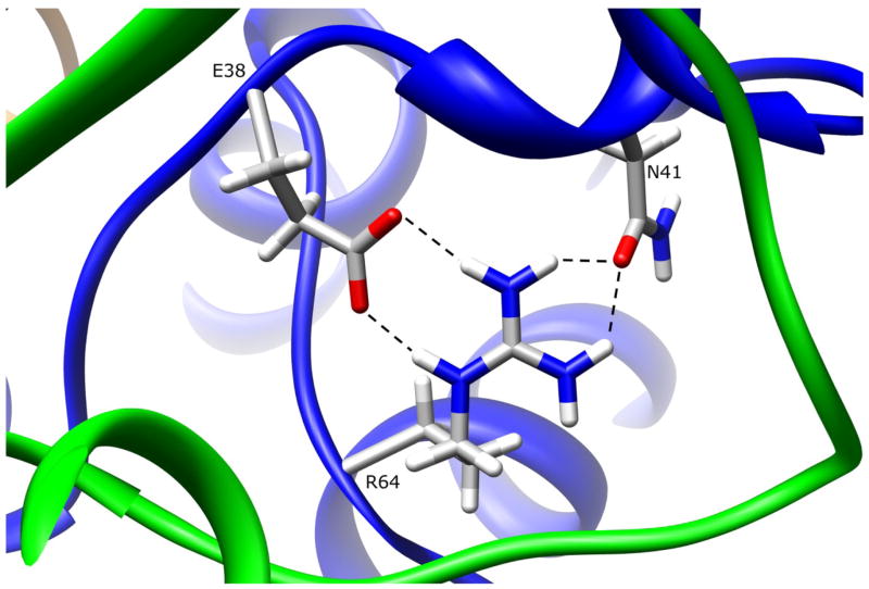
NAT1 SNP Residue Arginine 64. Residue R64 is partially surface exposed in domain I. The arginine side-chain makes multiple hydrogen bonds to E38 and N41, both of which are also in domain I. These interactions are lost when R64 is mutated to tryptophan.
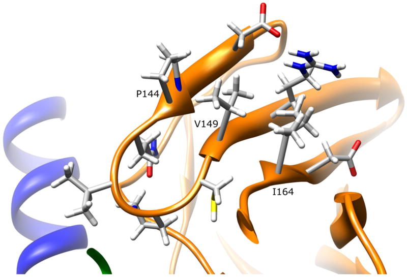
NAT1 SNP Residue Valine 149. Residue V149 is partially surface exposed on the domain II beta barrel. The valine side-chain has no apparent interactions with other residues. Mutating this residue to isoleucine is not expected to influence enzyme stability or function.
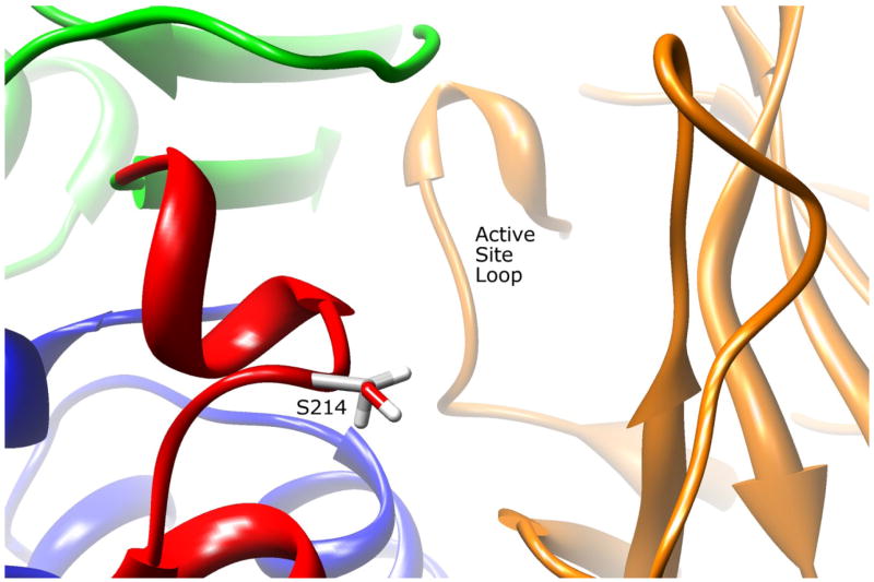
NAT2 SNP Residue Serine 214. Residue S214 is located adjacent to the active site pocket opening in the interdomain region. The serine side-chain has no apparent interactions with other residues. Mutating this residue to an alanine could influence AcCoA binding efficiency.
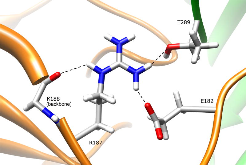
NAT1 SNP Residue Arginine 187. Residue R187 is located on the domain II beta barrel, and is
partially surface exposed both on the protein surface and the active site pocket. The arginine side-chain hydrogen bonds to E182 and K188, both in domain II, and to the domain III C-terminal residue T289. Mutating this residue to a glutamine results in loss of these interactions and could change the size/shape of the active site pocket.
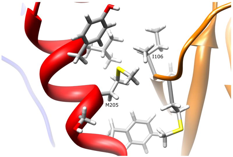
NAT1 SNP Residue Methionine 205. Residue M205 is partially surface exposed on the interdomain region helix. The methionine side-chain has no apparent interactions with other residues. Mutating M205 to a valine residue is not expected to influence enzyme stability or function.
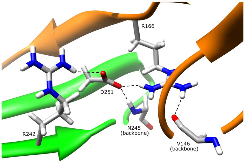
NAT1 SNP Residue Aspartate 251. Residue D251 is primarily in the protein core on the domain III
beta sheet. The aspartate side-chain hydrogen bonds to R242 and the backbone of N245 in domain III. The D251 side-chain also hydrogen bonds to the domain II loop residue R166, an interaction which may stabilize R166 hydrogen bonding interaction with domain II beta barrel residue V146. These interactions are lost when D251 is mutated to a valine residue.
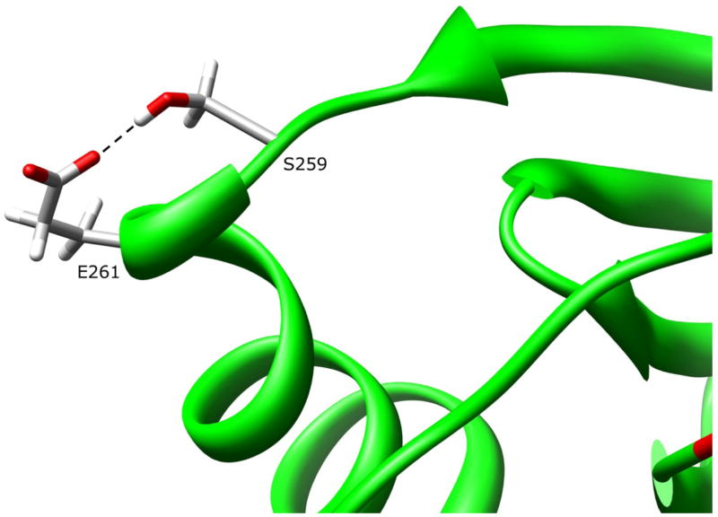
NAT1 SNP Residue Glutamate 261. Residue E261 is surface exposed on the domain III helix. The glutamate side-chain hydrogen bonds to S259. Mutating E261 to a lysine residue is not expected to influence enzyme stability or function.
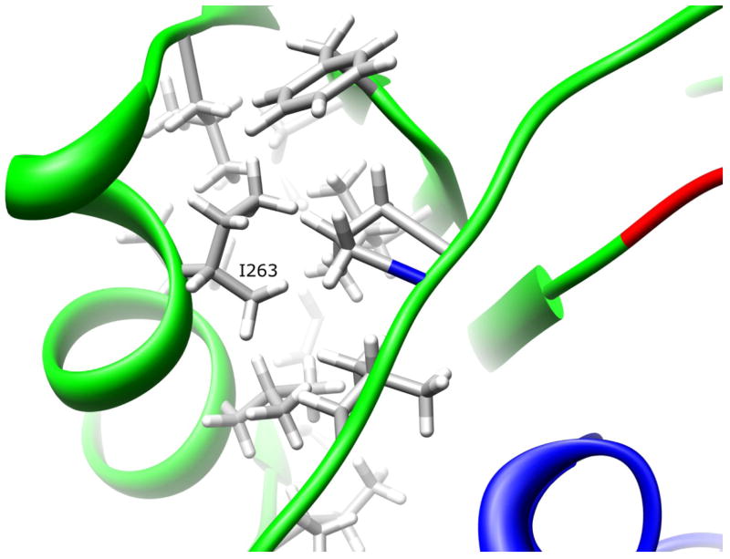
NAT1 SNP Residue Isoleucine 263. Residue I263 is located on the domain III helix. The isoleucine side-chain is part of a hydrophobic core that is partially responsible for the domain III tertiary structure. Mutating I263 to a valine residue is not expected to influence enzyme stability of function.
References
-
- Adam PJ, Berry J, Loader JA, Tyson KL, Craggs G, Smith P, De Belin J, Steers G, Pezzella F, Sachsenmeir KF, Stamps AC, Herath A, Sim E, O’Hare MJ, Harris AL, Terrett JA. Arylamine N-acetyltransferase-1 is highly expressed in breast cancers and conveys enhanced growth and resistance to etoposide in vitro. Mol Cancer Res. 2003;1:826–835. - PubMed
-
- Ambrosone CB, Abrams SM, Gorlewska-Roberts K, Kadlubar FF. Hair dye use, meat intake, and tobacco exposure and presence of carcinogen-DNA adducts in exfoliated breast ductal epithelial cells. Arch Biochem Biophys. 2007;464:169–175. - PubMed
-
- Bell DA, Stephens EA, Castranio T, Umbach DM, Watson M, Deakin M, Elder J, Hendrickse C, Duncan H, Strange RC. Polyadenylation polymorphism in the acetyltransferase 1 gene (NAT1) increases risk of colorectal cancer. Cancer Res. 1995;55:3537–3542. - PubMed
-
- Bieche I, Girault I, Urbain E, Tozlu S, Lidereau R. Relationship between intratumoral expression of genes coding for xenobiotic-metabolizing enzymes and benefit from adjuvant tamoxifen in estrogen receptor alpha-positive postmenopausal breast carcinoma. Breast Cancer Res. 2004;6:252–264. - PMC - PubMed
-
- Boukouvala S, Fakis G. Arylamine N-acetyltransferases: what we learn from genes and genomes. Drug Metab Rev. 2005;37:511–564. - PubMed
Publication types
MeSH terms
Substances
Grants and funding
LinkOut - more resources
Full Text Sources