. 2008 Mar;19(3):686-92.
doi: 10.1021/bc700376v. Epub 2008 Feb 9.

Noninvasive optical imaging of staphylococcus aureus bacterial infection in living mice using a Bis-dipicolylamine-Zinc(II) affinity group conjugated to a near-infrared fluorophore

Affiliations

Noninvasive optical imaging of staphylococcus aureus bacterial infection in living mice using a Bis-dipicolylamine-Zinc(II) affinity group conjugated to a near-infrared fluorophore

W Matthew Leevy et al. Bioconjug Chem. 2008 Mar.

Abstract

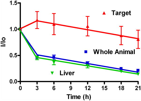
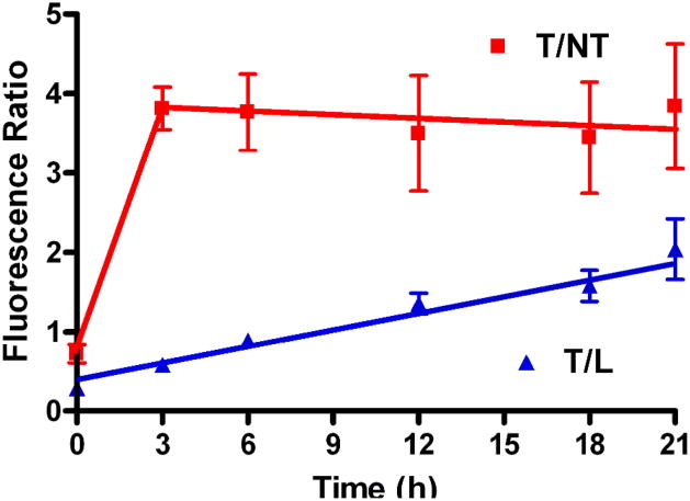
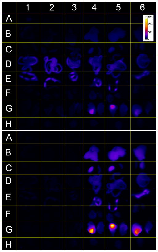
Optical imaging of bacterial infection in living animals is usually conducted with genetic reporters such as light-emitting enzymes or fluorescent proteins. However, there are many circumstances where genetic reporters are not applicable, and there is a need for exogenous synthetic probes that can selectively target bacteria. The focus of this study is a fluorescent imaging probe that is composed of a bacterial affinity group conjugated to a near-infrared dye. The affinity group is a synthetic zinc (II) coordination complex that targets the anionic surfaces of bacterial cells. The probe allows detection of Staphylococcus aureus infection (5 x 10 (7) cells) in a mouse leg infection model using whole animal near-infrared fluorescence imaging. Region of interest analysis showed that the signal ratio for infected leg to uninfected leg reaches 3.9 +/- 0.5 at 21 h postinjection of the probe. Ex vivo imaging of the organs produced a signal ratio of 8 for infected to uninfected leg. Immunohistochemical analysis confirmed that the probe targeted the bacterial cells in the infected tissue. Optimization of the imaging filter set lowered the background signal due to autofluorescence and substantially improved imaging contrast. The study shows that near-infrared molecular probes are amenable to noninvasive optical imaging of localized S. aureus infection.


Figures

Figure 1
Figure 1
Optical images of a mouse with a S. aureus infection in the left posterior leg. Images were acquired following intravenous injection of probe 1 (75 μL of a 1 mM stock) at 0 h (A), 3 h (B), 6 h (C), 12 h (D), 18 h (E), and 21 h (F). Calibration bar (a.u.) applies to all images in the montage. Frame A shows labels for each region of interest (ROI), Target (T), Non-Target (NT) and Liver (L).
Figure 2
Figure 2
Target to non-target (T/NT) and target to liver (T/L) fluorescence ratios at each imaging time point (n=4, error bars are standard error of the mean). The lines are provided to guide the eye.
Figure 3
Figure 3
Fluorescence emission time course for the infection target, whole animal, and liver ROIs (n=4, error bars are standard error of the mean). The lines are provided to guide the eye.
Figure 4
Figure 4
Optical images of a mouse with a S. aureus infection in the left posterior leg. Images were acquired following intravenous injection of a low dose of probe 1 (75 μL of a 0.2 mM stock) at 0 h (A), 3 h (B), 6 h (C), 12 h (D), 18 h (E), and 21 h (F). Calibration bar (a.u.) applies to all images in the montage.
Figure 5
Figure 5
Target to non-target (T/NT) and target to liver (T/L) fluorescence ratios at each imaging time point after injection with a 0.2 mM stock of 1 (n=4, error bars are standard error of the mean).
Figure 6
Figure 6
Optical images of four mice each with a S. aureus infection in the left posterior leg and acquired 21 h post injection of probe 1 (75 μL of a 1 mM stock). Left column: animals were imaged using an excitation of 755 nm and emission of 830 nm. Right column: the same animals were imaged using an excitation of 720 nm and emission of 790 nm. Calibration bar (a.u.) applies to all images in the montage.
Figure 7
Figure 7
Ex vivo image of mouse tissues from three uninfected mice (columns 1–3) and three infected mice that were injected with 1 (colums 4–6). The organs and blood are given in the following rows of Figure 5: (A) Blood, (B) Liver, (C) Kidney (left) and Spleen (right), (D) Small Intestine, (E) Large Intestine, (F) Lungs (left) and Heart (right), (G) Left and Right posterior leg tissue, and (H) Brain. Images were acquired with the 720/790 nm (top frame) and 755/830 nm (bottom) filter sets. Calibration bar (a.u.) set for both frames.
Figure 8
Figure 8
Ex vivo organ quantification of Figure 7 imaged with the 755/830 nm filter set (n=3, error bars are the standard error of the mean).
Figure 9
Figure 9
Histological section of infected leg muscle as viewed at 600X in the phase contrast (left), red (center) and NIR Cy7 (right) filter sets. The red box at left designates a patch of spots that mark the presence of bacteria. Infected tissue was sectioned 21 h post injection of 1. Scale bar equals 20 μM for each frame. Calibration bar is in arbitrary units.

References

    1. Leevy WM, Serazin N, Smith BD. Optical imaging of bacterial infection models. Drug Discovery Today: Disease Models. 2007 doi: 10.1016/j.ddmod.2007.07.001. - DOI - PMC - PubMed
    1. Hirose K, Marui A, Nomura T, Kaneda K, Kimura Y, Ikeda T, Fujita M, Mitsuyama M, Tabata Y, Komeda M. A novel approach to reduce catheter-related infection using sustained-release basic fibroblast growth factor for tissue regeneration in mice. Heart Vessels. 2007;22:261–267. - PubMed
    1. Embleton ML, Nair SP, Cookson BD, Wilson M. Selective lethal photosensitization of methicillin-resistand Staphylococcus aureus using a tin(IV) chlorin e6 conjugate. J Antimicrob Chemo. 2002;50:857–864. - PubMed
    1. Holm C, Jespersen L. A flow-cytometric gram-staining technique for milk-associated bacteria. Appl Environ Micro. 2003;69:2857–2863. - PMC - PubMed
    1. Qu LW, Luo PG, Taylor S, Lin Y, Huang WJ, Anyadike N, Tzeng TRJ, Stutzenberger F, Latour RA, Sun YPJ. Visualizing adhesion-induced agglutination of Escherichia coli with mannosylated nanoparticles. Nanosci Nanotechnol. 2005;5:319–322. - PubMed

Publication types

MeSH terms