. 2008 Feb 8:5:19.
doi: 10.1186/1742-4690-5-19.

Apoptosis resistance in HIV-1 persistently-infected cells is independent of active viral replication and involves modulation of the apoptotic mitochondrial pathway

Affiliations

Apoptosis resistance in HIV-1 persistently-infected cells is independent of active viral replication and involves modulation of the apoptotic mitochondrial pathway

Pablo N Fernández Larrosa et al. Retrovirology. .

Abstract

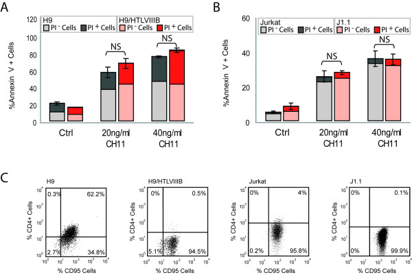
Background: HIV triggers the decline of CD4+ T cells and leads to progressive dysfunction of cell-mediated immunity. Although an increased susceptibility to cell death occurs during the acute phase of HIV infection, persistently-infected macrophages and quiescent T-cells seem to be resistant to cell death, representing a potential reservoir for virus production.

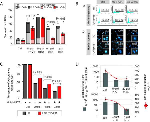
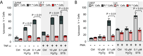
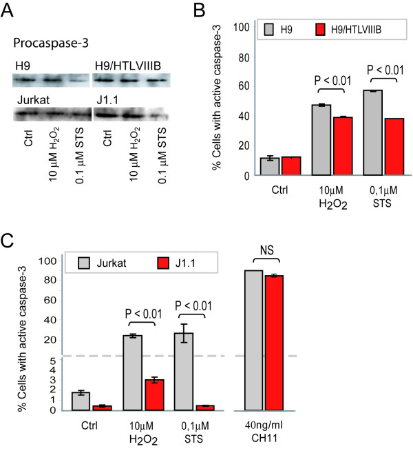
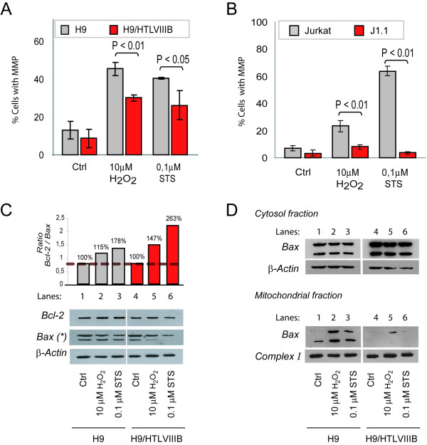
Results: Lymphoid (H9/HTLVIIIB and J1.1) and pro-monocytic (U1) HIV-1 persistently-infected cell lines were treated with hydrogen peroxide (H2O2) and staurosporine (STS) for 24 h, and susceptibility to apoptosis was evaluated and compared with uninfected counterparts (H9, Jurkat and U937 respectively). When exposed to different pro-apoptotic stimuli, all persistently-infected cell lines showed a dramatic reduction in the frequency of apoptotic cells in comparison with uninfected cells. This effect was independent of the magnitude of viral replication, since the induction of viral production in lymphoid or pro-monocytic cells by exposure to TNF-alpha or PMA did not significantly change their susceptibility to H2O2- or STS-induced cell death. A mechanistic analysis revealed significant diferences in mitochondrial membrane potential (MMP) and caspase-3 activation between uninfected and persistently-infected cells. In addition, Western blot assays showed a dramatic reduction of the levels of pro-apototic Bax in mitochondria of persistently-infected cells treated with H2O2 or STS, but not in uninfected cells.

Conclusion: This study represents the first evidence showing that resistance to apoptosis in persistently-infected lymphoid and monocytic cells is independent of active viral production and involves modulation of the mitochondrial pathway. Understanding this effect is critical to specifically target the persistence of viral reservoirs, and provide insights for future therapeutic strategies in order to promote complete viral eradication.


Figures

Figure 1
Figure 1
Apoptosis resistance in HIV persistently-infected H9/HTLVIIIBcells in comparison with non-infected H9 cells. A) H9 and H9/HTLVIIIB cells were treated with different concentrations of H2O2 or STS or complete medium as control. After 24 h, cells were harvested and annexin-V/PI staining was performed. The percentages of annexin-V+, PI- or PI+ cells are shown. B) (a) Analysis by APO-BrdU labeling by flow cytometry. The corresponding histograms and the percentages of APO-BrdU+ cells are shown; (b) Analysis of apoptosis with Hoechst 33324 by fluorescence microscopy. Micrographs (100×) of predominant Hoechst stained nuclei are depicted. C) H9 and H9/HTLVIIIB cells were treated with 0.1 μM STS or complete medium as control, and cells were harvested 24, 48 and 72 h post-treatment. Cell viabillity was analyzed by the MTT assay. Absorbances from treated samples were normalized to 100% of untreated controls. D) Cells treated with H2O2 or STS or complete medium for 24 h were pelleted and the supernatants were used to quantify infective viral (grey bar) and p24 antigen (red line) production.
Figure 2
Figure 2
Apoptosis resistance is independent of the magnitude of viral replication. A) Jurkat and J1.1 cells were incubated in the presence or absence of 1000 U/ml TNF-α for 48 h, and then treated with 10 μM H2O2 or 0.1 μM STS. The percentages of annexin-V+, PI- or PI+ cells are shown. B) U937 and U1 cells were incubated in the presence or absence of 100 ng/ml PMA for 24 h, and then exposed to 10 μM H2O2 or 0.1 μM STS. The percentages of annexin-V+, PI- or PI+ cells are shown.
Figure 3
Figure 3
Fas-mediated apoptosis in uninfected and HIV persistently-infected cells. H9 and H9/HTLVIIIB (A) and Jurkat and J1.1 (B) cells were incubated with 20 g/ml or 40 ng/ml of CH11, an anti-Fas activating antibody. After 24 h, cells were washed and stained with annexin-V/PI. The percentages of annexin-V+, PI- or PI+ cells are shown. C) Fas/CD95 and CD4 cell surface expression was analyzed by flow cytometry on H9, H9/HTLVIIIB, Jurkat and J1.1 cells.
Figure 4
Figure 4
Caspase-3 activation by H2O2 and STS treatment in uninfected and HIV persistently-infected lymphoid cell lines. A) H9, H9/HTLVIIIB, Jurkat and J1.1 cells were exposed to H2O2 and STS. After 24 h, cells were washed and lysed with RIPA buffer. Equal amounts of protein (30 μg/sample) were separated on a 10% SDS-PAGE and blotted onto nitrocellulose membranes. Blots were probed with anti-procaspase-3 for 1 h and revealed with a peroxidase-conjugated anti-IgG antibody and ECL (enhanced chemoluminiscence) Equal loading was checked by analyzing β-actin expression (data not shown). B) H9 and H9/HTLVIIIB cells were exposed to 10μM H2O2, 0.1 μM STS or complete medium for 24 h and collected to evaluate active caspase-3 by PE-conjugated monoclonal anti-active caspase-3 antibody by flow cytometry. C) Jurkat and J1.1 cells were exposed to 10 μM H2O2, 0.1 μM STS, 40 ng/ml CH11 or complete medium for 24 h and collected to evaluate active caspase-3 by PE-conjugated monoclonal anti-active caspase-3 antibody by flow cytometry as described in Materials and Methods
Figure 5
Figure 5
MMP induction and Bcl-2 and Bax expression in uninfected or HIV persistently-infected cell lines. H9 and H9/HTLVIIIB (A) and Jurkat and J1.1 (B) cells were exposed to 10 μM H2O2, 0.1 μM STS or complete medium for 24 h and harvested to evaluate mitochondrial membrane potential (ΔΨm) by JC-1 staining by flow cytometry. C) H9 and H9/HTLVIIIB cells were exposed to H2O2 and STS. After 24 h, cells were washed and lysed with RIPA buffer. Equal amounts of protein (30 μg/sample) were separated by 10% SDS-PAGE and blotted onto nitrocellulose membranes. The blots were probed with anti-Bcl-2 and anti-Bax antibody, revealed using a peroxidase-conjugated anti-IgG and developed using a chemiluminiscence Western blotting detection reagent. Equal loading was checked by analyzing β-actin expression. Films were analyzed with Scion image analysis software (Scion, Frederick, MD) and the Bcl-2/Bax ratio was depicted. D) H9 and H9/HTLVIIIB cells were exposed to H2O2 and STS and after 24 h lysates from cytosolic and mitochondrial fractions were prepared by differential centrifugation. Equal amounts of protein (30 μg/sample) were separated by 10% SDS-PAGE and blotted onto nitrocellulose membranes. Blots were then probed with an anti-Bax polyclonal antibody, incubated with a peroxidase-conjugated anti-rabbit secondary antibody and developed using ECL detection reagent. Equal loading was checked by analyzing β-actin (cytosol fraction) and Complex I (mitochondrial fraction) expression.

References

    1. Benedict CA, Norris PS, Ware CF. To kill or be killed: viral evasion of apoptosis. Nature immunology. 2002;3:1013–1018. doi: 10.1038/ni1102-1013. - DOI - PubMed
    1. He B. Viruses, endoplasmic reticulum stress, and interferon responses. Cell death and differentiation. 2006;13:393–403. doi: 10.1038/sj.cdd.4401833. - DOI - PubMed
    1. Brenchley JM, Price DA, Douek DC. HIV disease: fallout from a mucosal catastrophe? Nature immunology. 2006;7:235–239. doi: 10.1038/ni1316. - DOI - PubMed
    1. Hel Z, McGhee JR, Mestecky J. HIV infection: first battle decides the war. Trends in immunology. 2006;27:274–281. doi: 10.1016/j.it.2006.04.007. - DOI - PubMed
    1. Gougeon ML. To kill or be killed: how HIV exhausts the immune system. Cell death and differentiation. 2005;12 Suppl 1:845–854. doi: 10.1038/sj.cdd.4401616. - DOI - PubMed

Publication types

MeSH terms