

A novel feature-tracking echocardiographic method for the quantitation of regional myocardial function: validation in an animal model of ischemia-reperfusion
- PMID: 18261685
- PMCID: PMC3348770
- DOI: 10.1016/j.jacc.2007.10.029
A novel feature-tracking echocardiographic method for the quantitation of regional myocardial function: validation in an animal model of ischemia-reperfusion
Abstract
Objectives: The aim of this study was to validate a novel, angle-independent, feature-tracking method for the echocardiographic quantitation of regional function.
Background: A new echocardiographic method, Velocity Vector Imaging (VVI) (syngo Velocity Vector Imaging technology, Siemens Medical Solutions, Ultrasound Division, Mountain View, California), has been introduced, based on feature tracking-incorporating speckle and endocardial border tracking, that allows the quantitation of endocardial strain, strain rate (SR), and velocity.
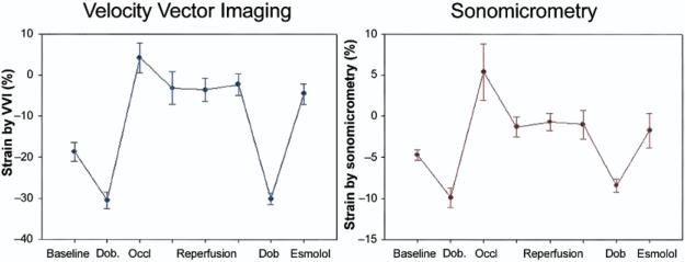
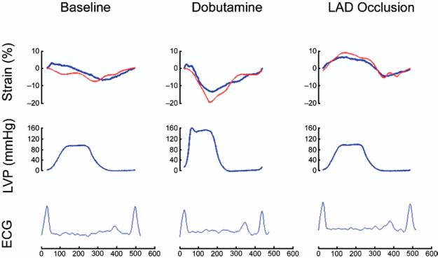
Methods: Seven dogs were studied during baseline, and various interventions causing alterations in regional function: dobutamine, 5-min coronary occlusion with reperfusion up to 1 h, followed by dobutamine and esmolol infusions. Echocardiographic images were acquired from short- and long-axis views of the left ventricle. Segment-length sonomicrometry crystals were used as the reference method.
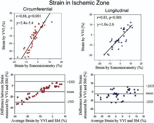
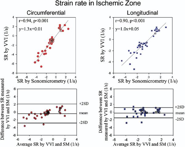
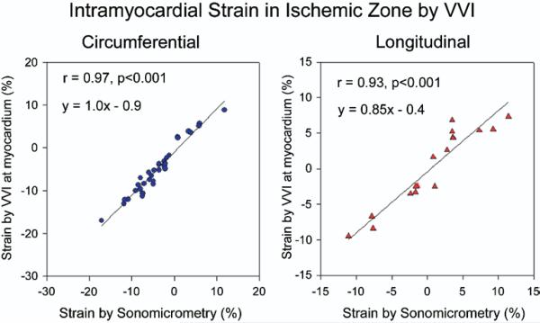
Results: Changes in systolic strain in ischemic segments were tracked well with VVI during the different states of regional function. There was a good correlation between circumferential and longitudinal systolic strain by VVI and sonomicrometry (r = 0.88 and r = 0.83, respectively, p < 0.001). Strain measurements in the nonischemic basal segments also demonstrated a significant correlation between the 2 methods (r = 0.65, p < 0.001). Similarly, a significant relation was observed for circumferential and longitudinal SR between the 2 methods (r = 0.94, p < 0.001 and r = 0.90, p < 0.001, respectively). The endocardial velocity relation to changes in strain by sonomicrometry was weaker owing to significant cardiac translation.
Conclusions: Velocity Vector Imaging, a new feature-tracking method, can accurately assess regional myocardial function at the endocardial level and is a promising clinical tool for the simultaneous quantification of regional and global myocardial function.
Figures


References
-
- Sutherland GR, Di Salvo SG, Claus P, D'hooge J, Bijnens B. Strain and strain rate imaging: a new clinical approach to quantifying regional myocardial function. J Am Soc Echocardiogr. 2004;17:788–802. - PubMed
-
- Greenberg NL, Firstenberg MS, Castro PL, et al. Doppler-derived myocardial systolic strain rate is a strong index of left ventricular contractility. Circulation. 2002;105:99–105. - PubMed
-
- Urheim S, Edvardsen T, Torp H, Angelsen B, Smiseth OA. Myocardial strain by Doppler echocardiography. Validation of a new method to quantify regional myocardial function. Circulation. 2000;102:1158–64. - PubMed
-
- Hanley HG, Lewis RM, Hartley CJ, Franklin D, Schwartz A. Effects of an inotropic agent, RO 2-2985 (X-537A), on regional blood flow and myocardial function in chronically instrumented conscious dogs and anesthetized dogs. Circ Res. 1975;37:215–25. - PubMed
-
- Pinsky WW, Lewis RM, Hartley CJ, Entman ML. Permanent changes of ventricular contractility and compliance in chronic volume overload. Am J Physiol. 1979;237:H575–83. - PubMed
Publication types
MeSH terms
Substances
Grants and funding
LinkOut - more resources
Full Text Sources
Other Literature Sources
Research Materials
