

Small GTPase protein Rac-1 is activated with maturation and regulates cell morphology and function in chondrocytes
- PMID: 18261726
- PMCID: PMC2288527
- DOI: 10.1016/j.yexcr.2007.12.029
Small GTPase protein Rac-1 is activated with maturation and regulates cell morphology and function in chondrocytes
Abstract
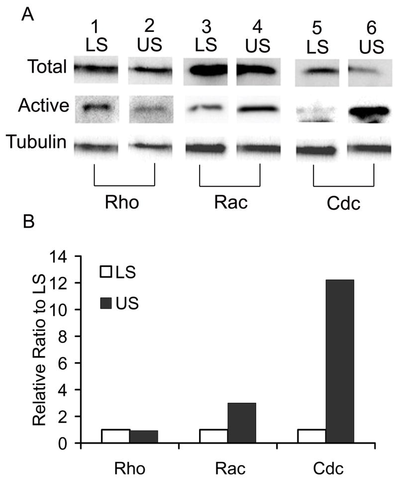
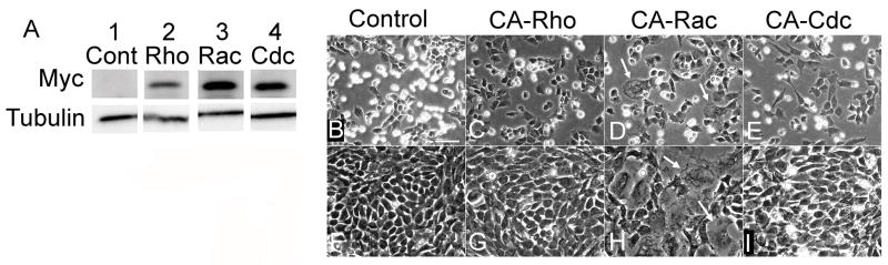
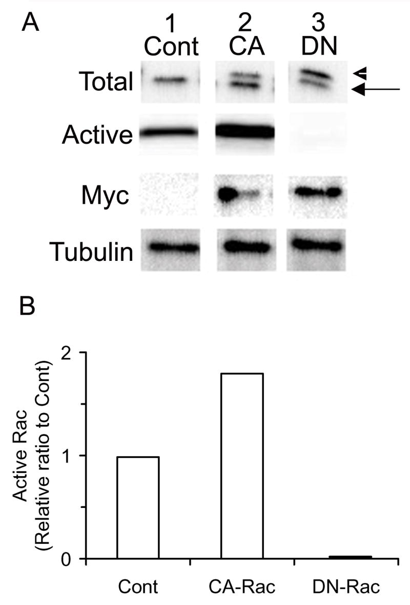
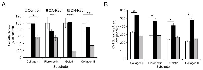
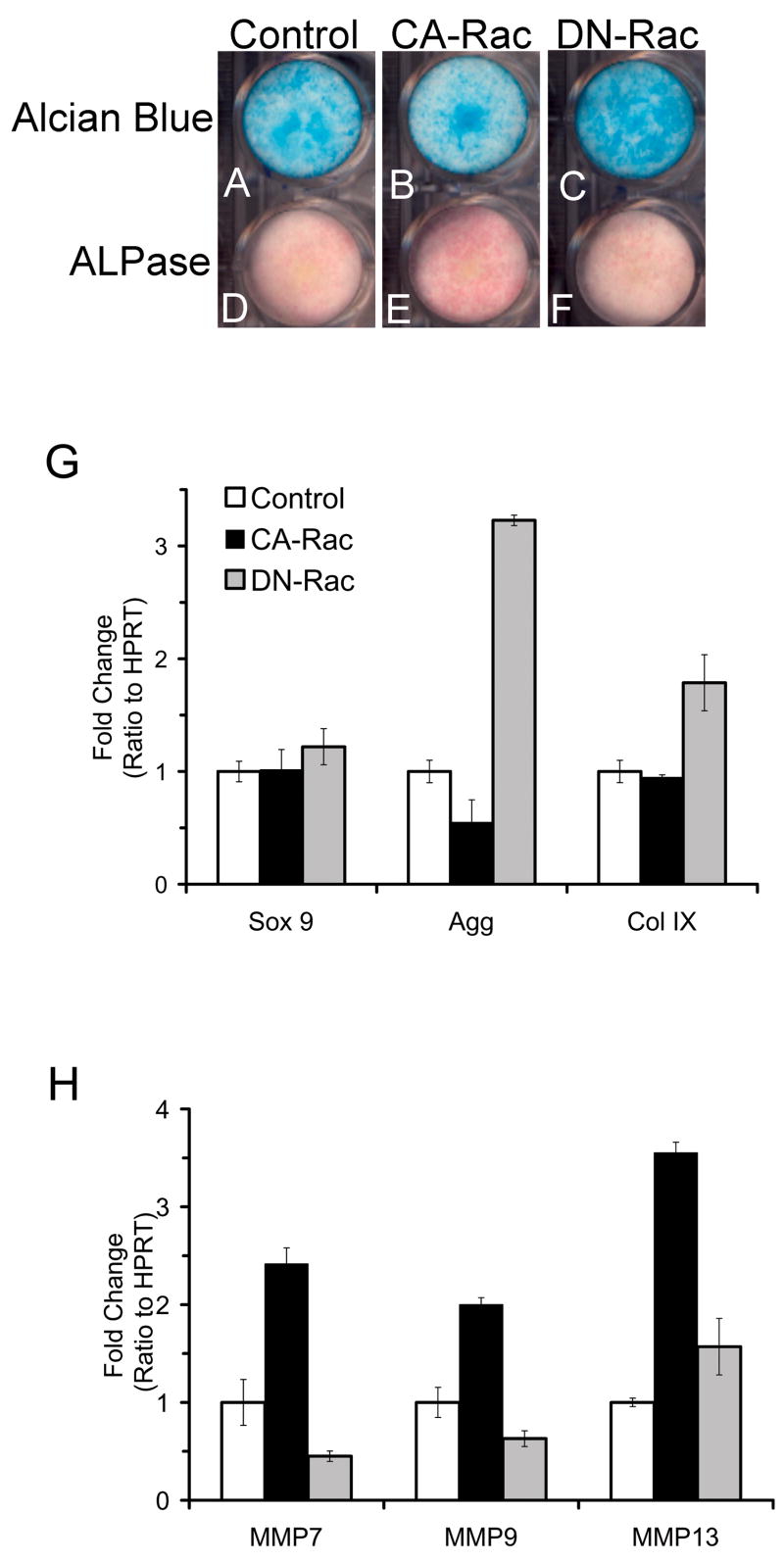
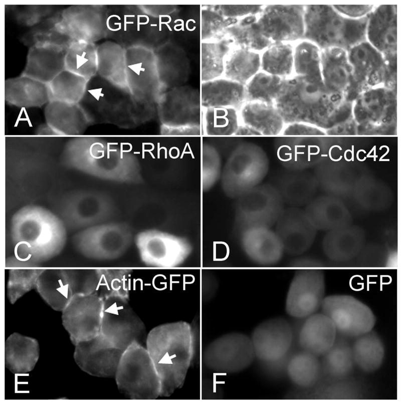
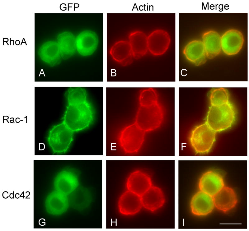
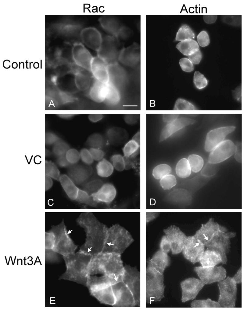
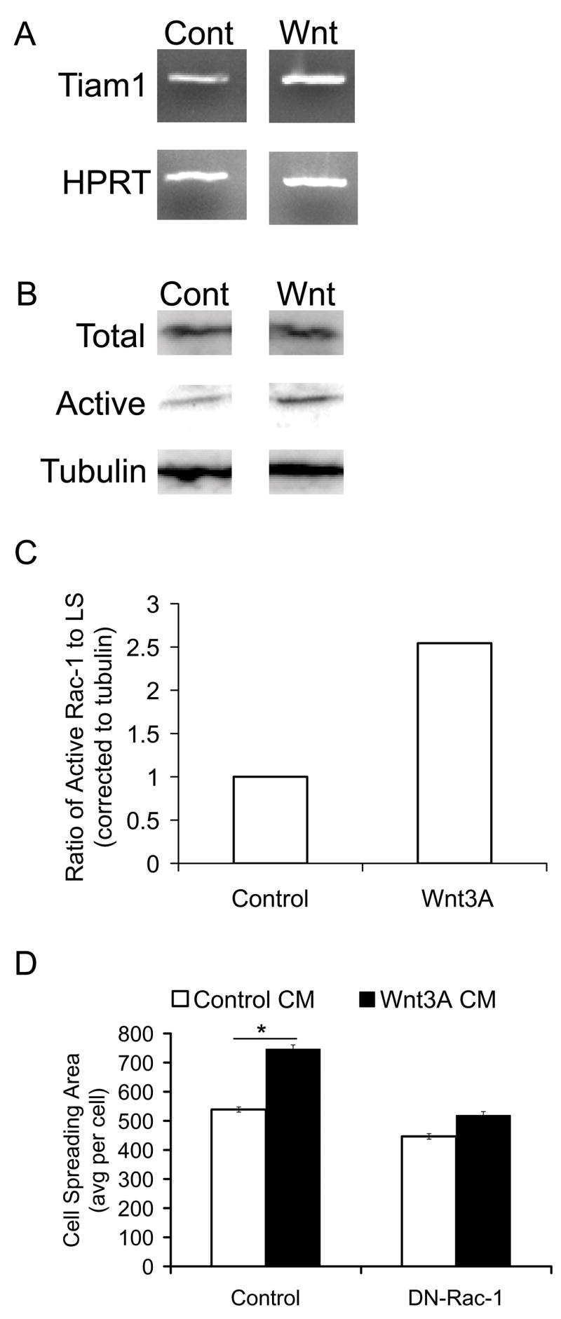
During maturation, chondrocytes undergo changes in morphology, matrix production, and gene expression; however, it remains unclear whether these are interrelated. In this study, we examined whether Rho GTPases were involved in these regulatory interplays. Levels of active Rho GTPases were assayed in immature and mature primary chondrocytes. We found that activation of Rac-1 and Cdc42 increased with maturation, whereas RhoA levels remained unchanged. GFP-tagged Rho GTPases tracked cellular localization. Rac-1 was enriched at the cell membrane where it co-localized with cortical actin, while RhoA and Cdc42 were cytoplasmic. To test the roles of Rac-1 in chondrocyte maturation, we force-expressed constitutively active or dominant negative forms of Rac-1 and assessed phenotypic consequences in primary chondrocytes. Activated Rac-1 expression induced chondrocyte enlargement and increased matrix metalloproteinase expression, which are characteristic of mature chondrocytes. Conversely, Rac-1 inactivation diminished adhesion, decreased alkaline phosphatase activity, and stimulated functions typical of immature chondrocytes. Exposure to a pro-maturation factor, Wnt3A, induced a flattened and enlarged morphology accompanied by peripheral Rac-1 re-arrangement. Wnt3A stimulated Tiam1 expression and Rac-1 activation, while DN-Rac-1 inhibited Wnt3A-induced cell spreading. Our data provide strong evidence that Rac-1 coordinates changes in chondrocyte phenotype and function and stimulates the maturation process essential for skeletal development.
Figures
Similar articles
-
Rac1/Cdc42 and RhoA GTPases antagonistically regulate chondrocyte proliferation, hypertrophy, and apoptosis.J Bone Miner Res. 2005 Jun;20(6):1022-31. doi: 10.1359/JBMR.050113. Epub 2005 Jan 31. J Bone Miner Res. 2005. PMID: 15883643
-
Mechanisms of guanine nucleotide exchange and Rac-mediated signaling revealed by a dominant negative trio mutant.J Biol Chem. 2004 Jan 30;279(5):3777-86. doi: 10.1074/jbc.M308282200. Epub 2003 Nov 3. J Biol Chem. 2004. PMID: 14597635
-
Heterotrimeric G protein betagamma subunits stimulate FLJ00018, a guanine nucleotide exchange factor for Rac1 and Cdc42.J Biol Chem. 2008 Jan 25;283(4):1946-53. doi: 10.1074/jbc.M707037200. Epub 2007 Nov 28. J Biol Chem. 2008. PMID: 18045877
-
Functions of Rho family of small GTPases and Rho-associated coiled-coil kinases in bone cells during differentiation and mineralization.Biochim Biophys Acta Gen Subj. 2017 May;1861(5 Pt A):1009-1023. doi: 10.1016/j.bbagen.2017.02.005. Epub 2017 Feb 8. Biochim Biophys Acta Gen Subj. 2017. PMID: 28188861 Review.
-
Regulation of phosphorylation pathways by p21 GTPases. The p21 Ras-related Rho subfamily and its role in phosphorylation signalling pathways.Eur J Biochem. 1996 Dec 1;242(2):171-85. doi: 10.1111/j.1432-1033.1996.0171r.x. Eur J Biochem. 1996. PMID: 8973630 Review.
Cited by
-
Rac1 is required for matrix metalloproteinase 13 production by chondrocytes in response to fibronectin fragments.Arthritis Rheum. 2013 Jun;65(6):1561-8. doi: 10.1002/art.37922. Arthritis Rheum. 2013. PMID: 23460186 Free PMC article.
-
Wnt and Rho GTPase signaling in osteoarthritis development and intervention: implications for diagnosis and therapy.Arthritis Res Ther. 2013 Jul 11;15(4):217. doi: 10.1186/ar4240. Arthritis Res Ther. 2013. PMID: 23856044 Free PMC article. Review.
-
Kindlin-3 mutation in mesenchymal stem cells results in enhanced chondrogenesis.Exp Cell Res. 2021 Feb 15;399(2):112456. doi: 10.1016/j.yexcr.2020.112456. Epub 2021 Jan 5. Exp Cell Res. 2021. PMID: 33417921 Free PMC article.
-
Increased CHST15 follows decline in arylsulfatase B (ARSB) and disinhibition of non-canonical WNT signaling: potential impact on epithelial and mesenchymal identity.Oncotarget. 2020 Jun 16;11(24):2327-2344. doi: 10.18632/oncotarget.27634. eCollection 2020 Jun 16. Oncotarget. 2020. PMID: 32595831 Free PMC article.
-
MicroRNA-140 Provides Robustness to the Regulation of Hypertrophic Chondrocyte Differentiation by the PTHrP-HDAC4 Pathway.J Bone Miner Res. 2015 Jun;30(6):1044-52. doi: 10.1002/jbmr.2438. J Bone Miner Res. 2015. PMID: 25529628 Free PMC article.
References
-
- Rosso SB, Sussman D, Wynshaw-Boris A, Salinas PC. Wnt Signaling Through Dishevelled, Rac and JNK Regulates Dendritic Development. Nature Neuroscience. 2005;8:34–42. - PubMed
-
- Poole AR, Kojima T, Yasuda T, Mwale F, Kobayashi M, Laverty S. Composition and Structure of Articular Cartilage. Clinical Orthopaedics and Related Research. 2001;391S:S26–33. - PubMed
-
- CE Farnum, WNJ . Growth Plate Cellular Function. In: Buckwalter J, Ehrlich M, Sandell L, Trippel S, editors. Skeletal Morphogenesis and Growth. American Academy of Orthopaedic Surgeons; Rosemont, IL: 1998. pp. 203–223.
-
- Lefebvre V, Smits P. Transcriptional Control of Chondrocyte Fate and Differentiation. Birth Defects Research (Part C) 2005;75:200–212. - PubMed
-
- Langelier E, Suetterlin R, Hoemann CD, Aebi U, Buschmann MD. The Chondrocyte Cytoskeleton in Mature Articular Cartilage: Structure and Distribution of Actin, Tubulin and Vimentin Filaments. The Journal of Histochemistry and Cytochemistry. 2000;48:1307–1320. - PubMed
Publication types
MeSH terms
Substances
Grants and funding
LinkOut - more resources
Full Text Sources
Research Materials
Miscellaneous
