Review
. 2008 Feb;18(1):106-11.
doi: 10.1016/j.sbi.2007.12.010. Epub 2008 Feb 7.

The functional duality of iron regulatory protein 1

Affiliations
Review

The functional duality of iron regulatory protein 1

Karl Volz. Curr Opin Struct Biol. 2008 Feb.

Abstract

Iron homeostasis in animal cells is controlled post-transcriptionally by the iron regulatory proteins IRP1 and IRP2. IRP1 can assume two different functions in the cell, depending on conditions. During iron scarcity or oxidative stress, IRP1 binds to mRNA stem-loop structures called iron responsive elements (IREs) to modulate the translation of iron metabolism genes. In iron-rich conditions, IRP1 binds an iron-sulfur cluster to function as a cytosolic aconitase. This functional duality of IRP1 connects the translational control of iron metabolizing proteins to cellular iron levels. The recently determined structures of IRP1 in both functional states reveal the large-scale conformational changes required for these mutually exclusive roles, providing new insights into the mechanisms of IRP1 interconversion and ligand binding.


Figures

Figure 1
Figure 1
Schematic of translational regulation by iron and IRPs. Left: conditions of low iron permit the IRPs to bind stem-loop IREs, resulting in either translational repression (top) or activation (bottom), depending on the position and context of the IREs. Right: high iron conditions cause release of the IRPs, reversing the effects on translation.
Figure 2
Figure 2
Structures of the two functional conformations of IRP1. Right: c-aconitase (PDB 2B3X and 2B3Y) [5λλ]. The Fe-S cluster is in the center of the molecule, hidden from view, accessible by solvent channels between domain 4 and the other domains. Center: cartoon of apo-IRP1, suggesting a dynamic, open conformation [–31]. Left: IRP1:ferritin H IRE RNA complex (PDB 2IPY) [29λλ]. Domains 3 and 4 open up by ~25 Å relative to c-aconitase to bind the IRE. Left and right figures done with PyMOL [44].
Figure 3
Figure 3
Figure 3a. Primary sequence alignment of known functional human IREs. Conventional base pairs are in regular upper case, lower case denotes unpaired bases, and italic denotes non-Watson-Crick base pairing. Numbering is for ferritin H IRE, and may differ for the other IREs due to deletions and/or insertions. Dashes represent true gaps in the alignment, while spaces are for formatting. The color scheme highlights the separate structure elements of the IRE motif: violet, lower helix; green, interhelical junction; red, highly conserved nucleotides; blue, upper helix; yellow, variable position 19 of the loop. Ftn H, ferritin H [37]; ftn L, ferritin L [38], eALAS, erythroid aminolevulinate synthtase [39]; mAco, mitochondrial aconitase [40]; FPN, ferroportin [41]; EPAS1, endothelial PAS domain protein 1 [20]; TfR C, transferrin receptor C [33]; DMT1, divalent metal ion transporter [42]; CDC14A, cell division cycle 14A [19]; and MRCKα, myotonic dystrophy kinase-related Cdc42-binding kinase α [18]. The functional succinate dehydrogenase IRE (not shown) [43] appears restricted to the Drosophila genus [15λ]. The first six IREs are located in the 5′ region of their mRNA transcripts; the bottom four have 3′ locations. Figure 3b. Secondary structure schematic and three-dimensional structure of ferritin H IRE (bullfrog) as bound to IRP1. Color scheme as above. Center figure done with PyMOL [44]. View is from the same side as figure 2, left.
Figure 3
Figure 3
Figure 3a. Primary sequence alignment of known functional human IREs. Conventional base pairs are in regular upper case, lower case denotes unpaired bases, and italic denotes non-Watson-Crick base pairing. Numbering is for ferritin H IRE, and may differ for the other IREs due to deletions and/or insertions. Dashes represent true gaps in the alignment, while spaces are for formatting. The color scheme highlights the separate structure elements of the IRE motif: violet, lower helix; green, interhelical junction; red, highly conserved nucleotides; blue, upper helix; yellow, variable position 19 of the loop. Ftn H, ferritin H [37]; ftn L, ferritin L [38], eALAS, erythroid aminolevulinate synthtase [39]; mAco, mitochondrial aconitase [40]; FPN, ferroportin [41]; EPAS1, endothelial PAS domain protein 1 [20]; TfR C, transferrin receptor C [33]; DMT1, divalent metal ion transporter [42]; CDC14A, cell division cycle 14A [19]; and MRCKα, myotonic dystrophy kinase-related Cdc42-binding kinase α [18]. The functional succinate dehydrogenase IRE (not shown) [43] appears restricted to the Drosophila genus [15λ]. The first six IREs are located in the 5′ region of their mRNA transcripts; the bottom four have 3′ locations. Figure 3b. Secondary structure schematic and three-dimensional structure of ferritin H IRE (bullfrog) as bound to IRP1. Color scheme as above. Center figure done with PyMOL [44]. View is from the same side as figure 2, left.
Figure 4
Figure 4
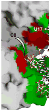
Overlap of the two ligand-binding sites of the IRP1 protein. The solvent-accessible-surface is of IRP1 with bound IRE RNA, and the IRE is shown as a stick model. The ‘footprint’ of the IRE is colored green, while the active site residues of c-aconitase are colored red. Most of the active site residues are within the IRE footprint. Orientation approximately the same as figure 3b. Figure done with Grasp [45].

References

    1. Wallander ML, Leibold EA, Eisenstein RS. Molecular control of vertebrate iron homeostasis by iron regulatory proteins. Biochim Biophys Acta. 2006;1763:668–689. λ This is the most current and comprehensive review of the molecular biology of vertebrate iron metabolism. - PMC - PubMed
    1. Gruer MJ, Artymiuk PJ, Guest JR. The aconitase family: three structural variations on a common theme. Trends Biochem Sci. 1997;22:3–6. - PubMed
    1. Baughn AD, Malamy MH. A mitochondrial-like aconitase in the bacterium Bacteroides fragilis: implications for the evolution of the mitochondrial Krebs cycle. Proc Natl Acad Sci USA. 2002;99:4662–4667. - PMC - PubMed
    1. Artymiuk PJ, Green J. The double life of aconitase. Structure. 2006;14:2–4. - PubMed
    1. Dupuy J, Volbeda A, Carpentier P, Darnault C, Moulis J-M, Fontecilla-Camps JC. Crystal structure of human iron regulatory protein 1 as cytosolic aconitase. Structure. 2006;14:129–139. λλ Dupuy, et al. present the first report of a structure of a mammalian cytosolic aconitase. - PubMed

Publication types