. 2008 Jun;29(6):1096-107.
doi: 10.1093/carcin/bgn026. Epub 2008 Feb 7.

FAK and IGF-IR interact to provide survival signals in human pancreatic adenocarcinoma cells

Affiliations

FAK and IGF-IR interact to provide survival signals in human pancreatic adenocarcinoma cells

Weiguo Liu et al. Carcinogenesis. 2008 Jun.

Abstract

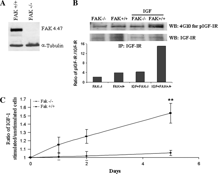
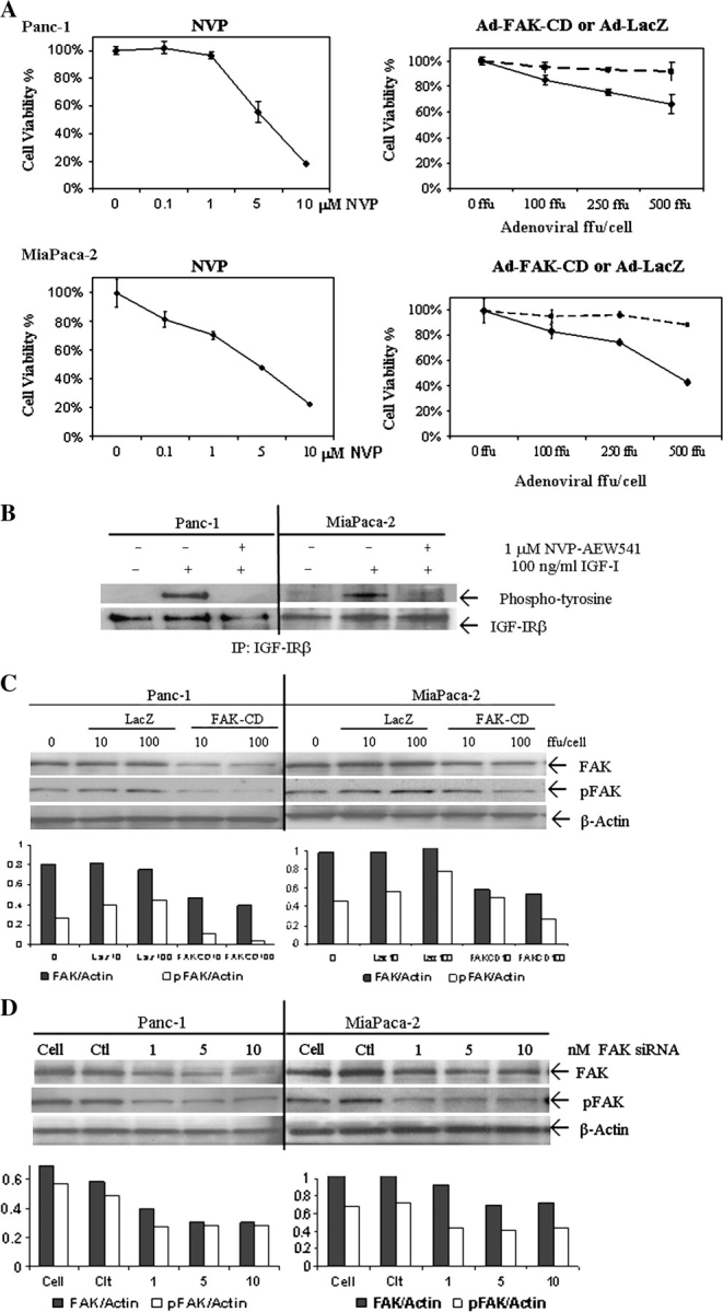
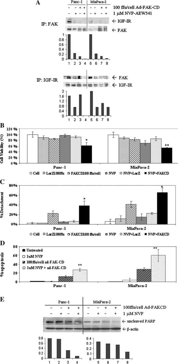
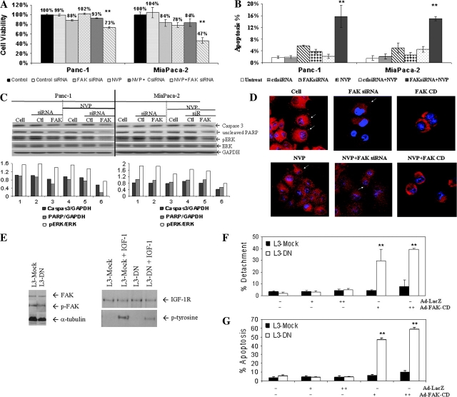
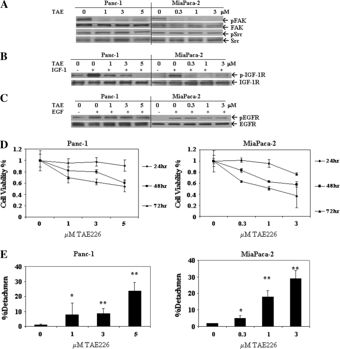
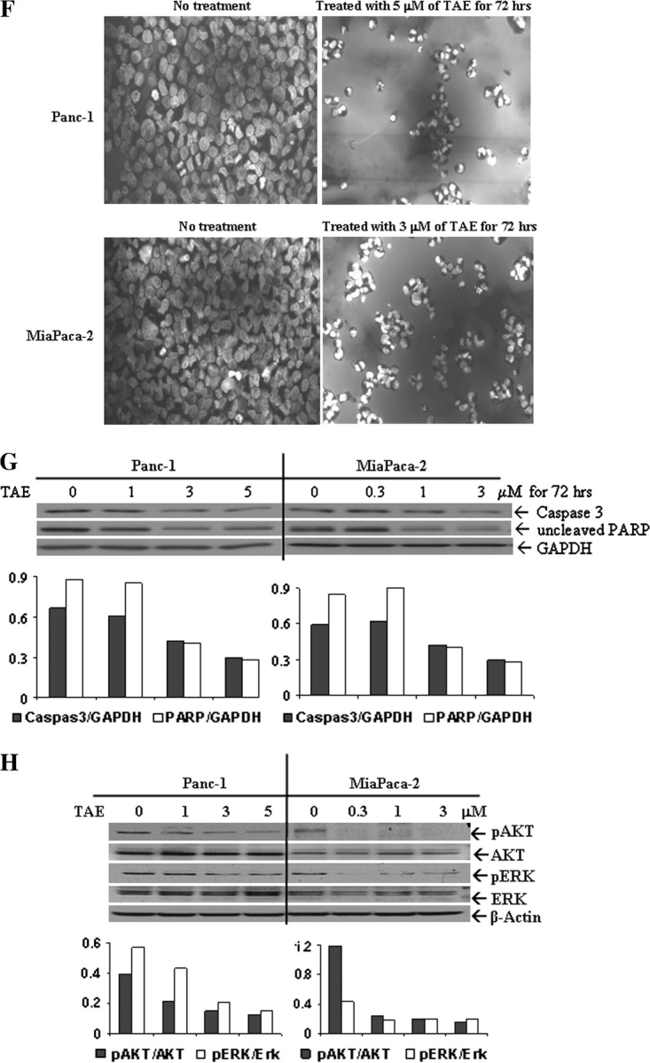
Pancreatic cancer is a lethal disease accounting for the fourth leading cause of cancer death in USA. Focal adhesion kinase (FAK) and the insulin-like growth factor-I receptor (IGF-1R) are tyrosine kinases that activate common pathways, leading to increased proliferation and cell survival. Sparse information is available regarding their contribution to the malignant behavior of pancreatic cancer. We analyzed the relationship between FAK and IGF-1R in human pancreatic cancer cells, determined which downstream signaling pathways are altered following kinase inhibition or downregulation and studied whether dual kinase inhibition represents a potential novel treatment strategy in this deadly disease. Using immunoprecipitation and confocal microscopy, we show for the first time that FAK and IGF-1R physically interact in pancreatic cancer cells and that inhibition of tyrosine phosphorylation of either kinase disrupts their interaction. Decreasing phosphorylation of either FAK or IGF-1R alone resulted in little inhibition of cell viability or increased apoptosis. However, dual inhibition of FAK, using either a dominant-negative construct (FAK-CD) or small interfering RNA, and IGF-1R, using a specific small molecule tyrosine kinase inhibitor (AEW-541) or stable expression of a truncated, mutated IGF-1R, led to a synergistic decrease in cell proliferation and phosphorylation of extracellular signal-regulated kinase (ERK) and increase in cell detachment and apoptosis compared with inhibition of either pathway alone. Dual kinase inhibition with FAK-CD and AEW-541 resulted in a marked increase in apoptosis when FAK was displaced from the focal adhesions. Inhibition of both tyrosine kinase activities via a novel single small molecular inhibitor (TAE 226), at low doses specific for FAK and IGF-1R, resulted in significant inhibition of cell viability, decrease in phosphorylation of ERK and Akt and increase in apoptosis accompanied by cleavage of Poly (ADP-ribose) polymerase (PARP) and activation of caspase-3 in pancreatic cancer cells. Thus, simultaneous inhibition of both tyrosine kinases represents a potential novel therapeutic approach in human pancreatic adenocarcinoma.


Figures

Fig. 1.
Fig. 1.
IGF-1 stimulation of mouse embryo fibroblast proliferation is dependent on FAK. (A) Whole-cell extracts from untreated mouse embryo fibroblasts were analyzed by western blot for FAK expression. (B) Untreated and treated (100 ng/ml IGF-1) mouse embryo fibroblasts were harvested and whole-cell lysates were immunoprecipitated with IGF-1R antibody and analyzed by western blot with IGF-1R and phospho-tyrosine antibody. Densitometry results shown under blot. (C) FAK+/+ and FAK−/− mouse embryo fibroblast cells were treated with human recombinant (hr) IGF-1, or control, for up to 5 days. At the indicated time points, cell viability was determined by CellTiter 96® AQueous. Cell viability was plotted as a ratio of hrIGF-I-stimulated cells to control treated cells. Results are shown from three experiments with six replicates (*P < 0.001).
Fig. 2.
Fig. 2.
FAK and IGF-1R physically interact and colocalize in the focal adhesions. (A) Panc 1 and Mia Paca-2 pancreatic cancer cell lines were analyzed by western blot for expression of FAK and IGF-1R. (B) FAK or IGF-1R was immunoprecipitated from whole-cell extracts. The immunoprecipitated complexes were analyzed for the presence of FAK and IGF-1R by western blot. FAK was present in the samples immunoprecipitated with the IGF-1R antibody (top panel) and IGF-1R was present in the samples immunoprecipitated with the FAK antibody (bottom panel). (C) The localization of FAK and IGF-1R in Panc-1 cells were analyzed by confocal microscopy. IGF-1R was labeled with fluorescein isothiocyanate (FITC), FAK was labeled with Texas red and the nucleus was stained with 4′6-diamidino-2-phenylindole (DAPI). The areas of colocalization appear yellow in the merged and cross-sectional images.
Fig. 3.
Fig. 3.
Inhibition of IGF-1R or FAK. (A) Panc-1 (top two panels) and MiaPaca-2 cells (bottom two panels) were treated with various doses of NVP-AEW541, Ad-FAK-CD or Ad-LacZ for 72 h. Cell proliferation was determined by CellTiter 96® AQueous. Ad-FAK-CD (solid line) caused a significant inhibition (P < 0.05) of cell proliferation in both cell lines compared with Ad-LacZ (dashed line). Results are shown for three experiments done in triplicate. (B) Cells were serum starved for 16 h then treated with 1 μM NVP-AEW541 for 10 min prior to induction with 100 ng/ml IGF-1 for 10 min. Whole-cell lysates were immunoprecipitated with IGF-1R antibody and analyzed by western blot for phospho-IGF-1R with anti-phospho-tyrosine antibodies. The blots were then stripped and reprobed with IGF-1R antibodies to demonstrate equal amounts of IGF-IR in each lane. (C) Cells were treated with Ad-LacZ or Ad-FAK-CD for 48 h and harvested to analyze the changes in phospho-FAK and total FAK by western blot. Densitometry results shown under blot. (D) Cells were treated with control siRNA (10 nM) or different concentrations of FAK siRNA for 48 h and harvested to analyze the changes in phospho-FAK and total FAK by western blot. β-Actin was used for loading control. Densitometry results shown under blot.
Fig. 4.
Fig. 4.
Dual inhibition of FAK and IGF-1R disrupts binding and has synergistic effects on cell viability, detachment and apoptosis. (A) To determine if inhibitors of FAK or IGF-1R phosphorylation altered binding, Panc-1 or MiaPaca-2 cells were treated with 100 FFU per cell of Ad-FAK-CD and/or 1 μM NVP-AEW541 for 48 h. Immunoprecipitation was performed from cell lysates and probed for FAK and IGF-1R. (B) Panc-1 and MiaPaca-2 cells were treated with 100 FFU per cell of Ad-FAK-CD or 1 μM of NVP-AEW541 or the combination of the two treatments for 72 h. Hundred FFU per cell Ad-LacZ was used as the control for Ad-FAK-CD. Cell viability was determined by CellTiter 96® AQueous. Results are shown for three experiments done in triplicate. (*P < 0.05, **P < 0.01 comparing Ad-FAK-CD alone to combined treatment). (C) After the same treatment as above, attached and detached cells were harvested and counted using a hemocytometer and the percent detachment was calculated. Results are shown for three experiments done in triplicate (*P < 0.05, Ad-FAK-CD alone to combined treatment). (D and E) After the same treatment as above, the whole cell population was harvested and subjected to analysis for apoptosis by using TUNEL assay (D) or western blot (probing for full-length PARP) (E). β-Actin was used for loading control. (**P < 0.01 comparing Ad-FAK-CD alone to combined treatment).
Fig. 5.
Fig. 5.
Dual inhibition of FAK and IGF-1R through different methods alters focal adhesions, decreases cell viability and increases apoptosis through downregulation of ERK. (A and B) Cells were transfected with 10 nM of control siRNA or FAK siRNA for 24 h and then 1 μM of NVP-AEW541 was added to the cells. After another 48 h, the whole cell populations were harvested and subjected to analysis for cell viability by MTT assay (A) and apoptosis by Hoechst staining (B) and western blot (probing for full-length PARP and caspase-3) (C). Cell extracts from the same treatment were used for western blot analysis for phospho-ERK and total ERK. GAPDH was used for loading control (C). (**P < 0.01 comparing FAK siRNA alone to combined treatment). Densitometry is shown below the blot. (D) Cells were treated with 1 μM NVP-AEW541 for 72 h, 100 FFU per cell Ad-FAK-CD for 48 h or 10 nM of FAK siRNA for 48 h alone or the combination of 1 μM NVP-AEW541 with either 100 FFU per cell Ad-FAK-CD or 10 nM FAK siRNA for 72 h. Cells were then harvested and stained with Texas red for FAK and DAPI for the nucleus. Focal adhesions were analyzed by confocal microscopy. In the presence of Ad-FAK-CD, FAK is displaced from the focal adhesions (white arrows), while they are still present with FAK siRNA treatment. (E) L3 pancreatic cancer cells stably expressing a truncated, mutated dominant-negative IGF-1R (L3-DN) were analyzed by western blot for FAK, p-FAK and total IGF-1R and compared with mock-transfected cells (L3-Mock). Of note, following stimulation by 100 ng/ml of IGF-1, phosphorylation of IGF-1R is increased in L3-Mock compared with L3-DN. (F) L3 pancreatic cancer cells stably expressing a truncated, mutated dominant-negative IGF-1R (L3-DN) are more sensitive to FAK inhibition than mock-transfected cells (L3-Mock). L3-DN and L3-Mock cells were treated with increasing doses of Ad-LacZ or Ad-FAK-CD for 72 h. Attached and detached cells were counted using a hemocytometer and the percent detachment was calculated. (G) Apoptosis was determined by TUNEL assay. Results are shown for three experiments done in triplicate (*P < 0.001, L3-DN + Ad-FAK-CD versus L3-Mock + Ad-FAK-CD).
Fig. 6.
Fig. 6.
Effect of TAE226 on phosphorylation of tyrosine kinases, cell viability, apoptosis and signaling to ERK and Akt. (A) Cells were treated with different doses of TAE226 for 1 h and harvested. Cell extracts were analyzed by western blot for the changes in phospho-FAK, total FAK, phospho-Src and total Src. (B) Cells were treated with different doses of TAE226 for 1 h prior to induction with 100 ng/ml IGF-1 for 10 min. Whole-cell lysates were immunoprecipitated with IGF-1R antibody and analyzed by western blot for phospho-IGF-1R with anti-phospho-tyrosine antibodies. The blots were then stripped and reprobed with IGF-1R antibodies to demonstrate equal amounts of IGF-1R in each lane. (C) Cells were treated with different doses of TAE226 for 1 h prior to exposure to 40 ng/ml epidermal growth factor (EGF) for 10 min. Whole-cell lysates were analyzed by western blot for the changes in phospho-EGFR and total EGFR. (D) Cells were treated with different concentrations of TAE226 for up to 3 days. At the indicated time points, cell viability was determined by CellTiter 96® AQueous. Results are shown for three experiments done in triplicate. (E) Cells were treated with different concentrations of TAE226 for 72 h. Attached and detached cells were then harvested and counted using a hemocytometer, and the percent cellular detachment was calculated. Results are shown for three experiments done in triplicate (*P < 0.05, **P < 0.01 comparing to no treatment). (F) Cells treated with 3 or 5 μM of TAE266 for 72 h were harvested for Hoechst staining. Bright areas indicate condensed apoptotic nuclei. (G) Cells treated with different concentrations of TAE226 for 72 h were harvested. Whole-cell lysates were analyzed for apoptosis by western blot, probing for full-length caspase-3 and PARP. GAPDH was used for loading control. Densitometry is shown under the blot. (H) Cells treated with different concentrations of TAE226 for 1 h were harvested. Whole-cell lysates were analyzed by western blot, probing for phospho-Akt and phospho-ERK. β-Actin was used for loading control. Densitometry is shown under the blot.
Fig. 6.
Fig. 6.
Effect of TAE226 on phosphorylation of tyrosine kinases, cell viability, apoptosis and signaling to ERK and Akt. (A) Cells were treated with different doses of TAE226 for 1 h and harvested. Cell extracts were analyzed by western blot for the changes in phospho-FAK, total FAK, phospho-Src and total Src. (B) Cells were treated with different doses of TAE226 for 1 h prior to induction with 100 ng/ml IGF-1 for 10 min. Whole-cell lysates were immunoprecipitated with IGF-1R antibody and analyzed by western blot for phospho-IGF-1R with anti-phospho-tyrosine antibodies. The blots were then stripped and reprobed with IGF-1R antibodies to demonstrate equal amounts of IGF-1R in each lane. (C) Cells were treated with different doses of TAE226 for 1 h prior to exposure to 40 ng/ml epidermal growth factor (EGF) for 10 min. Whole-cell lysates were analyzed by western blot for the changes in phospho-EGFR and total EGFR. (D) Cells were treated with different concentrations of TAE226 for up to 3 days. At the indicated time points, cell viability was determined by CellTiter 96® AQueous. Results are shown for three experiments done in triplicate. (E) Cells were treated with different concentrations of TAE226 for 72 h. Attached and detached cells were then harvested and counted using a hemocytometer, and the percent cellular detachment was calculated. Results are shown for three experiments done in triplicate (*P < 0.05, **P < 0.01 comparing to no treatment). (F) Cells treated with 3 or 5 μM of TAE266 for 72 h were harvested for Hoechst staining. Bright areas indicate condensed apoptotic nuclei. (G) Cells treated with different concentrations of TAE226 for 72 h were harvested. Whole-cell lysates were analyzed for apoptosis by western blot, probing for full-length caspase-3 and PARP. GAPDH was used for loading control. Densitometry is shown under the blot. (H) Cells treated with different concentrations of TAE226 for 1 h were harvested. Whole-cell lysates were analyzed by western blot, probing for phospho-Akt and phospho-ERK. β-Actin was used for loading control. Densitometry is shown under the blot.

References

    1. Schlaepfer DD, et al. Multiple Grb2-mediated integrin-stimulated signaling pathways to ERK2/mitogen-activated protein kinase: summation of both c-Src- and focal adhesion kinase-initiated tyrosine phosphorylation events. Mol. Cell. Biol. 1998;18:2571–2585. - PMC - PubMed
    1. Schlaepfer DD, et al. Integrin signalling and tyrosine phosphorylation: just the FAKs? Trends Cell Biol. 1998;8:151–157. - PubMed
    1. Frisch SM, et al. Control of adhesion-dependent cell survival by focal adhesion kinase. J. Cell Biol. 1996;134:793–799. - PMC - PubMed
    1. Ilic D, et al. Extracellular matrix survival signals transduced by focal adhesion kinase suppress p53-mediated apoptosis. J. Cell Biol. 1998;143:547–560. - PMC - PubMed
    1. Cary LA, et al. Stimulation of cell migration by overexpression of focal adhesion kinase and its association with Src and Fyn. J. Cell Sci. 1996;109:1787–1794. - PubMed

Publication types

MeSH terms