. 2008 Apr 4;283(14):9023-30.
doi: 10.1074/jbc.M800150200. Epub 2008 Feb 8.

Human Pso4 is a metnase (SETMAR)-binding partner that regulates metnase function in DNA repair

Affiliations

Human Pso4 is a metnase (SETMAR)-binding partner that regulates metnase function in DNA repair

Brian D Beck et al. J Biol Chem. .

Abstract

Metnase, also known as SETMAR, is a SET and transposase fusion protein with an undefined role in mammalian DNA repair. The SET domain is responsible for histone lysine methyltransferase activity at histone 3 K4 and K36, whereas the transposase domain possesses 5'-terminal inverted repeat (TIR)-specific DNA binding, DNA looping, and DNA cleavage activities. Although the transposase domain is essential for Metnase function in DNA repair, it is not clear how a protein with sequence-specific DNA binding activity plays a role in DNA repair. Here, we show that human homolog of the ScPSO4/PRP19 (hPso4) forms a stable complex with Metnase on both TIR and non-TIR DNA. The transposase domain essential for Metnase-TIR interaction is not sufficient for its interaction with non-TIR DNA in the presence of hPso4. In vivo, hPso4 is induced and co-localized with Metnase following ionizing radiation treatment. Cells treated with hPso4-siRNA failed to show Metnase localization at DSB sites and Metnase-mediated stimulation of DNA end joining coupled to genomic integration, suggesting that hPso4 is necessary to bring Metnase to the DSB sites for its function(s) in DNA repair.


Figures

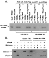
FIGURE 1.
FIGURE 1.
hPso4 is a novel Metnase-binding partner. A, SDS-PAGE analysis of Metnase and associated proteins. Whole cell extracts prepared from control 293 cells (lane 1) or cells stably expressing V5-Metnase (lanes 2 and 3) were incubated with 3 μg of V5-monoclonal antibody at 4 °C for 2 h and further incubated overnight following the addition of 100 μl of protein G-agarose beads. The complex was washed four times with a buffer containing 250 mm NaCl prior to running on a 10% SDS-PAGE and staining with Coomassie Blue. Lane M represents molecular weight markers. Proteins 1, 2, and 3 were identified as PRP19 gene product of S. cerevisiae (PRP19/Pso4), a cleavage product of Metnase (SETMAR), and the human homolog of Spf27, respectively. B, Western blot (WB) analysis of cells transfected with vector alone or vector expressing either V5-Metnase- or FLAG-hPso4. Ku80 was used as an internal loading control. C, interaction of Metnase with hPso4. Whole cell extracts (100 μg) from 293 cells overexpressing FLAG-Pso4 (lanes 1 and 5), V5-Metnase (lanes 2 and 6), or both (lanes 3, 4, 7, and 8) were incubated for 2 h with either anti-FLAG (lanes 1-4) or anti-V5 (lanes 5-8) antibody and then for an additional 2 h with protein G-agarose. In lanes 3 and 7, human 293 cells overexpressing FLAG-Pso4 were transfected with a plasmid expressing V5-Metnase, whereas in lanes 4 and 8, cells overexpressing V5-Metnase were transfected with a plasmid expressing FLAG-Pso4. Following immunoprecipitation (IP), the proteins were run on a 10% SDS-PAGE, transferred to membrane, and probed with either anti-V5 monoclonal (top panel) or anti-Pso4 polyclonal (bottom panel) antibody.
FIGURE 2.
FIGURE 2.
Interaction of Metnase or hPso4 with DNA. A, SDS-PAGE of immunoaffinity purified FLAG-Metnase (lane 1) and FLAG-hPso4 (lane 2) used in this study. B, DNA sequence of the TIR and the mutant (MAR3M). The bipartite Metnase binding core site (19-mer) is shaded gray. In MAR3M, mutation sites are indicated as bold type with underlines. C, interaction of Metnase or hPso4 with TIR32 or non-TIR (MAR3M) DNA. The reaction mixtures (20 μl) containing indicated amount of Metnase (or hPso4) and 5′-32P-labeled DNA (200 fmol; ∼3,000cpm/fmol) were incubated for 15 min at 25 °C. After incubation, the samples were analyzed by 5% native PAGE. D, interaction of hPso4 with dsDNA and not single-stranded DNA. Reaction mixtures (20 μl) containing increasing amounts of hPso4 and 200 fmol of 5′-32P-labeled single-stranded DNA (top strand of MAR3M) or dsDNA (MAR3M duplex DNA) were incubated for 15 min at 25 °C and analyzed by 5% PAGE.
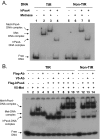
FIGURE 3.
FIGURE 3.
Metnase associates with non-TIR DNA in the presence of hPso4. A, V5-wt-Metnase and/or FLAG-wt-hPso4 were immunoprecipitated with cell extracts (100 μg) overexpressing V5-Metnase (lanes 3, 6, 9, and 12), FLAG-Pso4 (lanes 5, 8, 11, and 14), or both (lanes 4, 7, 10, 13) using either anti-V5 (lanes 3-5 and 9-11) or -FLAG (lanes 6-8 and 12-14) monoclonal antibody. Following immunoprecipitation, either 5′-32P-labeled TIR32 (lanes 3-8) or 32P-MAR3M (lanes 9-14) was added to the mixtures for interaction of Metnase/hPso4 with 32P-labeled DNA. Following 10% SDS-PAGE, gel was dried and exposed to x-ray film. In control lanes (lanes 1 and 2), purified FLAG-Metnase (100 ng) was incubated with either TIR32 or MAR3M prior to immunoprecipitation using an anti-FLAG antibody. B, purified FLAG-wt-Metnase and/or FLAG-wt-hPso4 were incubated for 15 min prior to addition of either biotin-labeled TIR (lanes 3-6) or MAR3M (lanes 7-10). Following the addition of Streptavidin-agarose beads, the protein-DNA complexes were precipitated, washed, and analyzed by 10% SDS-PAGE. Western blotting was done using an anti-FLAG antibody.
FIGURE 4.
FIGURE 4.
Metnase forms a stable complex with non-TIR DNA via interaction with hPso4. A, interaction of Metnase and/or hPso4 with TIR32 (lanes 1-4) or non-TIR (MAR3M) DNA (lanes 5-8). Reaction mixtures (20 μl) containing Metnase (0.2 μg) and/or hPso4 (0.2 μg), and 200 fmol of 5′-32P-labeled DNA were incubated for 15 min at 25 °C and analyzed by 5% native PAGE. B, interaction of V5-Metnase and/or FLAG-hPso4 with DNA in the presence of V5 or FLAG antibody. Reaction mixtures (20 μl) containing Metnase (0.2 μg) and/or hPso4 (0.2 μg) were incubated with 1 μg of V5 or FLAG antibody for 30 min at 25 °C prior to the addition of 5′-32P-labeled DNA (200 fmol). After 15 min of incubation, samples were analyzed by 5% native PAGE. The antibody-protein-DNA complexes (supershift) are indicated as asterisks.
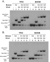
FIGURE 5.
FIGURE 5.
Formation of the Metnase-hPso4 complex on TIR and non-TIR DNA. Reaction mixtures (20 μl) were the same as those indicated in Fig. 2C, except that varying amounts of hPso4 (A) or wt-Metnase (B) were included. Individual protein-DNA complexes are marked on the left side of the figure.
FIGURE 6.
FIGURE 6.
The transposase domain is not sufficient for formation of the Metnase-hPso4 complex on DNA. A, wt-Metnase or a mutant lacking the SET domain (Met:dSET) was incubated with 32P-TIR (lanes 1-6) or non-TIR (32P-MAR3M, lanes 7-12) in the absence or the presence of hPso4. Following 15 min of incubation, protein-DNA complexes were analyzed by 5% nondenaturing PAGE. Individual protein-DNA complexes are marked on the left side of the figure.
FIGURE 7.
FIGURE 7.
hPso4 is necessary for formation of Metnase foci at DSB sites. A, Western blot analysis of hPso4 expression following IR treatment. Proliferating cell nuclear antigen was used as a loading control. B, co-localization of Metnase with hPso4 following DSB damage in vivo. Human 293 cells overexpressing FLAG-Metnase were treated with 0 Gy (top panels) or 10 Gy (bottom panels) of IR. Following 8 h of incubation, the cells were fixed and examined for cellular localization of Metnase and hPso4 using anti-FLAG monoclonal (Sigma) and anti-hPso4 polyclonal (Calbiochem) antibodies. The images were obtained following double labeling of fixed cells with polyclonal antibody to hPso4 and monoclonal antibody to FLAG epitope with two different fluorochromes, Texas Red-conjugated anti-rabbit antibody, and fluorescein-conjugated anti-mouse antibody. A co-localization of Metnase and hPso4 was examined using a Zeiss LSM-510 confocal microscope. C, effect of hPso4 on formation of discrete nuclear foci of Metnase at DSB sites. Human 293 cells stably expressing FLAG-Metnase were treated with a control (Cont.) siRNA or hPso4-siRNA (5′-ACCACAGGCUGGCCUCAUUTT-3′, 5′-AAUGAGGCCAGCCUGUGGUTT-3′) for 48 h. The cells were then treated with 0 or 10 Gy of IR and examined for formation of Metnase and/or Nbs-1 foci. D, kinetic analysis of Metnase foci formation following IR treatment. Human 293 cells expressing FLAG-wt-Metnase were treated with IR (10 Gy) and harvested samples at various times for nuclear foci formation of Metnase and Nbs-1. The cells were fixed, labeled with an anti-FLAG or anti-Nbs-1 antibody, and images were collected using a Zeiss LSM-510 confocal microscope. E, cells (%) with IR-induced Metnase foci were calculated from the experiment in Fig. 7D. For each time point, three samples were analyzed.
FIGURE 8.
FIGURE 8.
A targeted inhibition of hPso4 expression abolished Metnase-mediated stimulation of DNA end joining coupled to genomic integration. Top panels, effect of Metnase- or hPso4-specific siRNA on protein expression. Human 293 cells were treated with mock (lane 1), scrambled siRNA (lane 2), or Metnase- or hPso4-specific siRNA (lane 3). After 48 h, the cells were harvested and analyzed for Metnase or hPso4 expression by Western blotting. Proliferating cell nuclear antigen was used as a loading control. Bottom panel, effect of Metnase or hPso4 siRNA on DNA end joining coupled to genomic integration. Human 293 cells (1.0 × 10⁵/plate) stably transfected with pFLAG2 (mock) or pFLAG2-Metnase were treated with control-siRNA, Metnase-siRNA, or hPso4-siRNA. Forty-eight hours later, the cells were transfected with indicated amounts (2 and 4 μg) of KpnI-linearized pRNA/U6.Hygro plasmid. Following incubation at 37 °C for 14 days, the number of Hygr colonies were measured for assessment of the in vivo end joining efficiency (1). For statistical analysis, six plates/sample were analyzed using the paired t test.

Similar articles

Cited by

References

    1. Lee, S. H., Oshige, M., Durant, S. T., Rasila, K. K., Williamson, E. A., Ramsey, H., Kwan, L., Nickoloff, J. A., and Hromas, R. (2005) Proc. Natl. Acad. Sci. U. S. A. 102 18075-18080 - PMC - PubMed
    1. Hamamoto, R., Furukawa, Y., Morita, M., Iimura, Y., Silva, F. P., Li, M., Yagyu, R., and Nakamura, Y. (2004) Nat. Cell Biol. 6 731-740 - PubMed
    1. Krogan, N. J., Kim, M., Tong, A., Golshani, A., Cagney, G., Canadien, V., Richards, D. P., Beattie, B. K., Emili, A., Boone, C., Shilatifard, A., Buratowski, S., and Greenblatt, J. (2003) Mol. Cell Biol. 23 4207-4218 - PMC - PubMed
    1. Xiao, T., Hall, H., Kizer, K. O., Shibata, Y., Hall, M. C., Borchers, C. H., and Strahl, B. D. (2003) Genes Dev. 17 654-663 - PMC - PubMed
    1. Zhang, X., Yang, Z., Khan, S. I., Horton, J. R., Tamaru, H., Selker, E. U., and Cheng, X. (2003) Mol. Cell 12 177-185 - PMC - PubMed

Publication types

MeSH terms