Review
. 2008 Mar;466(3):622-33.
doi: 10.1007/s11999-007-0112-4. Epub 2008 Feb 10.

Biological augmentation of rotator cuff tendon repair

Affiliations
Review

Biological augmentation of rotator cuff tendon repair

David Kovacevic et al. Clin Orthop Relat Res. 2008 Mar.

Abstract

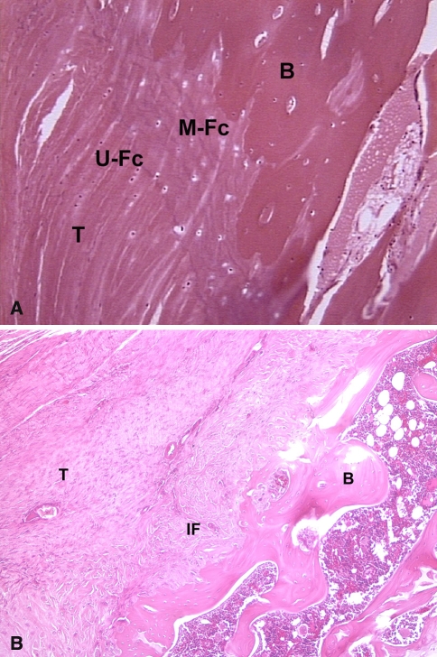
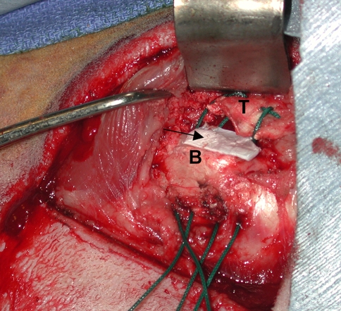
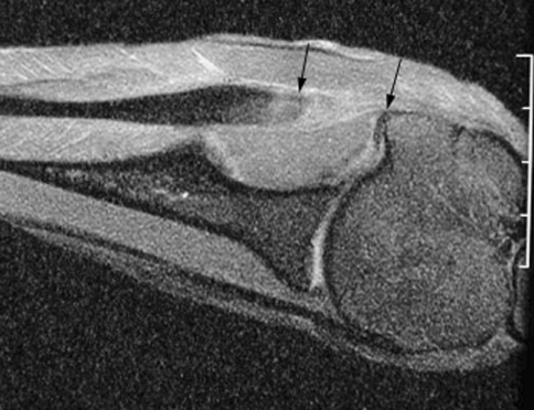
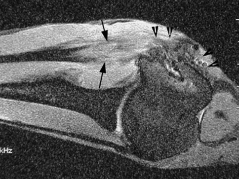
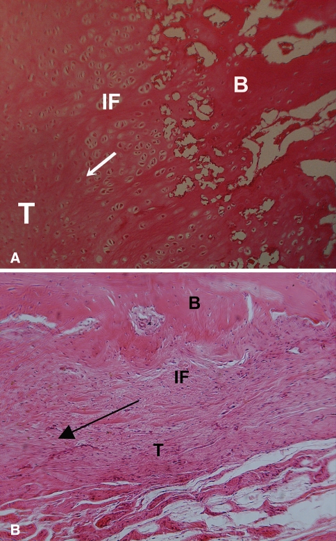
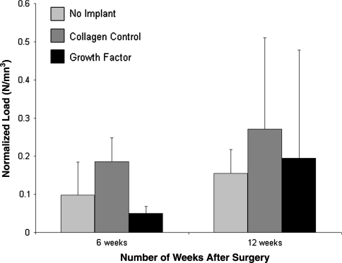
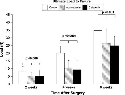
A histologically normal insertion site does not regenerate following rotator cuff tendon-to-bone repair, which is likely due to abnormal or insufficient gene expression and/or cell differentiation at the repair site. Techniques to manipulate the biologic events following tendon repair may improve healing. We used a sheep infraspinatus repair model to evaluate the effect of osteoinductive growth factors and BMP-12 on tendon-to-bone healing. Magnetic resonance imaging and histology showed increased formation of new bone and fibrocartilage at the healing tendon attachment site in the treated animals, and biomechanical testing showed improved load-to-failure. Other techniques with potential to augment repair site biology include use of platelets isolated from autologous blood to deliver growth factors to a tendon repair site. Modalities that improve local vascularity, such as pulsed ultrasound, have the potential to augment rotator cuff healing. Important information about the biology of tendon healing can also be gained from studies of substances that inhibit healing, such as nicotine and antiinflammatory medications. Future approaches may include the use of stem cells and transcription factors to induce formation of the native tendon-bone insertion site after rotator cuff repair surgery.


Figures

Fig. 1A–B
Fig. 1A–B
(A) Histologic section of a normal supraspinatus tendon insertion site demonstrates the four zones of a direct insertion: tendon (T), unmineralized fibrocartilage (U-Fc), mineralized fibrocartilage (M-Fc), and bone (B). (B) This histologic section of the tendon-bone attachment site 4 weeks after supraspinatus tendon repair in a rat model shows the site is characterized by a fibrovascular scar tissue interface (IF) without formation of an intermediate zone of fibrocartilage between tendon (T) and bone (B).
Fig. 2
Fig. 2
A collagen sponge (arrow) is used as a carrier vehicle to deliver growth factors to the tendon-bone attachment site in a sheep infraspinatus repair model (T = tendon, B = bone).
Fig. 3
Fig. 3
MR image of the healed tendon-bone interface 6 weeks after infraspinatus tendon repair to the greater tuberosity in a sheep model reveals the clear gap between the end of the repaired tendon and bone (between arrows). The low signal intensity native tendon is easily distinguished from the fibrovascular scar tissue interface.
Fig. 4
Fig. 4
MR image shows robust formation of new soft tissue (between arrows) in the tendon-bone interface and new bone formation (arrow heads) at the greater tuberosity 12 weeks after repair with a mixture of osteoinductive growth factors applied at the tendon attachment site on a collagen sponge carrier. Compare to the healed attachment site in an untreated repair in Fig. 3.
Fig. 5A–B
Fig. 5A–B
A histologic section shows fibrovascular tissue in the interface (IF) between tendon and bone at 6 weeks, with a more robust fibrocartilage interface zone between the bone (B) and tendon (T) in the (A) growth-factor treated animals compared to (B) controls that received the collagen sponge alone. The arrow indicates the direction of pull of the tendon.
Fig. 6
Fig. 6
The ultimate load-to-failure was higher in the animals treated with the osteoinductive growth factors; however, when the data were normalized by tissue volume there were no differences between groups. Figure reprinted with permission and © The Journal of Bone and Joint Surgery, Inc. from Rodeo SA, Potter HG, Kawamura S, Turner AS, Kim HJ, Atkinson BL. Biologic augmentation of rotator cuff tendon-healing with use of a mixture of osteoinductive growth factors. J Bone Joint Surg Am. 2007; 89:2485–2497.
Fig. 7
Fig. 7
Ultimate load-to-failure was lower (p < 0.001) in animals treated with NSAIDs.

References

    1. Alvarez CM, Litchfield R, Jackowski D, Griffin S, Kirkley A. A prospective, double-blind, randomized clinical trial comparing subacromial injection of betamethasone and xylocaine to xylocaine alone in chronic rotator cuff tendinosis. Am J Sports Med. 2005;33:255–262. - DOI - PubMed
    1. Asou Y, Nifuji A, Tsuji K, Shinomiya K, Olson EN, Koopman P, Noda M. Coordinated expression of scleraxis and Sox9 genes during embryonic development of tendons and cartilage. J Orthop Res. 2002;20:827–833. - DOI - PubMed
    1. Aspenberg P, Forslund C. Enhanced tendon healing with GDF 5 and 6. Acta Orthop Scand. 1999;70:51–54. - PubMed
    1. Aspenberg P, Forslund C. Bone morphogenetic proteins and tendon repair. Scand J Med Sci Sports. 2000;10:372–375. - DOI - PubMed
    1. Balasubramaniam P, Prathap K. The effect of injection of hydrocortisone into rabbit calcaneal tendons. J Bone Joint Surg Br. 1972;54:729–734. - PubMed

MeSH terms

Substances