CT scan method accurately assesses humeral head retroversion
- PMID: 18264854
- PMCID: PMC2505224
- DOI: 10.1007/s11999-007-0089-z
CT scan method accurately assesses humeral head retroversion
Abstract
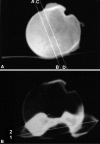
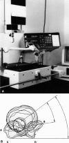
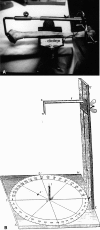
Humeral head retroversion is not well described with the literature controversial regarding accuracy of measurement methods and ranges of normal values. We therefore determined normal humeral head retroversion and assessed the measurement methods. We measured retroversion in 65 cadaveric humeri, including 52 paired specimens, using four methods: radiographic, computed tomography (CT) scan, computer-assisted, and direct methods. We also assessed the distance between the humeral head central axis and the bicipital groove. CT scan methods accurately measure humeral head retroversion, while radiographic methods do not. The retroversion with respect to the transepicondylar axis was 17.9 degrees and 21.5 degrees with respect to the trochlear tangent axis. The difference between the right and left humeri was 8.9 degrees. The distance between the central axis of the humeral head and the bicipital groove was 7.0 mm and was consistent between right and left humeri. Humeral head retroversion may be most accurately obtained using the patient's own anatomic landmarks or, if not, identifiable retroversion as measured by those landmarks on contralateral side or the bicipital groove.
Figures


Similar articles
-
[Torsional malalignment of the humerus].Unfallchirurg. 2018 Mar;121(3):199-205. doi: 10.1007/s00113-017-0453-8. Unfallchirurg. 2018. PMID: 29305619 Review. German.
-
Relationship of bicipital groove rotation with humeral head retroversion: a three-dimensional computed tomographic analysis.J Bone Joint Surg Am. 2013 Apr 17;95(8):719-24. doi: 10.2106/JBJS.J.00085. J Bone Joint Surg Am. 2013. PMID: 23595070
-
Linear relationship between lateralization of the bicipital groove and humeral retroversion and its link with the biepicondylar humeral line. Anatomical study of seventy cadaveric humerus scans.Int Orthop. 2017 Jul;41(7):1431-1434. doi: 10.1007/s00264-017-3495-1. Epub 2017 May 11. Int Orthop. 2017. PMID: 28497165
-
Determining humeral retroversion with computed tomography.J Bone Joint Surg Am. 2002 Oct;84(10):1753-62. doi: 10.2106/00004623-200210000-00003. J Bone Joint Surg Am. 2002. PMID: 12377904
-
[Three-dimensional geometry of the proximal humerus and rotator cuff attachment and its utilization in shoulder arthroplasty].Acta Chir Orthop Traumatol Cech. 2006 Apr;73(2):77-84. Acta Chir Orthop Traumatol Cech. 2006. PMID: 16735003 Czech.
Cited by
-
[Torsional malalignment of the humerus].Unfallchirurg. 2018 Mar;121(3):199-205. doi: 10.1007/s00113-017-0453-8. Unfallchirurg. 2018. PMID: 29305619 Review. German.
-
Is Anterior Bridge Plating for Mid-Shaft Humeral Fractures a Suitable Option for Patients Predominantly Involved in Overhead Activities? A Functional Outcome Study in Athletes and Manual Laborers.Clin Orthop Surg. 2016 Dec;8(4):358-366. doi: 10.4055/cios.2016.8.4.358. Epub 2016 Nov 4. Clin Orthop Surg. 2016. PMID: 27904716 Free PMC article.
-
Prediction of the rotational state of the humerus by comparing the contour of the contralateral bicipital groove: Method for intraoperative evaluation.Indian J Orthop. 2012 Nov;46(6):675-9. doi: 10.4103/0019-5413.104210. Indian J Orthop. 2012. PMID: 23325971 Free PMC article.
-
Correlation between the retroversion of the humeral head and the orientation of the intertubercular sulcus: a CT scan anatomical study.Surg Radiol Anat. 2015 May;37(4):357-61. doi: 10.1007/s00276-014-1354-y. Epub 2014 Aug 13. Surg Radiol Anat. 2015. PMID: 25113011
-
Ultrasonographic Measurement of Torsional Side Difference in Proximal Humerus Fractures and Humeral Shaft Fractures: Theoretical Background with Technical Notes for Clinical Implementation.Diagnostics (Basel). 2022 Dec 9;12(12):3110. doi: 10.3390/diagnostics12123110. Diagnostics (Basel). 2022. PMID: 36553117 Free PMC article.
References
-
- {'text': '', 'ref_index': 1, 'ids': [{'type': 'DOI', 'value': '10.1016/j.jse.2005.08.014', 'is_inner': False, 'url': 'https://doi.org/10.1016/j.jse.2005.08.014'}, {'type': 'PubMed', 'value': '16517364', 'is_inner': True, 'url': 'https://pubmed.ncbi.nlm.nih.gov/16517364/'}]}
- Balg F, Boulianne M, Boileau P. Bicipital groove orientation: considerations for the retroversion of a prosthesis in fractures of the proximal humerus. J Shoulder Elbow Surg. 2006;15:195–198. - PubMed
-
- {'text': '', 'ref_index': 1, 'ids': [{'type': 'PubMed', 'value': '2188343', 'is_inner': True, 'url': 'https://pubmed.ncbi.nlm.nih.gov/2188343/'}]}
- Bernageau J. Imaging of the shoulder in orthopedic pathology [in French]. Rev Prat. 1990;40:983–92. - PubMed
-
- {'text': '', 'ref_index': 1, 'ids': [{'type': 'DOI', 'value': '10.1067/mse.2002.124527', 'is_inner': False, 'url': 'https://doi.org/10.1067/mse.2002.124527'}, {'type': 'PubMed', 'value': '12378157', 'is_inner': True, 'url': 'https://pubmed.ncbi.nlm.nih.gov/12378157/'}]}
- Boileau P, Krishnan SG, Tinsi L, Walch G, Coste JS, Mole D. Tuberosity malposition and migration: reasons for poor outcomes after hemiarthroplasty for displaced fractures of the proximal humerus. J Shoulder Elbow Surg. 2002;11:401–412. - PubMed
-
- {'text': '', 'ref_index': 1, 'ids': [{'type': 'DOI', 'value': '10.1302/0301-620X.79B5.7579', 'is_inner': False, 'url': 'https://doi.org/10.1302/0301-620x.79b5.7579'}, {'type': 'PubMed', 'value': '9331050', 'is_inner': True, 'url': 'https://pubmed.ncbi.nlm.nih.gov/9331050/'}]}
- Boileau P, Walch G. The three-dimensional geometry of the proximal humerus. Implications for surgical technique and prosthetic design. J Bone Joint Surg Br. 1997;79:857–865. - PubMed
-
- {'text': '', 'ref_index': 1, 'ids': [{'type': 'PubMed', 'value': '1304634', 'is_inner': True, 'url': 'https://pubmed.ncbi.nlm.nih.gov/1304634/'}]}
- Boileau P, Walch G, Liotard JP. Radio-cinematographic study of active elevation of the prosthetic shoulder [in French]. Rev Chir Orthop. 1992;78:355–364. - PubMed

