Pelvic support osteotomy in the treatment of patients with excision arthroplasty
- PMID: 18264860
- PMCID: PMC2505230
- DOI: 10.1007/s11999-007-0094-2
Pelvic support osteotomy in the treatment of patients with excision arthroplasty
Abstract
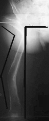
Resistant hip infection in adults can be a complicated problem that does not respond to surgical and medical treatment. In such cases, the only remaining option is excision arthroplasty. This line of treatment can eradicate the infection but also is associated with poor function. In some cases, conversion of excision arthroplasty to artificial joint replacement is associated with too great a risk because of local hip surgical risks or low immunity with risk of recurrent infection. Pelvic support osteotomy with the Ilizarov modification can present an alternative solution for such patients. This study included 11 patients with resistant hip infection who were treated using excision arthroplasty. Pelvic support osteotomy then was used to improve hip stability and abductor muscle function. The Ilizarov modification was applied to correct mechanical alignment of the limb and the limb length discrepancy. Harris hip scores improved in all patients: the average score preoperatively was 43.5 (range, 31-50), whereas at final followup, the average score was 70.9 (range, 65-80). Pelvic support osteotomy, along with the Ilizarov modification, can provide an alternative treatment to improve function in patients previously managed with excision hip arthroplasty and Girdlestone surgery.
Figures


Similar articles
-
Total hip replacement fifteen years after pelvic support osteotomy (PSO): a case report and review of the literature.Musculoskelet Surg. 2012 Aug;96(2):141-7. doi: 10.1007/s12306-011-0178-8. Epub 2012 Jan 12. Musculoskelet Surg. 2012. PMID: 22237840 Review.
-
Recurrent dislocations and complete necrosis: the role of pelvic support osteotomy.J Pediatr Orthop. 2013 Jul-Aug;33 Suppl 1:S45-55. doi: 10.1097/BPO.0b013e318281216b. J Pediatr Orthop. 2013. PMID: 23764793 Review.
-
One-third of Hips After Periacetabular Osteotomy Survive 30 Years With Good Clinical Results, No Progression of Arthritis, or Conversion to THA.Clin Orthop Relat Res. 2017 Apr;475(4):1154-1168. doi: 10.1007/s11999-016-5169-5. Clin Orthop Relat Res. 2017. PMID: 27905061 Free PMC article.
-
Reconstruction of Unstable Hips with Ilizarov Technique. Role of Pelvic Support and Distal Lengthening Realignment Osteotomy.Ortop Traumatol Rehabil. 2015 Oct;17(5):481-7. doi: 10.5604/15093492.1186823. Ortop Traumatol Rehabil. 2015. PMID: 26751748
-
Temporary External Fixation Can Stabilize Hip Transposition Arthroplasty After Resection of Malignant Periacetabular Bone Tumors.Clin Orthop Relat Res. 2019 Aug;477(8):1892-1901. doi: 10.1097/CORR.0000000000000764. Clin Orthop Relat Res. 2019. PMID: 30985613 Free PMC article.
Cited by
-
Revision arthroplasty with megaprosthesis after Girdlestone procedure for periprosthetic joint infection as an option in massive acetabular and femoral bone defects.Acta Biomed. 2022 Mar 10;92(S3):e2021531. doi: 10.23750/abm.v92iS3.12160. Acta Biomed. 2022. PMID: 35604274 Free PMC article.
-
Ilizarov hip reconstruction without external fixation: a new technique.J Child Orthop. 2010 Jun;4(3):259-66. doi: 10.1007/s11832-010-0256-8. Epub 2010 Apr 29. J Child Orthop. 2010. PMID: 21629378 Free PMC article.
-
Subacute Bacterial Infection Mimicking a Slipped Capital Femoral Epiphysis in an Obese Adolescent Male - A Case Report and Review of Literature.J Orthop Case Rep. 2023 May;13(5):14-19. doi: 10.13107/jocr.2023.v13.i05.3628. J Orthop Case Rep. 2023. PMID: 37255638 Free PMC article.
-
Total hip replacement fifteen years after pelvic support osteotomy (PSO): a case report and review of the literature.Musculoskelet Surg. 2012 Aug;96(2):141-7. doi: 10.1007/s12306-011-0178-8. Epub 2012 Jan 12. Musculoskelet Surg. 2012. PMID: 22237840 Review.
-
Hip reconstruction osteotomy by Ilizarov method as a salvage option for abnormal hip joints.Biomed Res Int. 2014;2014:835681. doi: 10.1155/2014/835681. Epub 2014 May 6. Biomed Res Int. 2014. PMID: 24895616 Free PMC article.
References
-
- {'text': '', 'ref_index': 1, 'ids': [{'type': 'PubMed', 'value': '10842880', 'is_inner': True, 'url': 'https://pubmed.ncbi.nlm.nih.gov/10842880/'}]}
- Aksoy MC, Musdal Y. Subtrochanteric valgus-extension osteotomy for neglected congenital dislocation of the hip in young adults. Acta Orthop Belg. 2000;66:181–186. - PubMed
-
- {'text': '', 'ref_index': 1, 'ids': [{'type': 'DOI', 'value': '10.1097/00003086-200302000-00019', 'is_inner': False, 'url': 'https://doi.org/10.1097/00003086-200302000-00019'}, {'type': 'PubMed', 'value': '12567138', 'is_inner': True, 'url': 'https://pubmed.ncbi.nlm.nih.gov/12567138/'}]}
- Charlton WP, Hozack WJ, Teloken MA, Rao R, Bissett GA. Complications associated with reimplantation after Girdlestone arthroplasty. Clin Orthop Relat Res. 2003;407:119–126. - PubMed
-
- {'text': '', 'ref_index': 1, 'ids': [{'type': 'PubMed', 'value': '16459858', 'is_inner': True, 'url': 'https://pubmed.ncbi.nlm.nih.gov/16459858/'}]}
- El-Mowafi H. Outcome of pelvic support osteotomy with the Ilizarov method in the treatment of the unstable hip joint. Acta Orthop Belg. 2005;71:686–691. - PubMed
-
- {'text': '', 'ref_index': 1, 'ids': [{'type': 'DOI', 'value': '10.1007/s00264-002-0374-0', 'is_inner': False, 'url': 'https://doi.org/10.1007/s00264-002-0374-0'}, {'type': 'PMC', 'value': 'PMC3620990', 'is_inner': False, 'url': 'https://pmc.ncbi.nlm.nih.gov/articles/PMC3620990/'}, {'type': 'PubMed', 'value': '12378361', 'is_inner': True, 'url': 'https://pubmed.ncbi.nlm.nih.gov/12378361/'}]}
- Emara KM. Hemi-corticotomy in the management of chronic osteomyelitis of the tibia. Int Orthop. 2002;26:310–313. - PMC - PubMed
-
- {'text': '', 'ref_index': 1, 'ids': [{'type': 'DOI', 'value': '10.1007/s001040170040', 'is_inner': False, 'url': 'https://doi.org/10.1007/s001040170040'}, {'type': 'PubMed', 'value': '11766659', 'is_inner': True, 'url': 'https://pubmed.ncbi.nlm.nih.gov/11766659/'}]}
- Esenwein SA, Robert K, Kollig E, Ambacher T, Kutscha-Lissberg F, Muhr G. Long-term results after resection arthroplasty according to Girdlestone for treatment of persisting infections of the hip joint. Chirurg. 2001;72:1336–1413. - PubMed
MeSH terms
LinkOut - more resources
Full Text Sources

