Review
. 2008 Jun;27(2):123-35.
doi: 10.1007/s10555-008-9127-x.

The type 2C phosphatase Wip1: an oncogenic regulator of tumor suppressor and DNA damage response pathways

Affiliations
Review

The type 2C phosphatase Wip1: an oncogenic regulator of tumor suppressor and DNA damage response pathways

Xiongbin Lu et al. Cancer Metastasis Rev. 2008 Jun.

Abstract

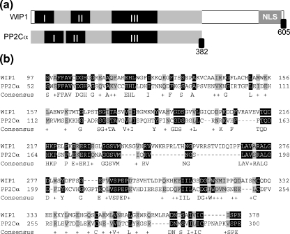
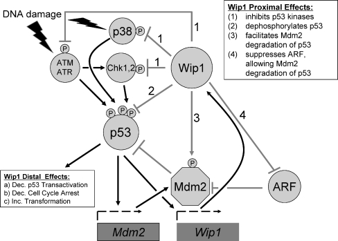
The Wild-type p53-induced phosphatase 1, Wip1 (or PPM1D), is unusual in that it is a serine/threonine phosphatase with oncogenic activity. A member of the type 2C phosphatases (PP2Cdelta), Wip1 has been shown to be amplified and overexpressed in multiple human cancer types, including breast and ovarian carcinomas. In rodent primary fibroblast transformation assays, Wip1 cooperates with known oncogenes to induce transformed foci. The recent identification of target proteins that are dephosphorylated by Wip1 has provided mechanistic insights into its oncogenic functions. Wip1 acts as a homeostatic regulator of the DNA damage response by dephosphorylating proteins that are substrates of both ATM and ATR, important DNA damage sensor kinases. Wip1 also suppresses the activity of multiple tumor suppressors, including p53, ATM, p16(INK4a) and ARF. We present evidence that the suppression of p53, p38 MAP kinase, and ATM/ATR signaling pathways by Wip1 are important components of its oncogenicity when it is amplified and overexpressed in human cancers.


Figures

Fig. 1
Fig. 1
Protein sequence alignment of human Wip1 and human PP2Cα. (a) The overall structures of Wip1 (top) and PP2Cα (bottom) show significant similarity. The conserved type 2C phosphatase domain is shaded in Wip1. The regions of more highly conserved sequences labeled I, II, and III are shown as black blocks. The C-terminal non-catalytic domain that is present only in Wip1 molecules (and is also well conserved among mammalian Wip1 orthologues) is indicated by the white block. A putative nuclear localization signal (NLS) is also indicated near the C-terminus of Wip1. (b) Primary amino acid sequence alignment between human Wip1 and human PP2Cα phosphatase domains is shown. Identical amino acids are highlighted with a black background while conservative amino acid substitutions are indicated with a gray background. The phosphatase domains of the two molecules show 30% identity and 45% similarity
Fig. 2
Fig. 2
Wip1 inhibits p53 activity by multiple mechanisms. When a cell is stressed by DNA damage, ATM, ATR, and p38 MAP kinase can phosphorylate p53 directly or through intermediary proteins such as Chk1 and Chk2. Phosphorylated p53 localizes to the nucleus and transactivates a battery of anti-proliferative genes. In addition, two p53 autoregulatory genes are activated, Mdm2 and Wip1. Mdm2 is an E3 ubiquitin ligase that promotes p53 degradation. However, early after the DNA damage response ATM (and possibly ATR) phosphorylate Mdm2 and this promotes Mdm2 degradation and prevents Mdm2 mediated p53 degradation. Activated p53 also upregulates Wip1 expression and after DNA damage is repaired, the accumulated Wip1 phosphatase inhibits a battery of proteins that activate p53. Wip1 dephosphorylates the upstream kinases that phosphorylate p53 (ATM, p38, Chk1, Chk2) and p53 itself (at Ser15). In addition, Wip1 dephosphorylates Mdm2 at Ser395 and this results in Mdm2 stabilization and Mdm2 mediated p53 degradation. Finally, increased Wip1 levels suppresses ARF which in turn results in increased Mdm2 activity and p53 proteolysis. The resulting destabilization of p53 helps return the normal cell to a pre-stress state after cellular damage is repaired. However, if Wip1 becomes amplified or overexpressed during tumor cell progression, this could result in chronic suppression of p53 activity and promote tumorigenesis. In the figure, proteins are indicated by circles or octagons and genes are indicated by rectangles. Small circles marked with P indicate phosphorylation sites. Black lines indicate early events in the DNA damage response and gray lines show later homeostatic events in the damage response

References

    1. Abraham, R. T. (2001). Cell cycle checkpoint signaling through the ATM and ATR kinases. Genes & Development, 15, 2177–2196. - DOI - PubMed
    1. Shiloh, Y. (2003). ATM and related protein kinases: safeguarding genome integrity. Nature Reviews Cancer, 3, 155–168. - DOI - PubMed
    1. Kim, S. T., Lim, D. S., Canman, C. E., & Kastan, M. B. (1999). Substrate specificities and identification of putative substrates of ATM kinase family members. Journal of Biological Chemistry, 274, 37538–37543. - DOI - PubMed
    1. Matsuoka, S., Ballif, B. A., Smogorzewska, A., McDonald 3rd, E. R., Hurov, K. E., Luo, J., et al. (2007). ATM and ATR substrate analysis reveals extensive protein networks responsive to DNA damage. Science, 316, 1160–1166. - DOI - PubMed
    1. Gorgoulis, V. G., Vassiliou, L. V., Karakaidos, P., Zacharatos, P., Kotsinas, A., Liloglou, T., et al. (2005). Activation of the DNA damage checkpoint and genomic instability in human precancerous lesions. Nature, 434, 907–913. - DOI - PubMed

Publication types