Review
. 2008 Jun;27(2):205-14.
doi: 10.1007/s10555-008-9114-2.

PTPL1: a large phosphatase with a split personality

Affiliations
Review

PTPL1: a large phosphatase with a split personality

Ogan D Abaan et al. Cancer Metastasis Rev. 2008 Jun.

Abstract

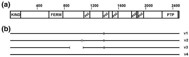
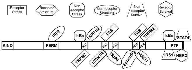
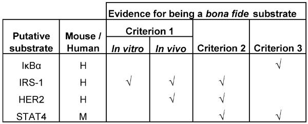
Protein tyrosine phosphatase, PTPL1, (also known as PTPN13, FAP-1, PTP-BAS, PTP1E) is a non-receptor type PTP and, at 270 kDa, is the largest phosphatase within this group. In addition to the well-conserved PTP domain, PTPL1 contains at least 7 putative macromolecular interaction domains. This structural complexity indicates that PTPL1 may modulate diverse cellular functions, perhaps exerting both positive and negative effects. In accordance with this idea, while certain studies suggest that PTPL1 can act as a tumor-promoting gene other experimental studies have suggested that PTPL1 may function as a tumor suppressor. The role of PTPL1 in the cancer cell is therefore likely to be both complex and context dependent with possible roles including the modulation of growth, stress-response, and cytoskeletal remodeling pathways. Understanding the nature of molecular complexes containing PTPL1, its interaction partners, substrates, regulation and subcellular localization are key to unraveling the complex personality of this protein phosphatase.


Figures

Fig. 1
Fig. 1
Homology between different PTPL1 homologues. Protein sequences from H. sapiens (NP_006255.1 v4), C. familiaris (XP_860003.1), M. musculus (NP_035334.1), R. norvegicus (XP_213997.4) and X. laevis (AAR97566) were entered into MatGAT (Matrix Global Alignment Tool) to calculate sequence similarity and identity (http://bitincka.com/ledion/matgat/). Values are percent identity (upper triangle) and percent similarity (lower triangle)
Fig. 2
Fig. 2
Schematic representation of PTPL1 structure and identified splice variants. (a) FERM, PDZ and PTP domains of PTPL1 were identified by using PFAM (http://www.sanger.ac.uk/Software/Pfam/) where the sequence from the longest variant of PTPL1, variant 4, was used. The KIND domains is not recognized by PFAM, therefore its location is arbitrary. (b) Different variants of PTPL1 are depicted with the straight lines where each gap in the sequence represents a splice site
Fig. 3
Fig. 3
Schematic representation of PTPL1 interacting molecules and substrates. Each interacting molecule was assigned to a category respective of its main function in the cell as described in the text. The molecules can be of any group: a receptor molecule in stress-response pathways, a receptor molecule in cytoskeletal/motility pathways, a non-receptor molecule in stress-response pathways, a non-receptor molecule in cell cytoskeletal/motility pathway, a non-receptor molecule in cell survival pathways, or a receptor molecule in cell survival pathways. Each molecule is placed on PTPL1 to where they are described to bind as in the text
Fig. 4
Fig. 4
Putative substrates of PTPL1. According to the guidelines suggested by Tiganis and Bennett [35] for the identification of bona fide PTP substrates, the evidence of a substrate has to be confirmed by three experimental criteria. Criterion 1 states that the presence of an enzyme-substrate complex has to be demonstrated both in vivo and in vitro. Criterion 2 states that the substrate tyrosine levels could be modulated by introducing substrate-trapping mutants of the PTP or altering endogenous PTP protein levels. Criterion 3 states that the phospho-substrate could be dephosphorylated in vitro by the PTP. The experimental evidence provided that fulfills any of these criteria for each putative PTPL1 substrate described in the text has been marked

References

    1. Alonso A, Sasin J, Bottini N, Friedberg I, Osterman A, Godzik A, et al. Protein tyrosine phosphatases in the human genome. Cell. 2004;117(6):699–711. - PubMed
    1. Dube N, Tremblay ML. Involvement of the small protein tyrosine phosphatases TC-PTP and PTP1B in signal transduction and diseases: from diabetes, obesity to cell cycle, and cancer. Biochimica et Biophysica Acta. 2005;1754(1-2):108–117. - PubMed
    1. Mohi MG, Neel BG. The role of Shp2 (PTPN11) in cancer. Current Opinion in Genetics & Development. 2007;17(1):23–30. - PubMed
    1. Ostman A, Hellberg C, Bohmer FD. Protein-tyrosine phosphatases and cancer. Nature Reviews Cancer. 2006;6(4):307–320. - PubMed
    1. Tonks NK. Protein tyrosine phosphatases: from genes, to function, to disease. Nature Reviews. Molecular Cell Biology. 2006;7(11):833–846. - PubMed

MeSH terms

Substances