. 2008 Mar;15(1):29-39.
doi: 10.1080/13506120701815324.

Biochemical and aggregation analysis of Bence Jones proteins from different light chain diseases

Affiliations

Biochemical and aggregation analysis of Bence Jones proteins from different light chain diseases

Laura A Sikkink et al. Amyloid. 2008 Mar.

Abstract

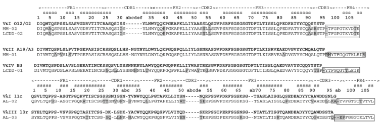
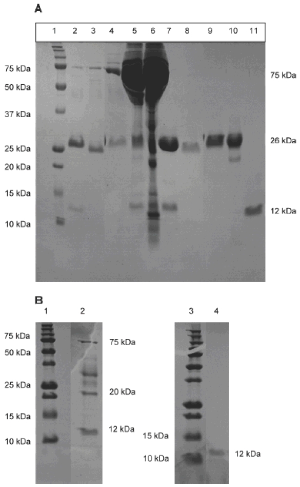
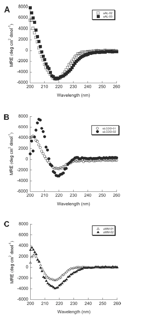
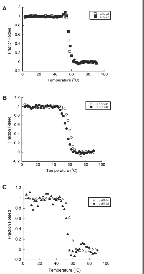
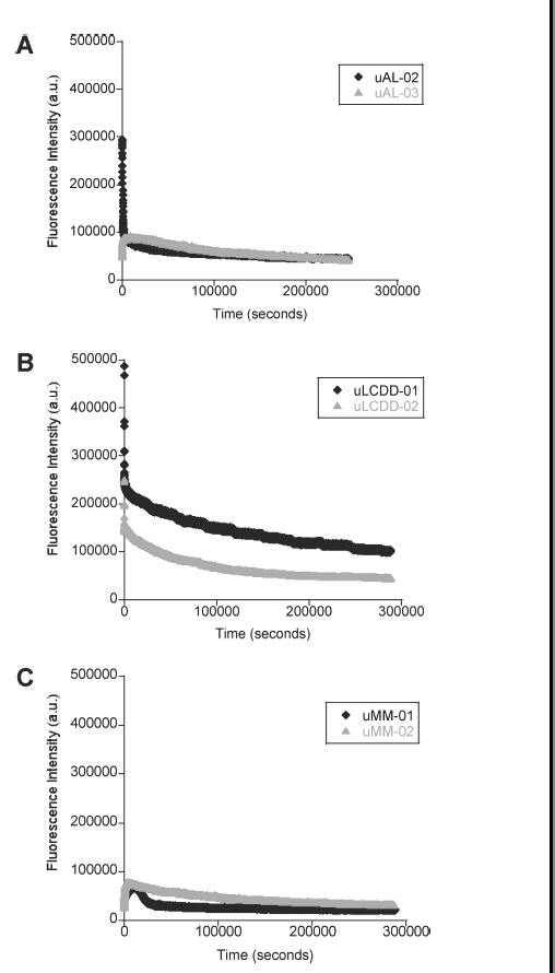
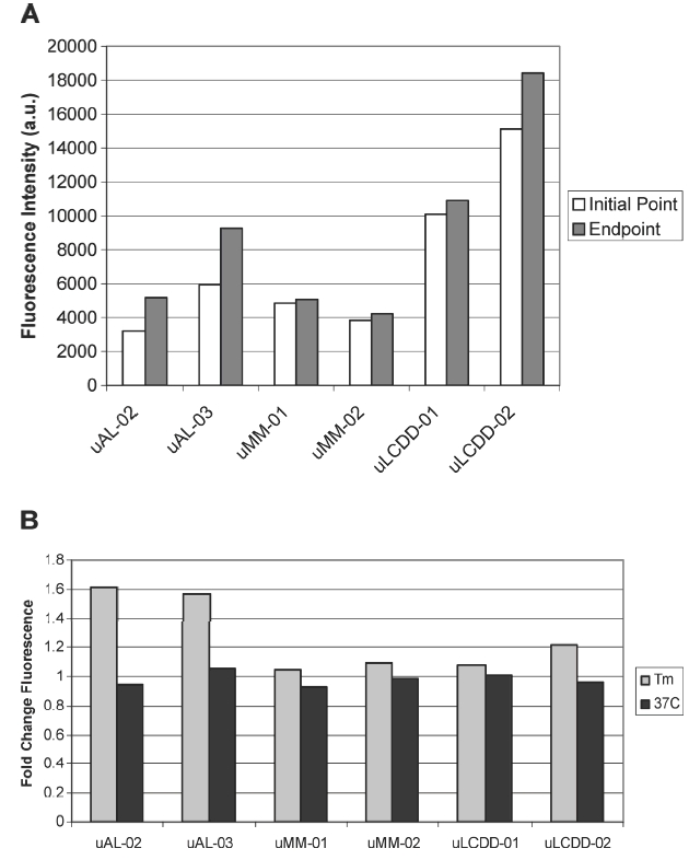
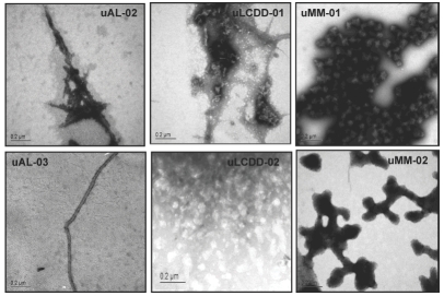
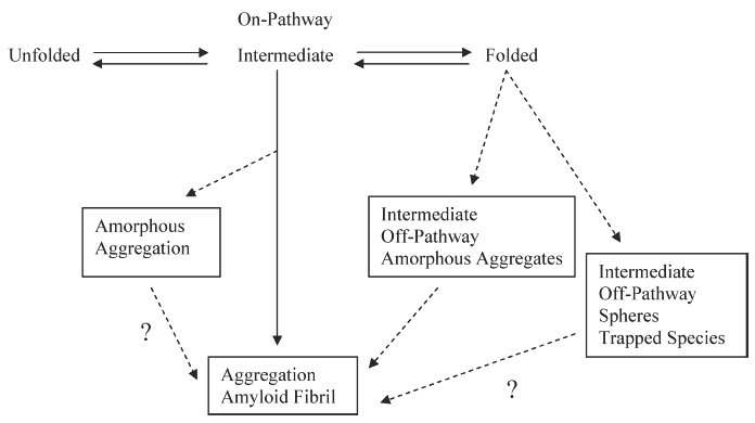
Deposition of immunoglobulin light chains is a result of clonal proliferation of monoclonal plasma cells that secrete free immunoglobulin light chains, also called Bence Jones proteins (BJP). These BJP are present in circulation in large amounts and excreted in urine in various light chain diseases such as light chain amyloidosis (AL), light chain deposition disease (LCDD) and multiple myeloma (MM). BJP from patients with AL, LCDD and MM were purified from their urine and studies were performed to determine their secondary structure, thermodynamic stability and aggregate formation kinetics. Our results show that LCDD and MM proteins have the lowest free energy of folding while all proteins show similar melting temperatures. Incubation of the BJP at their melting temperature produced morphologically different aggregates: amyloid fibrils from the AL proteins, amorphous aggregates from the LCDD proteins and large spherical species from the MM proteins. The aggregates formed under in vitro conditions suggested that the various proteins derived from patients with different light chain diseases might follow different aggregation pathways.


Figures

Figure 1
Figure 1
Sequence alignment of the variable and junction domain portions of the BJP. Donor germline sequence for each Bence Jones protein (BJP) was determined by PCR and sequence analysis. DNA sequences were translated into the amino acid sequence. Mutations in each protein sequence when compared to the germline sequence are highlighted in gray. The junction domain is highlighted with a box. FR, framework region; CDR, complementarity determining region; s, β-strand. The constant domains for AL and MM proteins are not shown here. Kappa proteins (MM) have only one constant domain. The constant domain for AL-02 is IGLC1 and for AL-03 it is IGLC2.
Figure 2
Figure 2
SDS-PAGE of urine and purified protein for each sample shows either a 26 kDa protein band for MM and AL proteins or a 12 kDa protein band for LCDD proteins. (A) Lanes 2–6, urine samples; lanes 7–11, purified protein samples. Lane 1, molecular weight marker; lanes 2 and 7, uMM-01; lanes 3 and 8, uMM-02; lanes 4 and 9, uAL-02; lanes 5 and 10, uAL-03; lanes 6 and 11, uLCDD-01. (B) uLCDD-02 gels: lanes 1 and 3, molecular weight marker; lane 2, urine sample; lane 4, purified protein.
Figure 3
Figure 3
AL, LCDD and MM proteins have a β-sheet conformation by Far UV-CD. All of the BJP in this study display a β-sheet secondary structure based on the minimum ellipticity around 218 nm. (A) AL proteins, (B) LCDD proteins and (C) MM proteins. Experimental conditions: all Far UV-CD scans were performed at 4°C in a 0.2 cm path-length cuvette and in 10 mM Tris–HCl pH 7.4 buffer. Protein concentrations were determined to be between 5.5–25.6 μM.
Figure 4
Figure 4
AL, LCDD and MM proteins have similar two state thermal unfolding transitions. Thermal denaturations were followed by CD following ellipticity at a wavelength of 218 nm. Unfolding and refolding curves were followed for each BJP. (A) AL proteins, (B) LCDD proteins and (C) MM proteins. All the BJP in this study were all able to refold reversibly except for uAL-03. The Tm for the proteins are 57.2°C for uAL-02, 57.3°C for uAL-03, 59.6°C for uLCDD-01, 56.0°C for uLCDD-02, 56.6°C for uMM-01 and 57.8°C for uMM-02. Protein concentrations are between 5.5 and 25.6 μM in 10 mM Tris–HCl pH 7.4 buffer in a 0.2 cm path-length cuvette.
Figure 5
Figure 5
Aggregate formation followed by tryptophan fluorescence. (A) AL proteins, (B) LCDD proteins and (C) MM proteins. Experimental conditions: 10 mM Tris–HCl pH 7.4 buffer, 15 μM ThT, 5 μM protein; incubated at Tm (57°C for uAL-02, uAL-03, uMM-01 and uMM-02, 60°C for uLCDD-01 and uLCDD-02); stirring at 300 rpm for 72 h with a reading occurring every 25 s with an integration time of 0.5 s; excitation wavelength 294 nm and emission wavelength 350 nm.
Figure 6
Figure 6
Aggregate formation followed by thioflavine T fluorescence (ThT). (A) Initial and end-point ThT fluorescence readings at their Tm. (B) Fold change between initial and endpoint ThT fluorescence readings from samples incubated at either their Tm or at 37°C. Experimental conditions: 10 mM Tris–HCl pH 7.4 buffer, 15 μM ThT, 5 μM protein; incubated at Tm (57°C for uAL-02, uAL-03, uMM-01 and uMM-02, 60°C for uLCDD-01 and uLCDD-02) with stirring at 300 rpm for 72 h with a reading occurring every 25 s with an integration time of 0.5 s.
Figure 7
Figure 7
AL proteins show amyloid fibrils. LCDD proteins show amorphous aggregates and MM proteins show spherical species. uAL-03 and uLCDD-02 were stained with 4% uranyl acetate. uAL-02, uLCDD-01, uMM-01 and uMM-02 were stained with 1% phosphotungstic acid. The negatively stained grids were examined using a JEOL 1200 EX transmission electron microscope. Scale bars are located in the lower left corner of each image.
Figure 8
Figure 8
Model for protein folding and misfolding where aggregation may occur through an on or off pathway intermediate.

Similar articles

Cited by

References

    1. Roussel A, Spinelli S, Deret S, Navaza J, Aucouturier P, Cambillau C. The structure of an entire noncovalent immunoglobulin kappa light-chain dimer (Bence-Jones protein) reveals a weak and unusual constant domains association. Eur J Biochem. 1999;26:192–199. - PubMed
    1. Gertz MA, Lacy MQ, Dispenzieri A, Amyloidosis Hematol Oncol Clin North Am. 1999;13:1211–1233. - PubMed
    1. Sezer O, Eucker J, Schmid P, Possinger K. New therapeutic approaches in primary systemic AL amyloidosis. Ann Hematol. 2000;79:1–6. - PubMed
    1. Roye B, Arnulf B, Martinez F, Roy L, Flageul B, Etienne I, Ronco P, Brouet JC, Fermand JP. High dose chemotherapy in light chain or light and heavy chain deposition disease. Kidney Int. 2004;65:642–648. - PubMed
    1. Wetzel R. Domain stability in immunoglobulin light chain deposition disorders. Adv Protein Chem. 1997;50:183–242. - PubMed

Publication types

MeSH terms

Substances