. 2008 Feb 8;4(2):e21.
doi: 10.1371/journal.ppat.0040021.

Brucella control of dendritic cell maturation is dependent on the TIR-containing protein Btp1

Affiliations

Brucella control of dendritic cell maturation is dependent on the TIR-containing protein Btp1

Suzana P Salcedo et al. PLoS Pathog. .

Abstract

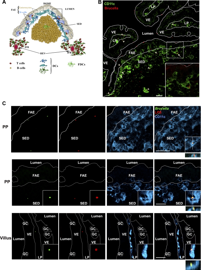
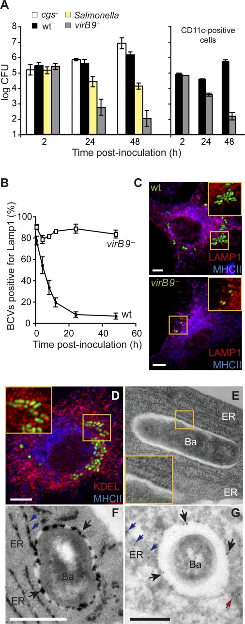
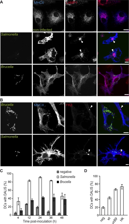
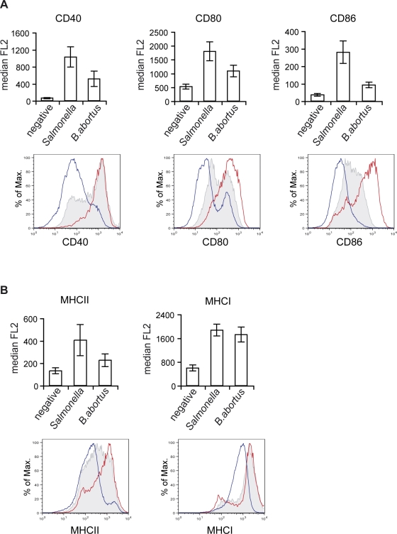
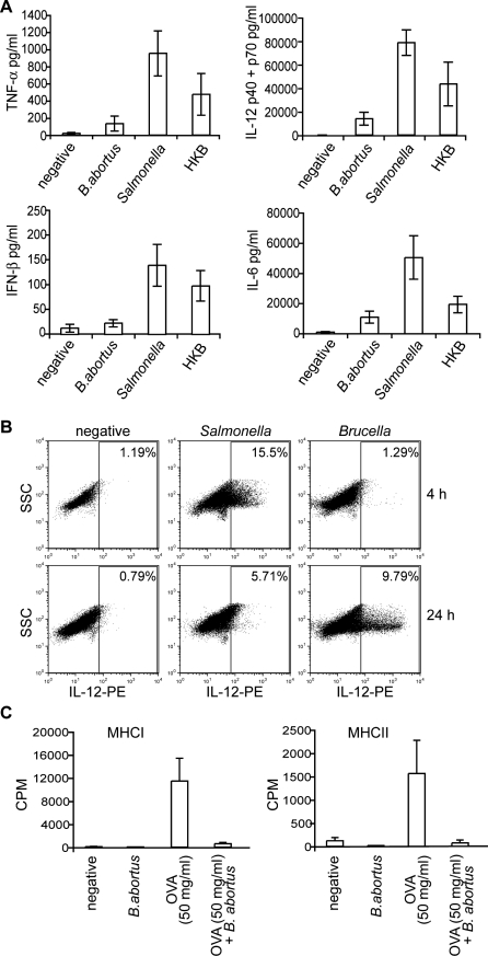
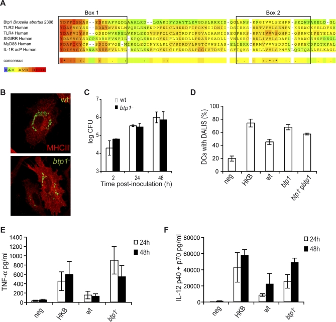
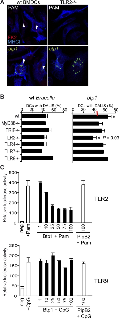
Brucella is an intracellular pathogen able to persist for long periods of time within the host and establish a chronic disease. We show that soon after Brucella inoculation in intestinal loops, dendritic cells from ileal Peyer's patches become infected and constitute a cell target for this pathogen. In vitro, we found that Brucella replicates within dendritic cells and hinders their functional activation. In addition, we identified a new Brucella protein Btp1, which down-modulates maturation of infected dendritic cells by interfering with the TLR2 signaling pathway. These results show that intracellular Brucella is able to control dendritic cell function, which may have important consequences in the development of chronic brucellosis.


Conflict of interest statement

Competing interests. The authors have declared that no competing interests exist.

Figures

Figure 1
Figure 1. Infection of DCs from the Sub-epithelial Dome Region of Ileal Peyer's Patches with B. abortus
(A) Cellular organization of a Peyer's patch dome. DCs are enriched in the Sub-Epithelial Dome (SED) underlying the Follicle-Associated Epithelium (FAE) and in the Inter-Follicular Region (IFR) in vicinity of the High Endothelial Venules (HEV). (B) Sixty minutes after intestinal loop infection, B. abortus (red) adhered on both villi epithelium (VE) and FAE surfaces. However, only few of them were observed in villi lamina propria (LP). These were always observed in association with CD11c+ cells (green, arrowhead). Most B. abortus bacteria reached the under-epithelial compartment of the gut through the FAE. Some of these were observed inside CD11c+ cells (arrows and insert), while others were in close association with CD11c+ dendrites (arrowheads). (C) High magnification field of a Peyer's patch infected with GFP-expressing Brucella. Images in the first and the second row, corresponding to area depicted by the squared box in (A), show either Brucella (GFP in green and LPS in red) internalized by CD11+ cells (blue, representative of 29 out of 118 B. abortus-associated cells) or the close association of one B. abortus expressing GFP with dendrites from a CD11c+ cell of the SED region (representative of 30 out of 118 B. abortus-associated cells), respectively. The image in the last row shows one of the rare B. abortus-associated cells (all CD11c+) from the villus (GC, goblet cells). Close association or internalization was always confirmed by the combined xy, xz, and/or yz views as shown in inserts. Bars, 20 μm.
Figure 2
Figure 2. Survival and Replication of B. abortus in DCs
(A) BMDCs infected with wild-type B. abortus, cgs mutant, virB9 mutant or the wild-type S. typhimurium were lysed and intracellular CFUs enumerated at different times after inoculation. Graph on the right corresponds to bacterial CFU counts performed in CD11c-positive cells infected with either wild-type B. abortus or virB9 mutant. Statistical significance was obtained at 24 and 48 h between wild-type and the virB mutant (p < 0.002) and also between wild-type Brucella and Salmonella at 24 h (p = 0.047) and 48 h (p < 0.002). (B) Quantification of the percentage of wild-type or virB9 mutant BCVs that contain LAMP1 by confocal immunofluorescence microscopy. The difference between wild-type and the virB mutant were always statistically significant (p = 0.045 at 30 min and 4 h; p < 0.002 for other timepoints). (C) Representative confocal images of DCs infected for 24 h with wild-type B. abortus or virB9 mutant expressing GFP, labelled with antibodies against MHC II (blue) and LAMP1 (red). (D) Confocal image of DCs infected for 24 h with wild-type B. abortus expressing GFP, labelled with antibodies against MHC II (blue) and KDEL (red). Samples were also processed for conventional electron microscopy (E), cytochemistry for glucose 6 phosphatase detection (F) or immunogold labelling with an anti-calnexin antibody (G). Bars: 5 μm (C and D); 0.5 μm (F and G). For (A) and (B) data are means ± standard errors of four independent experiments.
Figure 3
Figure 3. B. abortus Interferes with DC Maturation
DCs were infected with different B. abortus strains or with Salmonella and labelled with the anti-MHC II and anti-LAMP1 antibodies (A) or FK2 antibody (B) to detect MVBs or DALIS, respectively. (C) Comparison between the percentages of DCs infected with either wild-type B. abortus (black bars) or S. typhimurium (white bars) that contain DALIS. The negative control (grey bars) corresponds to mock-infected DCs with DALIS. The difference between Brucella and Salmonella infected cells at 24 h is statistically significant, p < 0.0001. (D) Quantification of the percentage of DCs with DALIS at 24 h after incubation with media (negative), wild-type B. abortus (wt), virB9 mutant or heat-killed B. abortus (HK). The differences between wild-type wt and either virB9 or HK are statistically significant (p = 0.0059 and p = 0.018, respectively). For (C) and (D) data represent means ± standard errors of at least three independent experiments. Bars: 10 μm (A); 5 μm (B).
Figure 4
Figure 4. B. abortus-infected DCs Have Reduced Expression of Co-stimulatory Molecules
(A and B) DCs were either incubated with media only (blue line) or infected with B. abortus (shaded grey) or S. typhimurium (red line) expressing GFP for 24 h. DCs were labelled with anti-CD11c conjugated with APC and anti-CD40, CD80, CD86, MHC II, and MHCI antibodies conjugated with PE. Representative histograms and values of median of the PE fluorescence (FL2) correspond to CD11c+ cells for the negative and CD11c+ GFP+ cells for the Brucella and Salmonella infected cells. Values of the median fluorescence correspond to four independent experiments.
Figure 5
Figure 5. B. abortus Interferes with DC Function
(A) and (B) B. abortus blocks secretion of pro-inflammatory cytokines. (A) Supernatants were obtained at 24 h from DCs inoculated with either media (negative), B. abortus, S. typhimurium, or heat-killed B. abortus (HKB) and the levels of cytokines were determined by ELISA. Values correspond to means ± standard errors of at least three independent experiments. A clear statistical difference was observed between Brucella and Salmonella for TNF (p = 0.014) and IL12 (p = 0.0098) and to a lesser extent for IL6 (p = 0.041) and IFNß (p = 0.040). (B) DCs were infected for 4 or 24 h with either media (negative), B. abortus or S. typhimurium constitutively expressing GFP. DCs were then treated for 1 h with brefeldin A, permeabilised and labelled with anti-CD11c and anti-IL12 (p40/p70) conjugated with APC and PE, respectively. Representative dot blots are shown for CD11c+ populations. (C) B. abortus reduces the capability of DCs to induce T cell proliferation. DCs were infected with wild-type B. abortus and the model antigen ovabulmin (OVA) was added after 30 min at a final concentration of 50 μg/ml. Cells were stimulated with OVA for 2 h and then washed to remove the antigen. Splenic CD4+ T cells prepared from OTI and OTII transgenic mice were then added at a 1:1 ratio and co-cultured for 48 h. IL-2 production in culture supernatants was measured by 3H-thymidine incorporation on the IL-2-dependent CTLL-2 cell line. Data are the means ± standard errors from triplicates of a representative experiment. A small statistical difference was observed between Ova and Ova+Brucella for both OT1 (p = 0.035) and OT2 (p = 0.045).
Figure 6
Figure 6. Btp1 Reduces DC Maturation
(A) Identification of Btp1 as bacterial member of TLR/IL-1R (TIR) family. Comparison of the predicted amino acid sequences of Btp1 with the human members of the TIR family: TLR2, TLR4, Single Immunoglobulin IL1-R related molecule (SIGIRR), Myeloid Differentiation protein 88 (MyD88), and IL-1 Receptor accessory protein (IL-1R acP). Significant sequence similarity is observed in Box 1 and Box 2, which are important signatures of all TIR domain proteins. The alignment was constructed with T-Coffee::advanced server from EMBnet (http://www.ch.embnet.org/). The color scheme is represented in the figure and is indicative of the reliability of the alignment, with red corresponding to highest probability of correct alignment. DCs infected with wild-type B. abortus or btp1 were either (B) fixed at 24 h and immunolabelled for MHC II (red) and Brucella (green) or (C) cells were lysed at 2, 24, and 48 h and CFU enumerated. (D) Quantification of the percentage of DCs containing DALIS. DCs were incubated for 24 h with media (negative), heat-killed B. abortus (HKB), wild-type B. abortus (wt), btp1 mutant strain or a btp1 mutant with a plasmid containing btp1 (btp1 pbtp1). There is a very significant statistical difference between the wt and btp1 (p = 0.0002) but only a small difference between btp1 and btp1 pbtp1 (p = 0.05). (E and F) Analysis of TNF-α and IL-12 (p40/p70) secretion measured by ELISA from the supernatant of DCs 24 and 48 h after inoculation. All the results correspond to the means ± standard errors of four independent experiments. A statistical difference was observed between wild-type Brucella and btp1 mutant at 24 h for TNF (p = 0.029) and IL12 (p = 0.045).
Figure 7
Figure 7. B. abortus Protein Btp1 Modulates TLR Signaling
(A) Representative images obtained by immunofluorescence microscopy of DCs from wild-type mice or TLR2 knockout mice labelled for MHC II (blue), DALIS (red), and in the case of lower panel also labelled for Brucella (green). Cell were either stimulated with PAM (500 ng/μl, upper panel) or infected with the Brucella wild-type and btp1 mutant strains (lower panel). (B) DCs obtained from wild-type mice or different knockout mice were infected for 24 h with either B. abortus wild-type or btp1 mutant strain. The percentage of DALIS in infected cells was then quantified and the data represent means ± standard errors of four independent experiments. The average value for the wild-type Brucella is indicated with a red line. For btp1 we observed a statistical difference (*) between the wt and TLR2−/− DCs (p = 0.031). (C) HEK293 cells were transiently transfected for 24 h with three vectors: a luciferase reporter vector and either empty vector, myc-Btp1 or myc-PipB2 along with either TLR2 (upper panel) or TLR9 (lower panel). The amounts of Btp plasmids are indicated in the graph (ng). Data represent the means ± standard errors of relative luciferase activity obtained from triplicates of a representative experiment. In the case of TLR2, cells were stimulated with PAM (500 ng/μl) and for TLR9 with CpG (1 μM) for 6 h before measuring the luciferase activity.

References

    1. Geijtenbeek TB, Van Vliet SJ, Koppel EA, Sanchez-Hernandez M, Vandenbroucke-Grauls CM, et al. Mycobacteria target DC-SIGN to suppress dendritic cell function. J Exp Med. 2003;197:7–17. - PMC - PubMed
    1. Skinner JA, Pilione MR, Shen H, Harvill ET, Yuk MH. Bordetella type III secretion modulates dendritic cell migration resulting in immunosuppression and bacterial persistence. J Immunol. 2005;175:4647–4652. - PubMed
    1. Svensson M, Johansson C, Wick MJ. Salmonella enterica serovar typhimurium-induced maturation of bone marrow-derived dendritic cells. Infect Immun. 2000;68:6311–6320. - PMC - PubMed
    1. Petrovska L, Aspinall RJ, Barber L, Clare S, Simmons CP, et al. Salmonella enterica serovar Typhimurium interaction with dendritic cells: impact of the sifA gene. Cell Microbiol. 2004;6:1071–1084. - PubMed
    1. Tobar JA, Gonzalez PA, Kalergis AM. Salmonella escape from antigen presentation can be overcome by targeting bacteria to Fc gamma receptors on dendritic cells. J Immunol. 2004;173:4058–4065. - PubMed

Publication types

MeSH terms