. 2008 Feb;4(2):e30.
doi: 10.1371/journal.pgen.0040030.

X chromosome activity in mouse XX primordial germ cells

Affiliations

X chromosome activity in mouse XX primordial germ cells

Susana M Chuva de Sousa Lopes et al. PLoS Genet. 2008 Feb.

Abstract

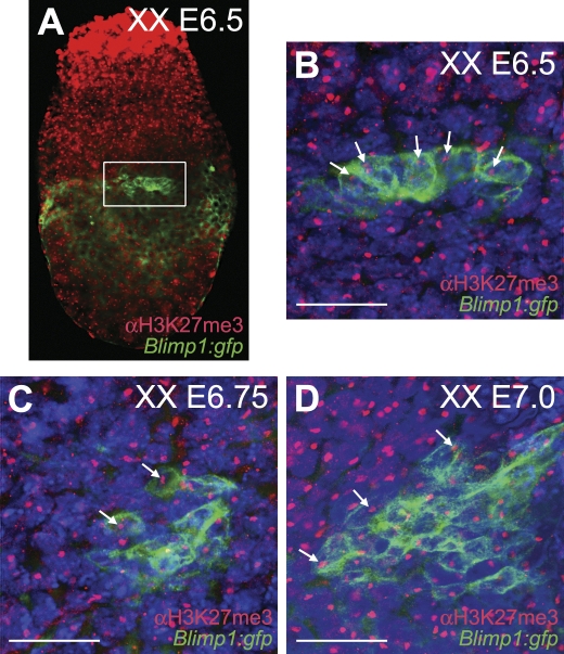
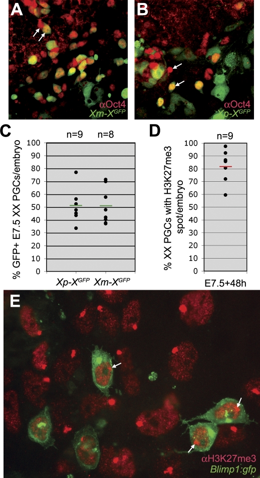
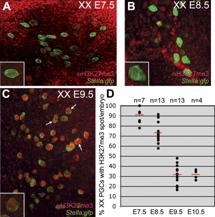
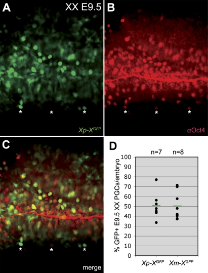
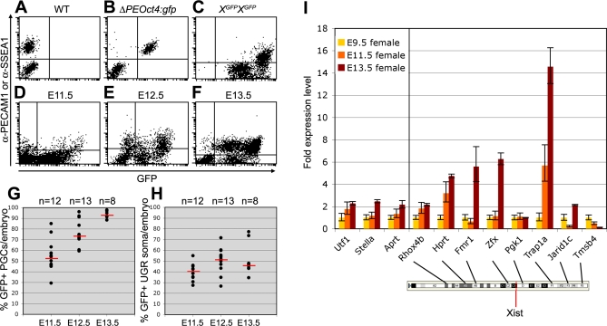
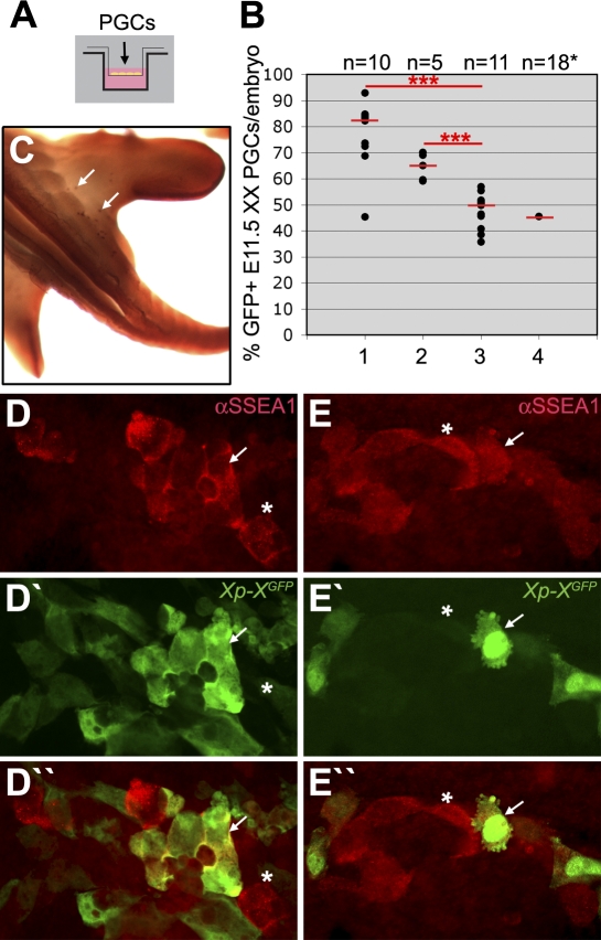
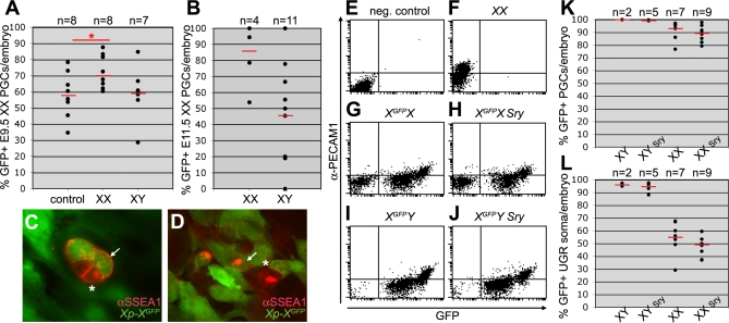
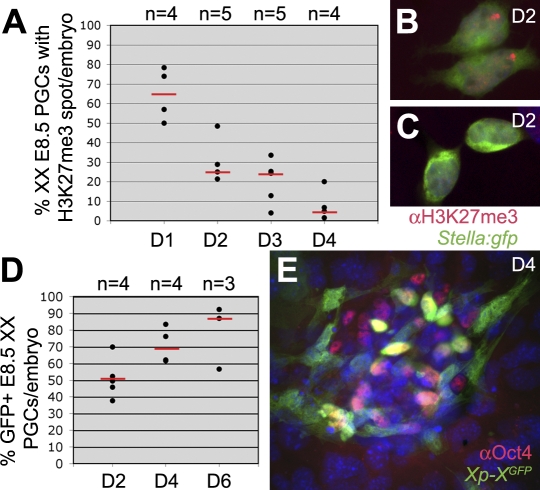
In the early epiblast of female mice, one of the two X chromosomes is randomly inactivated by a Xist-dependent mechanism, involving the recruitment of Ezh2-Eed and the subsequent trimethylation of histone 3 on lysine 27 (H3K27me3). We demonstrate that this random inactivation process applies also to the primordial germ cell (PGC) precursors, located in the proximal region of the epiblast. PGC specification occurs at about embryonic day (E)7.5, in the extraembryonic mesoderm, after which the germ cells enter the endoderm of the invaginating hindgut. As they migrate towards the site of the future gonads, the XX PGCs gradually lose the H3K27me3 accumulation on the silent X chromosome. However, using a GFP transgene inserted into the X chromosome, we observed that the XX gonadal environment (independently of the gender) is important for the substantial reactivation of the inactive X chromosome between E11.5 and E13.5, but is not required for X-chromosome reactivation during the derivation of pluripotent embryonic germ cells. We describe in detail one of the key events during female PGC development, the epigenetic reprogramming of the X chromosome, and demonstrate the role of the XX somatic genital ridge in this process.


Conflict of interest statement

Competing interests. The authors have declared that no competing interests exist.

Figures

Figure 1
Figure 1. PGC Precursors Show a Pronounced H3K27me3 Nuclear Accumulation Indicative of One Inactive X Chromosome
XX Blimp1:gfp mouse embryos were immunostained for H3K27me3. GFP under the control of the Blimp1 promoter was tagged to the cell membrane. (A) E6.5 whole embryo showing in the white box the Blimp1(GFP)-positive PGC progenitors. (B) Magnification of the Blimp1(GFP)-positive PGC progenitor cluster depicted in (A). White arrows indicate individual H3K27me3-positive nuclear accumulation in the PGC progenitors. Note that all somatic cells exhibit a H3K27me3 nuclear accumulation. (C) E6.75 and (D) E7.0 whole embryos similarly stained.
Figure 2
Figure 2. X-chromosome Inactivation in E7.5 PGCs Is Random
(A–C) The posterior part of XX E7.5 XGFP embryos, which inherited X-linked GFP either from the mother (A) or the father (B), were cultured for 48 hours and immunostained for Oct4 to identify the PGCs. White arrows depict PGCs that do or do not co-express GFP. The expression of GFP was analysed in the total number of PGCs per embryo (C). Green bars depict the median, n is the total number of embryos analysed. (D,E) The posterior part of XX E7.5 Blimp1:gfp embryos were cultured for 48 hours and immunostained for H3K27me3. The total number of Blimp1(GFP)-positive PGCs was screened for the presence of a H3K27me3-positive nuclear accumulation (D) Red bar depicts the median, n is the total number of embryos analysed. A representative image of the posterior cluster containing the Blimp1(GFP)-positive PGCs after 48 hours culture is shown in (E). White arrows show Blimp1(GFP)-positive PGCs that contain a prominent H3K27me3 nuclear accumulation.
Figure 3
Figure 3. During PGC Migration the H3K27me3 Nuclear Accumulation Is Gradually Lost
The total number of PGCs per embryo was analysed in XX Stella:gfp embryos after whole mount immunostaining for H3K27me3. Stella is a marker of specified PGCs. (A–C) E7.5 (A), E8.5 (B), and E9.5 (C) confocal projections showing all PGCs present in the depicted area. Magnification of single Stella(GFP)-positive PGCs containing a prominent nuclear H3K27me3 accumulation are depicted in the lower right corner of (A–C). Note that the overall levels of H3K27me3 in the nucleus increase during development (white arrows in C). (D) The percentage of PGCs containing a prominent H3K27me3 accumulation from E7.5 to E10.5. Red bars depict the median, n is the total number of embryos analysed.
Figure 4
Figure 4. X-chromosome Inactivation in E9.5 PGCs Is Still Random
The hindgut of XX E9.5 XGFP embryos, which inherited X-linked GFP either from the mother or the father, were immunostained whole mount for Oct4 to identify the PGCs. (A–C) White asterisks depict PGCs that do or do not co-express GFP. Note that the lumen of the hindgut shows aspecific Oct4 immunostaining. (D) The expression of GFP was analysed in the total number of PGCs per embryo. Green bars depict the median, n is the total number of embryos analysed. Red bar depicts the median, n is the total number of embryos analysed.
Figure 5
Figure 5. Xi Reactivates in XX PGCs between E11.5 and E13.5
(A,B) FACS-analysis of XX E13.5 WT (A) and ΔPEOct4:gfp (B) PGCs show that anti-PECAM1 is a suitable antibody to separate PGCs from the surrounding somatic tissue. Anti-SSEA1 was used to separate XX E11.5 and E12.5 PGCs from the surrounding somatic tissue. (C) FACS-analysis of XX E13.5 XGFP homozygous genital ridges containing 100% GFP-positive cells, used as positive control. (D–F) Representative dot-plots showing FACS-analysis of XX E11.5 (D), E12.5 (E), and E13.5 (F) genital ridges from individual Xp-XGFP embryos. (G,H) The percentage of FACS-analysed GFP-expressing PGCs (G) and surrounding somatic tissue (H) in the genital ridges of individual XX E11.5, E12.5, and E13.5 Xp-XGFP embryos. PGCs and somatic cells were respectively positive and negative for SSEA1 (E11.5, 12.5) or PECAM1 (E13.5). Red bars depict the median, n is the total number of embryos analysed. (I) Transcriptional levels of X-coded genes and autosomal genes at E9.5, E11.5, and E13.5. Shown are the relative transcription levels of each gene compared to the expression of that same gene observed at E9.5. The localization of the X-coded genes analysed and Xist are shown on the cartoon of the X chromosome (from Ensembl).
Figure 6
Figure 6. XX E11.5 PGCs Require Somatic XX UGR To Reactivate the Xi
(A) A transwell system was used to study the X-chromosome reactivation in XX E11.5 XGFP PGCs cultured in the top compartment for 48 hours. (B) The percentage of X-linked GFP-expressing PGCs in XX embryos. Genital ridges were dissociated and added to the top compartment (lane 1) or FACS-sorted for SSEA-1, with PGCs (SSEA1-positive) added to the top compartment and somatic tissue (SSEA1-negative) to the lower compartment (lane 2). Lane 3, FACS-sorted PGCs (SSEA1-positive) were added in the top compartment. Lane 4, the base of the tail, containing ectopic PGCs, was dissociated and cultured in the top compartment. Due to the low number of PGCs present ectopically, the results of 18 individual tails analysed were pooled. Red bars depict the median, n is the total number of embryos analysed. ***, p < 0.001. (C) E11.5 embryo showing ectopic PGCs, positive for alkaline phosphatase-activity (white arrows), in the base of the tail. (D,E) GFP-positive (white arrow) and GFP-negative (asterisk) Xp-XGFP PGCs (SSEA1-positive) cultured together with SSEA1-negative somatic tissue (D) or with culture medium alone (E).
Figure 7
Figure 7. XX, but Not XY, UGRs Induce Xi Reactivation in E9.5 and E11.5 XX PGCs
A transwell system was used to study the X-chromosome reactivation in E9.5 and E11.5 XGFP PGCs cultured for a period of 48 hours. (A) The percentage of X-linked GFP-expressing XX E9.5 PGCs after culture. Hindgut and the surrounding mesentery were dissociated and cultured in the top compartment with the lower compartment containing: medium only (control), one dissociated XX E11.5 UGR pair (XX), or one dissociated XY E11.5 UGR pair (XY). Red bars depict the median, n is the total number of transwell filter membranes (each containing 1.5× embryo) analysed. *, p < 0.05. (B) The percentage of X-linked GFP-expressing XX E11.5 PGCs after culture. FACS-sorted PGCs (SSEA1-positive) were cultured in the top compartment with FACS-sorted XX (XX) or XY (XY) E11.5 UGR somatic tissue (SSEA1-negative). Red bars depict the median, n is the total number of embryos analysed. (C,D) GFP-positive (white arrow) and GFP-negative (asterisk) Xp-XGFP E9.5 PGCs (SSEA1-positive) after culture. (E–J) Representative dot-plots showing FACS-analysis of E13.5 genital ridges from individual XX (E,F), XGFPX (G), XGFPX Sry (H), XGFPY (I), and XGFPY Sry (J) embryos. (K,L) The percentage of FACS-analysed GFP-expressing PGCs (K) and surrounding somatic tissue (L) in the genital ridges of individual XGFPX, XGFPX Sry, XGFPY, and XGFPY Sry E13.5 embryos. PGCs and somatic cells were respectively positive and negative for PECAM1. Red bars depict the median, n is the total number of embryos analysed.
Figure 8
Figure 8. Kinetics of Xi Reactivation during EGC Derivation from XX E8.5 PGCs
(A) The percentage of XX Stella:gfp PGCs containing a prominent H3K27me3 nuclear accumulation during the first 4 days (D1-D4) of EGC derivation. Red bars depict the median, n is the total number of embryos analysed. (B,C) Stella(GFP)-positive PGCs after 48 hours culture towards EGCs, showing a prominent H3K27me3 nuclear accumulation and a migratory phenotype (B) or rounding up and containing no H3K27me3 accumulation or nuclear staining (C). (D) The percentage of GFP-expressing XX E8.5 Xp-XGFP PGCs (Oct4-positive) per embryo during the first 6 days (D) of EGC derivation. Red bars depict the median, n is the total number of embryos analysed. (E) Oct4-positive PGCs after 4 days of culture showed a mixed population of GFP-positive and GFP-negative cells.

Similar articles

Cited by

References

    1. Mak W, Nesterova TB, de Napoles M, Appanah R, Yamanaka S, et al. Reactivation of the paternal X chromosome in early mouse embryos. Science. 2004;303:666–669. - PubMed
    1. Okamoto I, Otte AP, Allis CD, Reinberg D, Heard E. Epigenetic dynamics of imprinted X inactivation during early mouse development. Science. 2004;303:644–649. - PubMed
    1. Rastan S. Timing of X-chromosome inactivation in postimplantation mouse embryos. J Embryol Exp Morphol. 1982;71:11–24. - PubMed
    1. Rastan S, Kaufman MH, Handyside AH, Lyon MF. X-chromosome inactivation in extra-embryonic membranes of diploid parthenogenetic mouse embryos demonstrated by differential staining. Nature. 1980;288:172–173. - PubMed
    1. McMahon A, Fosten M, Monk M. Random X-chromosome inactivation in female primordial germ cells in the mouse. J Embryol Exp Morphol. 1981;64:251–258. - PubMed

Publication types

MeSH terms