

Prenatal alcohol exposure: foetal programming, the hypothalamic-pituitary-adrenal axis and sex differences in outcome
- PMID: 18266938
- PMCID: PMC8942074
- DOI: 10.1111/j.1365-2826.2008.01669.x
Prenatal alcohol exposure: foetal programming, the hypothalamic-pituitary-adrenal axis and sex differences in outcome
Abstract
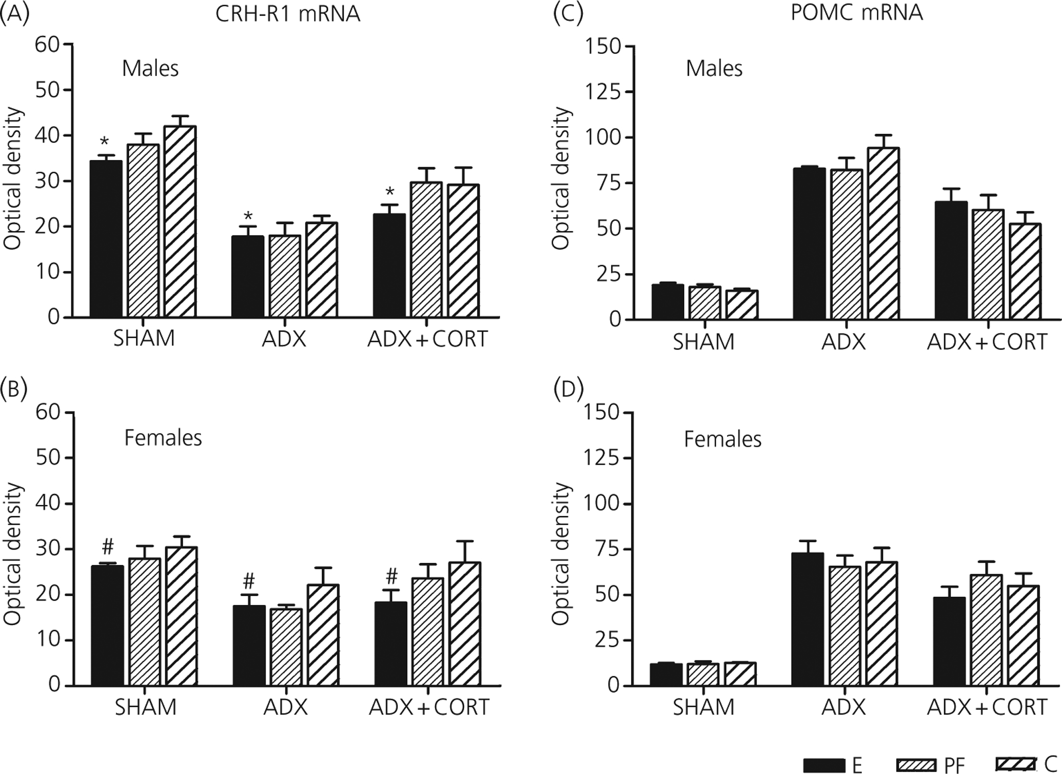
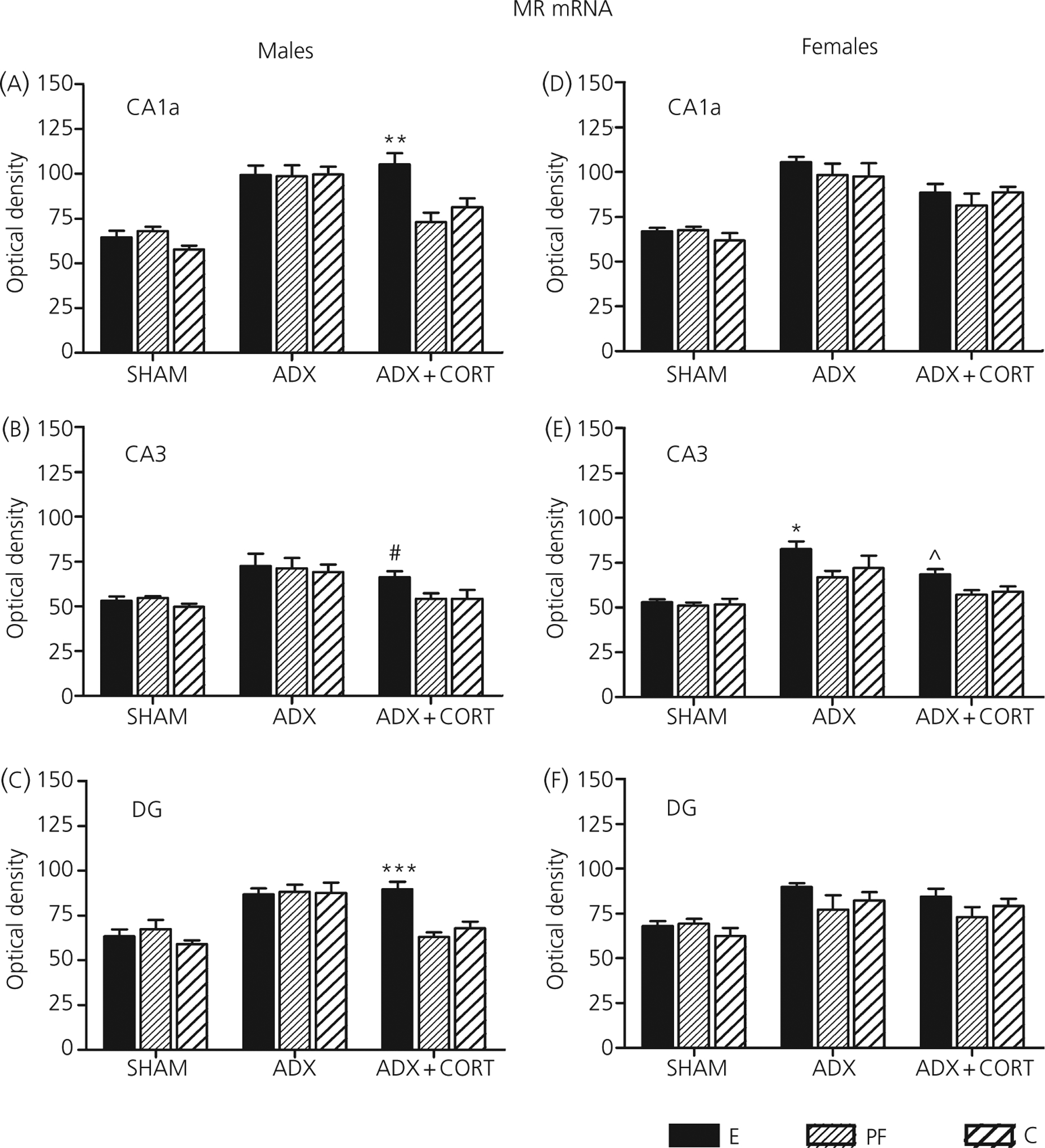
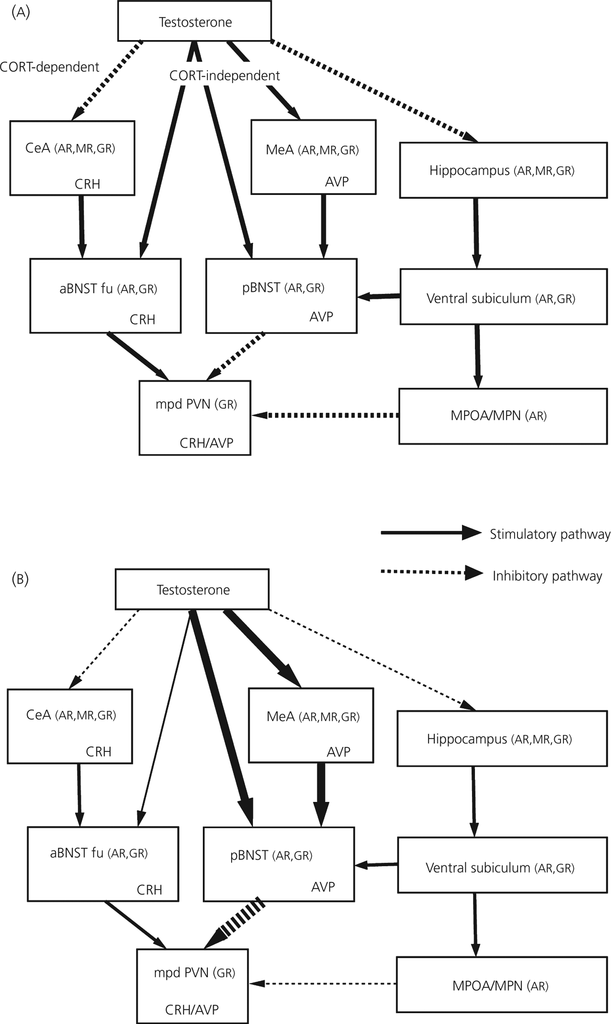
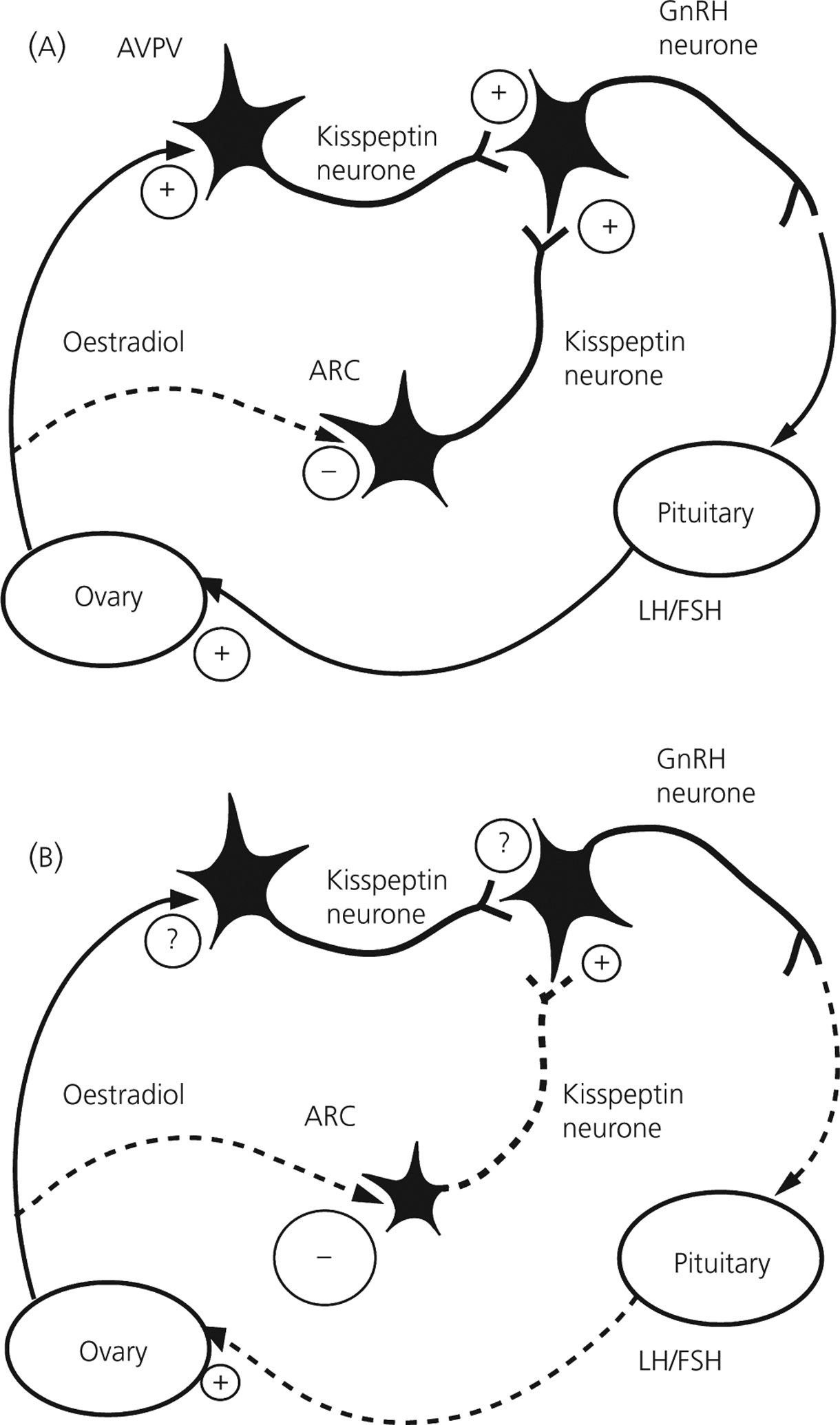
Prenatal exposure to alcohol has adverse effects on offspring neuroendocrine and behavioural functions. Alcohol readily crosses the placenta, thus directly affecting developing foetal endocrine organs. In addition, alcohol-induced changes in maternal endocrine function can disrupt the normal hormonal interactions between the pregnant female and foetal systems, altering the normal hormone balance and, indirectly, affecting the development of foetal metabolic, physiological and endocrine functions. The present review focuses on the adverse effects of prenatal alcohol exposure on offspring neuroendocrine function, with particular emphasis on the hypothalamic-pituitary-adrenal (HPA) axis, a key player in the stress response. The HPA axis is highly susceptible to programming during foetal and neonatal development. Here, we review data demonstrating that alcohol exposure in utero programmes the foetal HPA axis such that HPA tone is increased throughout life. Importantly, we show that, although alterations in HPA responsiveness and regulation are robust phenomena, occurring in both male and female offspring, sexually dimorphic effects of alcohol are frequently observed. We present updated findings on possible mechanisms underlying differential effects of alcohol on male and female offspring, with special emphasis on effects at different levels of the HPA axis, and on modulatory influences of the hypothalamic-pituitary-gonadal hormones and serotonin. Finally, possible mechanisms underlying foetal programming of the HPA axis, and the long-term implications of increased exposure to endogenous glucocorticoids for offspring vulnerability to illnesses or disorders later in life are discussed.
Figures
Similar articles
-
Prenatal alcohol exposure and fetal programming: effects on neuroendocrine and immune function.Exp Biol Med (Maywood). 2005 Jun;230(6):376-88. doi: 10.1177/15353702-0323006-05. Exp Biol Med (Maywood). 2005. PMID: 15956767 Review.
-
Prenatal ethanol exposure enhances the susceptibility to metabolic syndrome in offspring rats by HPA axis-associated neuroendocrine metabolic programming.Toxicol Lett. 2014 Apr 7;226(1):98-105. doi: 10.1016/j.toxlet.2014.01.023. Epub 2014 Jan 26. Toxicol Lett. 2014. PMID: 24472613
-
Hypothalamic-pituitary-adrenal responses to 5-HT1A and 5-HT2A/C agonists are differentially altered in female and male rats prenatally exposed to ethanol.Alcohol Clin Exp Res. 2007 Feb;31(2):345-55. doi: 10.1111/j.1530-0277.2006.00316.x. Alcohol Clin Exp Res. 2007. PMID: 17250628
-
Periconceptional ethanol exposure alters hypothalamic-pituitary-adrenal axis function, signalling elements and associated behaviours in a rodent model.Psychoneuroendocrinology. 2020 Dec;122:104901. doi: 10.1016/j.psyneuen.2020.104901. Epub 2020 Oct 7. Psychoneuroendocrinology. 2020. PMID: 33070024
-
Role of various neurotransmitters in mediating the long-term endocrine consequences of prenatal alcohol exposure.Ann N Y Acad Sci. 2008 Nov;1144:176-88. doi: 10.1196/annals.1418.015. Ann N Y Acad Sci. 2008. PMID: 19076376 Free PMC article. Review.
Cited by
-
Neurobehavioral Disorder Associated with Prenatal Alcohol Exposure (ND-PAE): Proposed DSM-5 Diagnosis.Child Psychiatry Hum Dev. 2016 Apr;47(2):335-46. doi: 10.1007/s10578-015-0566-7. Child Psychiatry Hum Dev. 2016. PMID: 26202432 Review.
-
Prenatal alcohol exposure and adolescent stress increase sensitivity to stress and gonadal hormone influences on cognition in adult female rats.Physiol Behav. 2015 Sep 1;148:157-65. doi: 10.1016/j.physbeh.2015.02.033. Epub 2015 Feb 21. Physiol Behav. 2015. PMID: 25707383 Free PMC article.
-
Hypothalamic-pituitary-adrenal axis and behavioral dysfunction following early binge-like prenatal alcohol exposure in mice.Alcohol. 2015 May;49(3):207-17. doi: 10.1016/j.alcohol.2015.01.005. Epub 2015 Jan 26. Alcohol. 2015. PMID: 25709101 Free PMC article.
-
Prenatal Adversity Alters the Epigenetic Profile of the Prefrontal Cortex: Sexually Dimorphic Effects of Prenatal Alcohol Exposure and Food-Related Stress.Genes (Basel). 2021 Nov 9;12(11):1773. doi: 10.3390/genes12111773. Genes (Basel). 2021. PMID: 34828381 Free PMC article.
-
The Placenta as a Target for Alcohol During Pregnancy: The Close Relation with IGFs Signaling Pathway.Rev Physiol Biochem Pharmacol. 2021;180:119-153. doi: 10.1007/112_2021_58. Rev Physiol Biochem Pharmacol. 2021. PMID: 34159446 Review.
References
-
- Morgan MY. Alcohol and nutrition. Br Med Bull 1982; 38: 21–29. - PubMed
-
- Adler RA. Clinical review 33: clinically important effects of alcohol on endocrine function. J Clin Endocrinol Metab 1992; 74: 957–960. - PubMed
-
- Glavas MM, Weinberg J. Stress alcohol and the hypothalamic–pituitary–adrenal axis. In: Yehuda S, Mostofsky D, eds. Stress, Nutrition and Medical Disorders. Totowa, NJ: Humana Press Inc., 2005: 165–184.
-
- Anderson RA Jr. Endocrine balance as a factor in the etiology of the fetal alcohol syndrome. Neurobehav Toxicol Teratol 1981; 3: 89–104. - PubMed
-
- Rudeen PK, Taylor JA. Fetal alcohol neuroendocrinopathies. In: Watson RR, ed. Alcohol and Neurobiology: Brain Development and Hormone Regulation. Boca Raton, FL: CRC Press, 1992: 109–138.
Publication types
MeSH terms
Substances
Grants and funding
LinkOut - more resources
Full Text Sources
Other Literature Sources
Medical
