. 2008 Dec;12(6B):2704-16.
doi: 10.1111/j.1582-4934.2008.00255.x. Epub 2008 Feb 4.

Acrolein: unwanted side product or contribution to antiangiogenic properties of metronomic cyclophosphamide therapy?

Affiliations

Acrolein: unwanted side product or contribution to antiangiogenic properties of metronomic cyclophosphamide therapy?

M Günther et al. J Cell Mol Med. 2008 Dec.

Abstract

Tumour therapy with cyclophosphamide (CPA), an alkylating chemotherapeutic agent, has been associated with reduced tumour blood supply and antiangiogenic effects when applied in a continuous, low-dose metronomic schedule. Compared to conventional high-dose scheduling, metronomic CPA therapy exhibits antitumoural activity with reduced side effects. We have studied potential antiangiogenic properties of acrolein which is released from CPA after hydroxylation. Acrolein adducts were found in tumour cells and tumour endothelial cells of CPA-treated mice, suggesting an in vivo relevance of acrolein. In vitro, acrolein inhibited endothelial cell proliferation, endothelial cell migration and tube formation. Moreover, acrolein caused disassembly of the F-actin cytoskeleton and inhibition of alphavbeta3 integrin clustering at focal adhesions points in endothelial cells. Acrolein treatment modulated expression of thrombospondin-1 (TSP-1), an endogenous inhibitor of angiogenesis known to be linked to antiangiogenic effects of metronomic CPA therapy. Further on, acrolein treatment of primary endothelial cells modified NF-(kappa)B activity levels. This is the first study that points at an antiangiogenic activity of acrolein in metronomically scheduled CPA therapy.


Figures

Fig. 1
Fig. 1
Metronomic cyclophosphamide (CPA) treatment of CT26 tumours. (A) Tumour growth delay by metronomically scheduled intraperitoneal application of CPA (40 mg/kg on two consecutive days followed by 2 days without treatment) in a subcutaneous CT26 tumour model. Treatment started at an average tumour volume of 40 mm3. Open symbols: control animals; full symbols: CPA-treated animals. Mean tumour volumes (±SD) are shown for each treatment group; (n= 10 in each group). (B) SCID mice bearing subcutaneous CT26 tumours were treated with metronomically scheduled CPA as indicated; intravenous injected Hoechst 33258 stain was used as a tracer for functional vasculature. After resection, tumours were cryosectioned and Hoechst 33258 fluorescence signal was quantified. From each tumour 3 × 3 sections of the indicated tumour region (central area, intermediate area and tumour peripheral region) were analysed. Open symbols: control tumours; filled symbols: CPA-treated tumours. Medianswere determined from slides of six animals in the control group and 10 animals in the CPA-treated group, including lower quartile, upper quartile as well as smallest and largest observation. n.s. = not significant, ***P < 0.001; **P < 0.01 compared to control tumours (Mann-Whitney U-test).
Fig. 2
Fig. 2
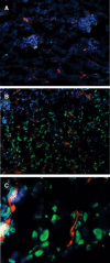
Acrolein adducts in CT26 tumours. Cryosections (5 μm) were stained with specific antibodies for endothelial cells (rat-anti-mouse CD31 (red)) and acrolein modified proteins (green). Cell nuclei are stained by intravenously applicated Hoechst 33258 dye (blue). Untreated control tumour (A); CPA-treated tumour (B) and (C). Fluorescence microscopy of stained cryosections was carried out using an Axiovert 200 fluorescence microscope equipped with a Zeiss Axiocam camera. Light was collected through a 20 × 0.4 NA (A, B) or 63 × 1.4 oil immersion objective (C).
Fig. 3
Fig. 3
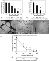
Influence of acrolein on survival of tumour cells and endothelial cells. Cell survival was determined by measuring metabolic activity with the MTT assay. Values are normalized to metabolic activity of untreated control cells. The upper x-axis in A-C indicates the concentration of total acrolein (and 4-OOH-CPA in A) added; the second x-axis at the bottom of the figure reflects the concentrations of free acrolein measured in the cell culture medium used immediately after addition of acrolein (acrolein peak concentration) as described in materials and methods. (A) Metabolic activity ofprimary endothelial cells (HUVECs) treated with indicated concentrations of 4-OOH-CPA (open symbols) or acrolein (full symbols) after a 3 day treatment period (mean ± SD for n= 3 replicates). (B) Metabolic activity of primary endothelial cells treated with indicated concentrations of acrolein for 24 hrs. Cell survival of HUVECs (open symbols) and PEC cells (full symbols) was determined (mean ±SD for n=3 replicates). (C) Metabolic activity of CT26 tumour cells treated with indicated concentrations of acrolein for 24 hrs (mean ±SD for n= 4 replicates).
Fig. 4
Fig. 4
Dose-dependent inhibition of migration (A and B) and tube formation (C and D) by acrolein in endothelial cells. The upper x-axis in A, B and D indicates the concentration of total acrolein added; the lower x-axis reflects the concentrations of free acrolein measured in the cell culture medium used immediately after addition of acrolein (acrolein peak concentration) as described in materials and methods. In C, total acrolein added is indicated by the first, acrolein peak concentration by the second number in the respective pictures. HUVEC (A) and PEC (B) were treated for 24 hrs with indicated concentrations of acrolein. A confluent cell layer of endothelial cells was scratched with a pipette tip and photographed. After 24 hrs of incubation with increasing concentrations of acrolein cells were fixed and photographed again. In the absence of acrolein HUVEC cells as well as PEC cells grow again to a confluent monolayer and migration capability was set to 100%. Migration capability was normalized on migration of untreated control cells and is presented as mean ± SD for n = 10 replicates. *P < 0.05, compared to untreated control cells (Mann-Whitney U-test). (C) and (D): Dose-dependent inhibition of tube formation. HUVEC cells were treated in the matrigel tube formation assay in the absence or in the presence of indicated concentrations of acrolein for four hours. Transmission light microscopy was performed using an Axiovert 200 microscope equipped with a Sony DSC-S75 digital camera. Light was collected through a Zeiss 10 × 0.25 NA objective and images were captured using phase contrast (C). The number of tubes without intersections per field were counted 4 hrs after seeding. Three fields were counted for each data point. Mean values ± SD are shown for three values per datapoint (D). *P < 0.05, compared to untreated control cells (Mann-Whitney U-test).
Fig. 5
Fig. 5
Disruption of the HUVEC cytoskeleton by acrolein. HUVEC were cultured on a collagen G coated glass surface in the presence of 10% foetal bovine serum (FBS) and 10 ng/ml bFGF. Cells were treated with the indicated concentration of acrolein for 12 hrs and thereafter stained with phal-loidin-FITC (A) or phalloidin-FITC (green) and anti- integrin αvβ3 (red) (B). Light was collected through a Zeiss 63 × 1.4 oil immersion objective. Total acrolein added is indicated by the first, acrolein peak concentration by the second number in the respective pictures.
Fig. 6
Fig. 6
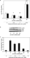
Modulation of NF-κB activity and thrombospondin-1 expression in HUVEC cells by acrolein. In (A) and (B) the upper x-axis indicates the concentration of total acrolein added; the second x-axis at the bottom of the figure reflects the concentration of free acrolein measured in the cell culture medium used immediately after addition of acrolein (acrolein peak concentration) as described in materials and methods. (A) HUVEC cells were transfected with pNF-κB-LUC or the control plasmid pCMV-LUC. 24 hrs after transfection, cells were treated with indicated concentrations of acrolein for 24 hrs and luciferase activity measurement was performed thereafter; additionally, protein content was determined. Ratios of luciferase activity obtained with pNF-κB-LUC and pCMV-LUC (open columns) and relative luciferase activity obtained with pNF-κB-LUC in correlation to protein content (filled columns) are shown after normalization to untreated control cells. n= 8 ± SD, *P < 0.05, *< 0.01 and ***P < 0.001, compared to untreated cells (Mann-Whitney U-test). (B) HUVEC cells were cultivated in the presence of indicated concentrations of added acrolein for 24 hrs before analysis. Levels of NF-κB p105/50 and NF-κB p65 were measured by Western blot analysis. (C) HUVEC were cultured in the presence of indicated concentrations of acrolein for 24 hrs. Thrombospondin-1 was quantified in the cell lysates by competitive ELISA and normalized to total protein content.

Similar articles

Cited by

  • Assessing the carcinogenic potential of low-dose exposures to chemical mixtures in the environment: the challenge ahead.
    Goodson WH 3rd, Lowe L, Carpenter DO, Gilbertson M, Manaf Ali A, Lopez de Cerain Salsamendi A, Lasfar A, Carnero A, Azqueta A, Amedei A, Charles AK, Collins AR, Ward A, Salzberg AC, Colacci A, Olsen AK, Berg A, Barclay BJ, Zhou BP, Blanco-Aparicio C, Baglole CJ, Dong C, Mondello C, Hsu CW, Naus CC, Yedjou C, Curran CS, Laird DW, Koch DC, Carlin DJ, Felsher DW, Roy D, Brown DG, Ratovitski E, Ryan EP, Corsini E, Rojas E, Moon EY, Laconi E, Marongiu F, Al-Mulla F, Chiaradonna F, Darroudi F, Martin FL, Van Schooten FJ, Goldberg GS, Wagemaker G, Nangami GN, Calaf GM, Williams G, Wolf GT, Koppen G, Brunborg G, Lyerly HK, Krishnan H, Ab Hamid H, Yasaei H, Sone H, Kondoh H, Salem HK, Hsu HY, Park HH, Koturbash I, Miousse IR, Scovassi AI, Klaunig JE, Vondráček J, Raju J, Roman J, Wise JP Sr, Whitfield JR, Woodrick J, Christopher JA, Ochieng J, Martinez-Leal JF, Weisz J, Kravchenko J, Sun J, Prudhomme KR, Narayanan KB, Cohen-Solal KA, Moorwood K, Gonzalez L, Soucek L, Jian L, D'Abronzo LS, Lin LT, Li L, Gulliver L, McCawley LJ, Memeo L, Vermeulen L, Leyns L, Zhang L, Valverde M, Khatami M, Romano MF, Chapellier M, Williams MA, Wade M, Manjili MH, Lleonart ME, Xia M, Gonzalez MJ, Karamouzis … See abstract for full author list ➔ Goodson WH 3rd, et al. Carcinogenesis. 2015 Jun;36 Suppl 1(Suppl 1):S254-96. doi: 10.1093/carcin/bgv039. Carcinogenesis. 2015. PMID: 26106142 Free PMC article. Review.
  • Post-transplant cyclophosphamide-induced cardiotoxicity: A comprehensive review.
    Alizadehasl A, Shahrami B, Rahbarghazi R, Yalameh Aliabadi A, Hosseini Jebelli SF, Afsari Zonooz Y, Hakimian H, Fathi F, Forati S, Rezabakhsh A. Alizadehasl A, et al. J Cardiovasc Thorac Res. 2024;16(4):211-221. doi: 10.34172/jcvtr.33230. Epub 2024 Dec 23. J Cardiovasc Thorac Res. 2024. PMID: 40027370 Free PMC article. Review.
  • Acrolein induces endoplasmic reticulum stress and causes airspace enlargement.
    Kitaguchi Y, Taraseviciene-Stewart L, Hanaoka M, Natarajan R, Kraskauskas D, Voelkel NF. Kitaguchi Y, et al. PLoS One. 2012;7(5):e38038. doi: 10.1371/journal.pone.0038038. Epub 2012 May 31. PLoS One. 2012. PMID: 22675432 Free PMC article.

References

    1. Kozekov ID, Nechev LV, Moseley MS, Harris CM, Rizzo CJ, Stone MP, Harris TM. DNA interchain cross-links formed by acrolein and crotonaldehyde. J Am Chem Soc. 2003;125:50–61. - PubMed
    1. Seiner DR, Labutti JN, Gates KS. Kinetics and Mechanism of Protein Tyrosine Phosphatase 1B Inactivation by Acrolein. Chem Res Toxicol. 2007;20:1315–20. - PMC - PubMed
    1. Wrabetz E, Peter G, Hohorst HJ. Does acrolein contribute to the cytotoxicity of cyclophosphamide? J Cancer Res Clin Oncol. 1980;98:119–26. - PubMed
    1. Munoz R, Shaked Y, Bertolini F, Emmenegger U, Man S, Kerbel RS. Anti-angiogenic treatment of breast cancer using metronomic low-dose chemotherapy. Breast. 2005;14:466–79. - PubMed
    1. Browder T, Butterfield CE, Kraling BM, Shi B, Marshall B, O'Reilly MS, Folkman J. Antiangiogenic scheduling of chemotherapy improves efficacy against experimental drug-resistant cancer. Cancer Res. 2000;60:1878–86. - PubMed

Publication types

MeSH terms