

The genomic response of the ipsilateral and contralateral cortex to stroke in aged rats
- PMID: 18266980
- PMCID: PMC3828887
- DOI: 10.1111/j.1582-4934.2008.00252.x
The genomic response of the ipsilateral and contralateral cortex to stroke in aged rats
Abstract
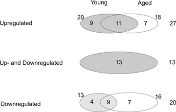
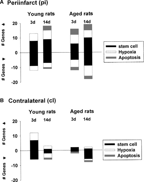
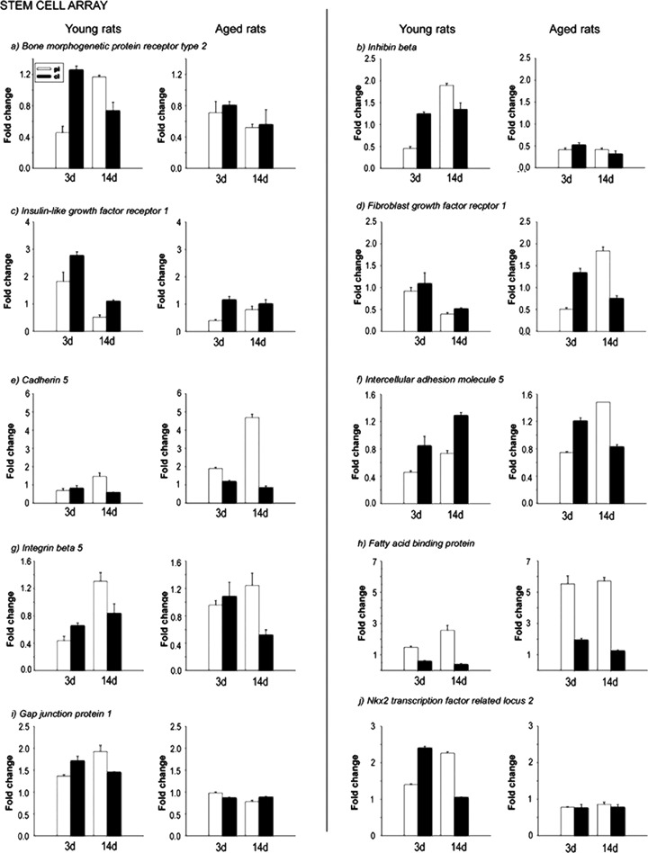
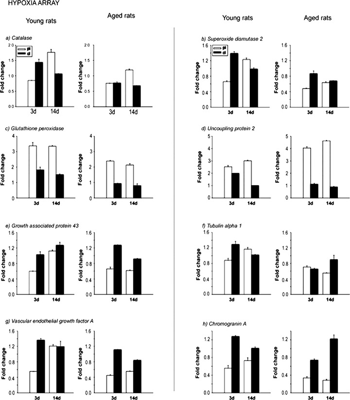
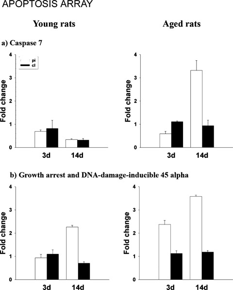
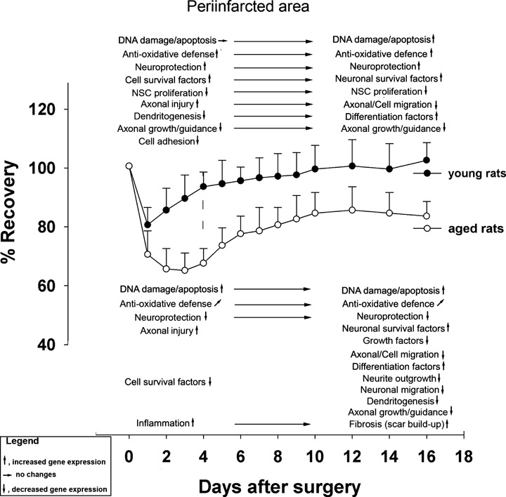
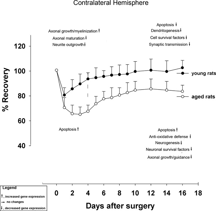
Aged rats recover poorly after unilateral stroke, whereas young rats recover readily possibly with the help from the contralateral, healthy hemisphere. In this study we asked whether anomalous, age-related changes in the transcriptional activity in the brains of aged rats could be one underlying factor contributing to reduced functional recovery. We analysed gene expression in the periinfarct and contralateral areas of 3-month- and 18-month-old Sprague Dawley rats. Our experimental end-points were cDNA arrays containing genes related to hypoxia signalling, DNA damage and apoptosis, cellular response to injury, axonal damage and re-growth, cell lineage differentiation, dendritogenesis and neurogenesis. The major transcriptional events observed were: (i) Early up-regulation of DNA damage and down-regulation of anti-apoptosis-related genes in the periinfarct region of aged rats after stroke; (ii) Impaired neurogenesis in the periinfarct area, especially in aged rats; (iii) Impaired neurogenesis in the contralateral (unlesioned) hemisphere of both young and aged rats at all times after stroke and (iv) Marked up-regulation, in aged rats, of genes associated with inflammation and scar formation. These results were confirmed with quantitative real-time PCR. We conclude that reduced transcriptional activity in the healthy, contralateral hemisphere of aged rats in conjunction with an early up-regulation of DNA damage-related genes and pro-apoptotic genes and down-regulation of axono- and neurogenesis in the periinfarct area are likely to account for poor neurorehabilitation after stroke in old rats.
Figures
References
-
- Barnett HJ. Stroke prevention in the elderly. Clin Exp Hypertens. 2002;24:563–71. - PubMed
-
- Popa-Wagner A, Schroder E, Walker LC, Kessler C. Beta-amyloid precursor protein and ss-amyloid peptide immunoreactivity in the rat brain after middle cerebral artery occlusion: effect of age. Stroke. 1998;29:2196–202. - PubMed
-
- Badan I, Buchhold B, Hamm A, Gratz M, Walker LC, Platt D, Kessler Ch, Popa-Wagner A. Accelerated glial reactivity to stroke in aged rats correlates with reduced functional recovery. J Cereb Blood Flow Metab. 2003;23:845–4. - PubMed
-
- Badan I, Dinca I, Buchhold B, Suofu Y, Walker L, Gratz M, Platt D, Kessler Ch, Popa-Wagner A. Accelerated accumulation of N- and C-terminal betaAPP fragments and delayed recovery of microtubule-associated protein 1B expression following stroke in aged rats. Eur J Neurosci. 2004;19:2270–80. - PubMed
Publication types
MeSH terms
LinkOut - more resources
Full Text Sources
Medical
