. 2008 Feb;14(2):227-38.
doi: 10.1016/j.devcel.2007.11.001.

piggyBac-based mosaic screen identifies a postmitotic function for cohesin in regulating developmental axon pruning

Affiliations

piggyBac-based mosaic screen identifies a postmitotic function for cohesin in regulating developmental axon pruning

Oren Schuldiner et al. Dev Cell. 2008 Feb.

Abstract

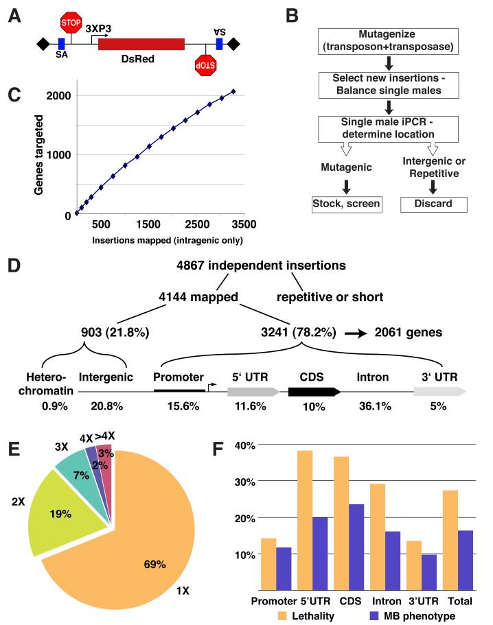
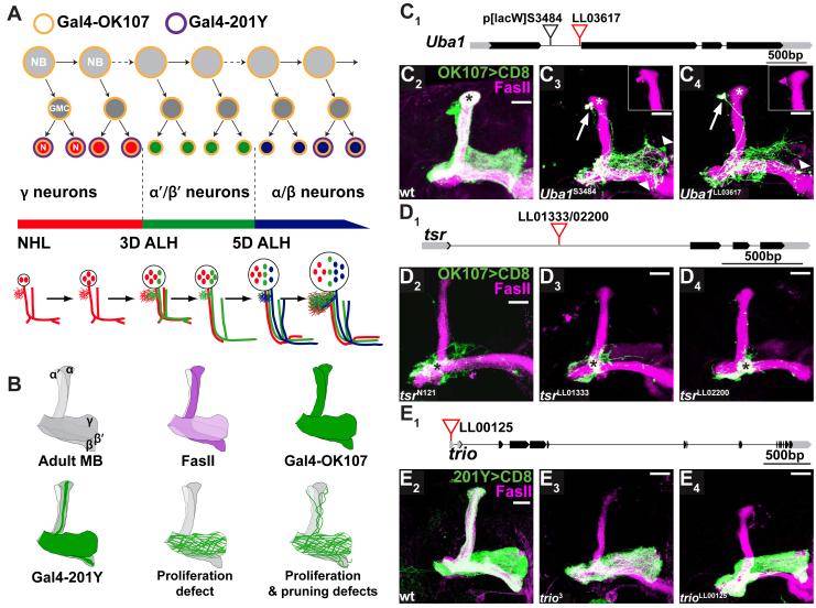
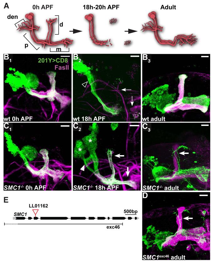
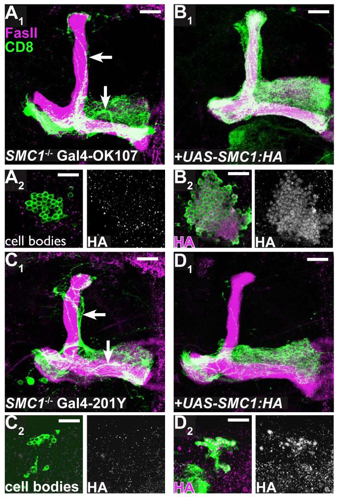
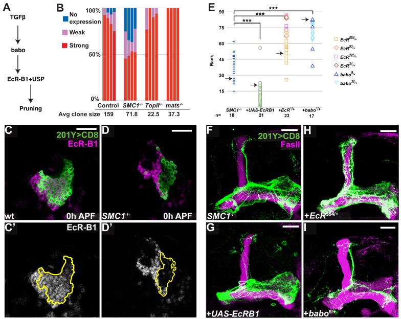
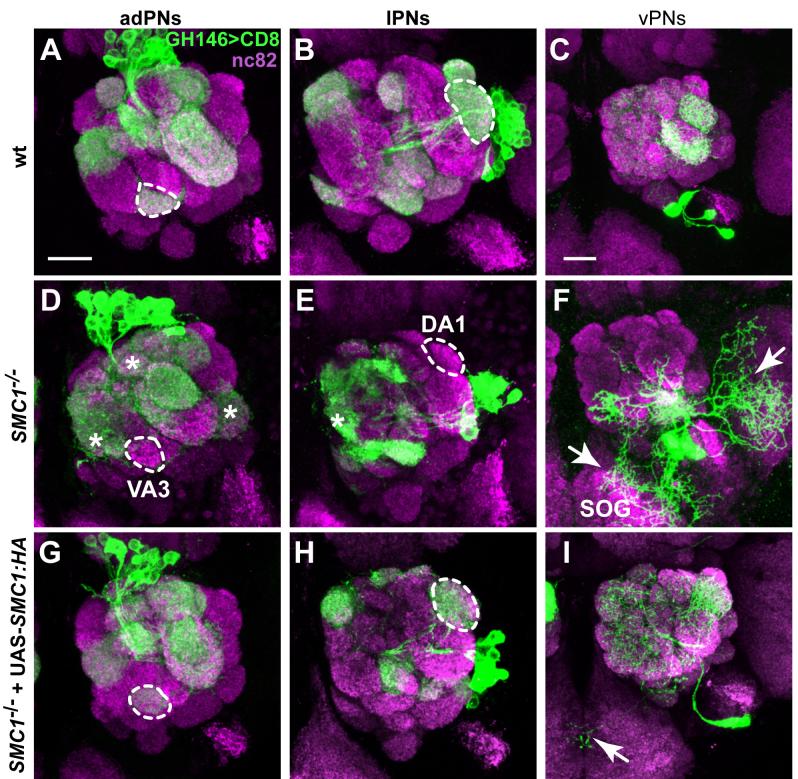
Developmental axon pruning is widely used to refine neural circuits. We performed a mosaic screen to identify mutations affecting axon pruning of Drosophila mushroom body gamma neurons. We constructed a modified piggyBac vector with improved mutagenicity and generated insertions in >2000 genes. We identified two cohesin subunits (SMC1 and SA) as being essential for axon pruning. The cohesin complex maintains sister-chromatid cohesion during cell division in eukaryotes. However, we show that the pruning phenotype in SMC1(-/-) clones is rescued by expressing SMC1 in neurons, revealing a postmitotic function. SMC1(-/-) clones exhibit reduced levels of the ecdysone receptor EcR-B1, a key regulator of axon pruning. The pruning phenotype is significantly suppressed by overexpressing EcR-B1 and is enhanced by a reduced dose of EcR, supporting a causal relationship. We also demonstrate a postmitotic role for SMC1 in dendrite targeting of olfactory projection neurons. We suggest that cohesin regulates diverse aspects of neuronal morphogenesis.


Figures

Figure 1
Figure 1. Overview of piggyBac-based Insertional Mutagenesis
(A) Our modified piggyBac mutator element contains, in both orientations, a splice acceptor (SA) followed by stop codons in all three reading frames; it is marked with a DsRed reporter. 3XP3 is a synthetic promoter expressed mainly in the eye (Sheng et al., 1997) and shown to effectively drive the expression of different fluorescent proteins as markers for piggyBac (Horn et al., 2000). (B) Mutant generation scheme; see text and Supplemental Experimental Procedures for details. (C) Number of genes targeted plotted against intragenic insertions mapped. (D) Distribution of piggyBac transposons with regard to a generic gene structure. (E) Frequency distribution of 3241 independent insertions that fall within transcriptional units of 2061 different genes. (F) Rates of lethality and MB mutant phenotype for insertions in different parts of the transcriptional unit.
Figure 2
Figure 2. Phenotypic Comparison of piggyBac and Previously Characterized Alleles
(A) Scheme of sequential generation of MB neurons. MB neurons are generated from four neuroblasts (NBs) per hemisphere. Each NB divides asymmetrically to generate another NB and a ganglion mother cell (GMC), which divides once more to generate two postmitotic neurons (N). MB neuroblasts sequentially give rise to γ neurons (red), α’/β’ (green) and α/β neurons (blue) according to depicted developmental timeline. NHL, newly hatched larvae; 3D ALH, 3 days after larval hatching. Gal4-OK107 is expressed in all MB neurons including dividing NBs (orange outline). Gal4-201Y is expressed in postmitotic γ and a subset of later born α/β neurons (purple outline). Schematic representation of the stereotypical projection of MB neurons at different developmental stages is shown in the lower part of the panel. Adapted from Lee et al., 1999. (B) Schematic drawing shows 1) Adult MB with 5 axon lobes contributed by γ, α’/β’ and α/β neurons; 2) anti-FasII labels the γ lobe weakly and the α/β lobes strongly; 3-4) Gal4-OK107 and Gal4-201Y expression patterns in the corresponding lobes; 5) MB MARCM neuroblast clones defective in proliferation, with axons only innervating the γ lobe; 6) MB neuroblast clones defective in proliferation and pruning (as is the case for Uba1-/- and SMC1-/- clones), with residual unpruned γ axons around the dorsal lobes. (C) piggyBac line LL03617 is inserted in the first intron of Uba1 (C1). Compared to wt MB clones (C2), homozygous UbaLL03617 MB neuroblast clones (C4) in adult exhibit neuroblast proliferation and axon pruning defects, and signs of axon degeneration, similar to the previously reported phenotype of the strong loss-of-function UbaS3484 (C3) (Watts et al., 2003). Arrows point to unpruned dorsal projections that are positive for FasII (insets). Arrowheads show blebbing and sparse axons; asterisks mark late born α/β neurons. (D) piggyBac lines LL01333 and LL02200 are inserted in opposite orientations at the same location in the first intron of twinstar (tsr) (D1). MB neuroblast clones homozygous for either insertion contain mostly axons that fail to extend beyond the branching point (asterisks, D3,4), similar to the axon growth phenotypes previously described for null mutation tsrN121 (asterisk, D2) (Ng and Luo, 2004). (E) piggyBac line LL00125 is inserted in the 5′ UTR of trio (E1). MB neuroblast clones homozygous for trioLL00125 do not contain late born α/β neurons (E4) as in wt (E2), indicating a neuroblast proliferation defect. This phenocopies a strong loss-of-function trio3 mutant clones (E3). Black blocks, CDS; gray blocks, UTRs; lines, introns. All images in this and subsequent figures are confocal z-stacks of MB neurons and their axons, unless otherwise stated. Scale bars, 20μm. Genotypes are described in Supplemental Information.
Figure 3
Figure 3. SMC1 is Required for Axon Pruning
(A) Scheme of developmental pruning of MB γ neurons. At 0 hr after puparium formation (0h APF), each γ neuron has a single process that gives off dendritic branches (den) near the cell body, continues an axon peduncle (p), and bifurcates to form a dorsal (d) and a medial (m) branch. The dorsal and medial axon branches, as well as dendrites, are pruned by 18h APF, leaving some fragmented axons at the tips of the lobes but an intact axon peduncle. Later, γ neurons extend axons only to the adult-specific medial lobe, with extensive branches within. Adapted from Watts et al. 2004. (B-D) Wt (B), SMC1LL01162 (C) and SMC1Δexc46 (D) MB neuroblast MARCM clones labeled with Gal4-201Y. At 0h APF, both wt (B1) and SMC1-/- (C1) γ neurons project their axons to the dorsal and medial lobes. The cell number within SMC1-/- neuroblast clone is significantly reduced. At 18h APF, wt γ neurons have pruned their medial and dorsal axon branches (dashed arrows in B2) as well as their dendrites (open arrowhead). SMC1-/- γ neurons retain most of their axons at this stage (arrows in C2), which persist to adulthood (arrow in C3, D). In addition, SMC1-/- γ neurons retain partially unpruned dendrites at 18h APF (arrowhead in C2). Asterisks in C2 indicate two independent neuroblast clones. (E) SMC1 gene structure. piggyBac line LL01162 is inserted in the third exon. The approximate borders of the null mutation SMC1Δexc46 (Scott Page & Scott Hawley, personal communication) are also depicted. Green, Gal4-201Y driven mCD8::GFP; magenta, anti-FasII. Scale bars, 20μm.
Figure 4
Figure 4. Rescue of Axon Pruning and Proliferation Defect by SMC1 transgene
(A-B) SMC1-/- MB neuroblast MARCM clones labeled with Gal4-OK107 exhibit unpruned γ neurons (arrows in A1) and reduced cell number (A2, also note the lack of α/β or α’/β’ lobes in A1). Expression of UAS-SMC1-HA transgene in mutant clones (B) rescues both phenotypes (B1, B2). SMC1-HA protein is predominantly located in the nuclei of neurons (B2 right panel). (C-D) SMC1-/- MB neuroblast MARCM clones labeled with Gal4-201Y exhibit unpruned γ neurons (arrows in C1) and reduced cell number (C2). Expression of UAS-SMC1-HA only in mutant clones by postmitotic Gal4 driver (Gal4-201Y) either rescues pruning completely (20/27) or shows only a single unpruned γ neuron (7/27, D1). Cell number remains unchanged (D2). Green, Gal4-OK107 (A-B) or Gal4-201Y (C-D) driven mCD8::GFP; magenta, anti-FasII (A1-D1) or anti-HA (A2-D2). Single confocal sections are shown for A2-D2. Scale bars, 20μm.
Figure 5
Figure 5. SMC1 Affects Pruning by Regulating the Levels of EcR-B1
(A) Scheme of TGFβ/EcR-B1 pathway regulating MB axon pruning. (B) Summary of EcR-B1 expression in MB neuroblasts at 0h APF. 4 neuroblast clones for each genotype were blindly analyzed; cells were classified as expressing high, low or no EcR-B1. Average clone size is also shown. (C, D) Expression of EcR-B1 (magenta in C, D; white in C’, D’) in wt (C) and SMC1-/- (D) MB neuroblast clones. Extent of the clone is depicted in C’, D’ by a yellow line. Single confocal sections are shown. (E) Summary of genetic interactions. To analyze the suppression or enhancement of the SMC1-/- pruning phenotype, 79 confocal Z projections from different genotypes were blindly ranked for the severity of the pruning defect. The severity was determined by comparing the unpruned dorsal γ axons to pruned γ axons in the adult-specific medial lobe; these pruned axons can be distinguished from unpruned medial axons as they branch extensively and are located at a more dorsal and posterior position. The ranks grouped by genotypes are shown. Symbols for different EcR and baboon alleles are shown on the right. Pair-wise Mann-Whitney U-tests were performed to determine significance: ***, p<0.001. Arrows indicate the examples shown in (F-I). (F-I) Genetic interactions between SMC1 and EcR-B1. SMC1-/- MB neuroblast clone (F) with additional expression of UAS-EcR-B1 in mutant clone (G); in an EcR heterozygous background (H); and in a babo heterozygous background (I). Green represents Gal4-201Y driven mCD8::GFP and magenta is anti-EcR-B1 (C, D) or anti-FasII (F-I). Scale bars, 20μm.
Figure 6
Figure 6. SMC1 Is Required for PN Dendrite Targeting
(A-C) Stereotypic dendrite projection pattern of wt neuroblast clones that give rise to adPNs (A), lPNs (B), and vPNs (C). (D-F) SMC1-/- PNs exhibit dendrite targeting defects. SMC1-/- adPNs do not innervate VA3 (9/12; dotted outline, D), but instead project to several inappropriate glomeruli in the medial and dorsal parts of the antennal lobe and VL2a (asterisks, D). SMC1-/- lPNs fail to target DA1 (12/14; dotted outline, E) but innervate additional medial glomeruli (asterisk, E). SMC1-/- vPNs show most severe targeting defects, with large portions of dendrites targeting to the SOG and lateral areas outside the antennal lobe (arrows, F). (G-I) Postmitotic expression of SMC1::HA in SMC1-/- PNs fully rescues dendrite targeting targeting in adPNs (G) and lPNs (H), respectively. vPN dendrites are either indistinguishable from wt or show rare dendrites wandering to the SOG (arrow, I). Dotted outlines represent selected glomeruli used for scoring the penetrance of SMC1 mutant phenotypes and corresponding rescues: VA3 for adPNs (in A, D and G), DA1 for lPNs (in B, E and H). Magenta, nc82 as a presynapic marker for all glomeruli; green, UAS-mCD8::GFP driven by GH146-Gal4. Scale bars, 20μm.

References

    1. Awasaki T, Ito K. Engulfing action of glial cells is required for programmed axon pruning during Drosophila metamorphosis. Curr Biol. 2004;14:668–677. - PubMed
    1. Bellen HJ, Levis RW, Liao G, He Y, Carlson JW, Tsang G, Evans-Holm M, Hiesinger PR, Schulze KL, Rubin GM, et al. The BDGP gene disruption project: single transposon insertions associated with 40% of Drosophila genes. Genetics. 2004;167:761–781. - PMC - PubMed
    1. Berdnik D, Chihara T, Couto A, Luo L. Wiring stability of the adult Drosophila olfactory circuit after lesion. J Neurosci. 2006;26:3367–3376. - PMC - PubMed
    1. Bishop DL, Misgeld T, Walsh MK, Gan WB, Lichtman JW. Axon branch removal at developing synapses by axosome shedding. Neuron. 2004;44:651–661. - PubMed
    1. Celniker SE, Wheeler DA, Kronmiller B, Carlson JW, Halpern A, Patel S, Adams M, Champe M, Dugan SP, Frise E, et al. Finishing a whole-genome shotgun: release 3 of the Drosophila melanogaster euchromatic genome sequence. Genome Biol. 2002;3 RESEARCH0079. - PMC - PubMed

Publication types

MeSH terms

LinkOut - more resources