

Lipid peroxidation-derived aldehyde-protein adducts contribute to trichloroethene-mediated autoimmunity via activation of CD4+ T cells
- PMID: 18267128
- PMCID: PMC2440665
- DOI: 10.1016/j.freeradbiomed.2008.01.012
Lipid peroxidation-derived aldehyde-protein adducts contribute to trichloroethene-mediated autoimmunity via activation of CD4+ T cells
Abstract
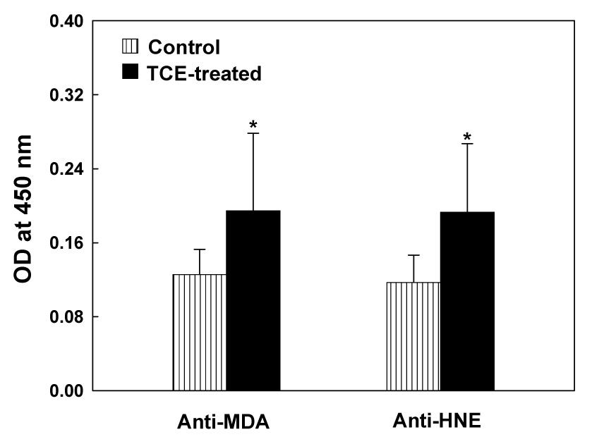
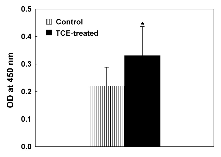
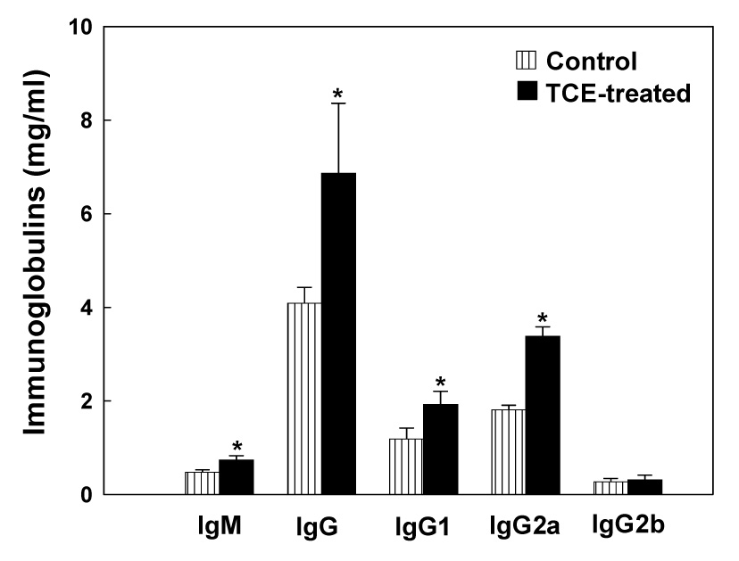
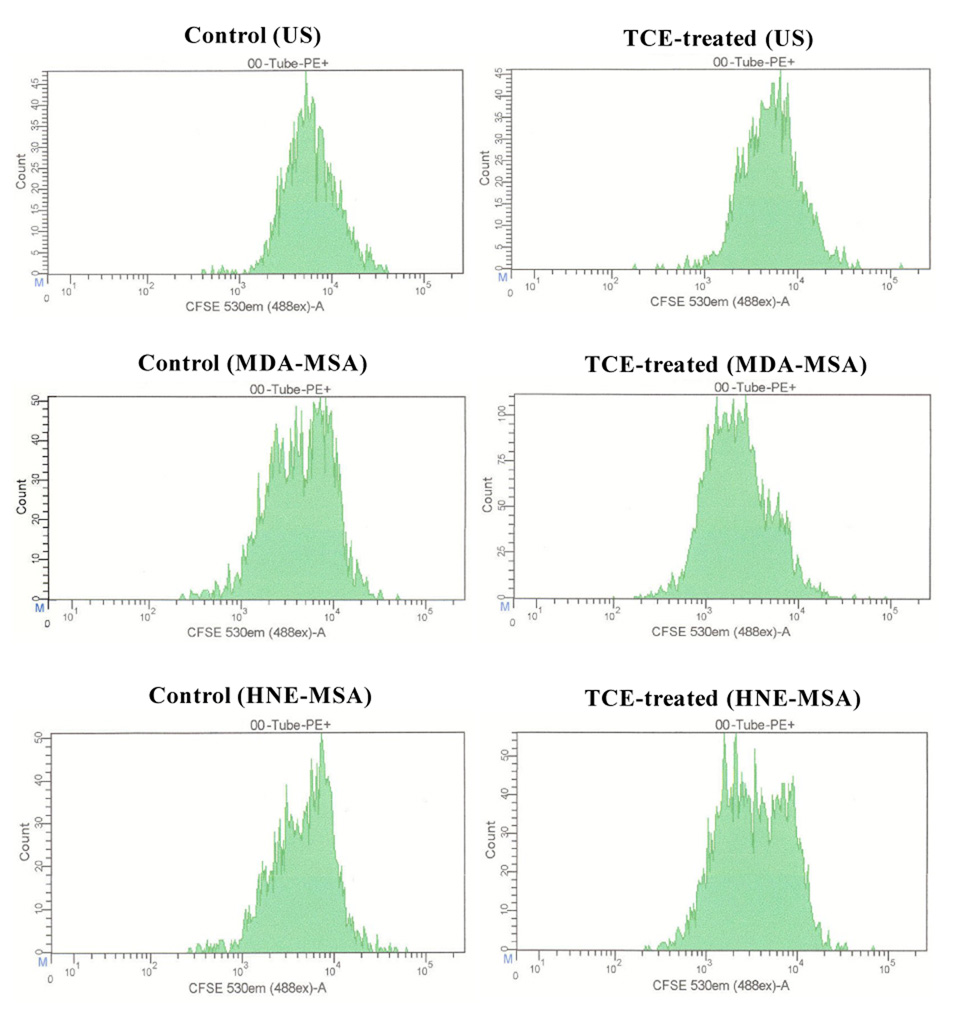
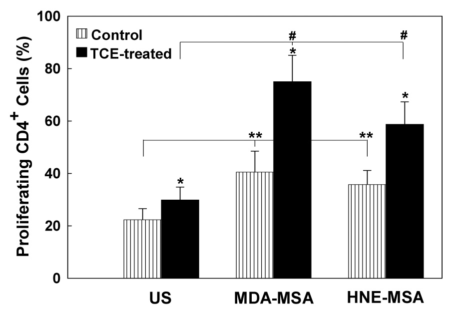
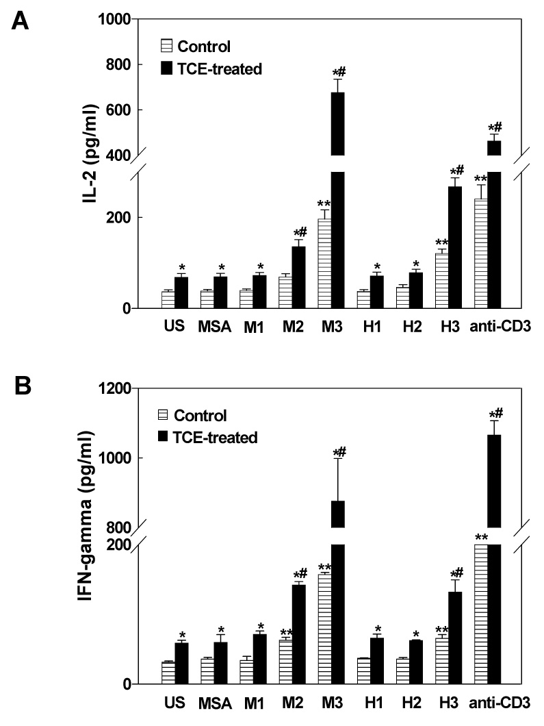
Lipid peroxidation is implicated in the pathogenesis of various autoimmune diseases. Lipid peroxidation-derived aldehydes such as malondialdehyde (MDA) and 4-hydroxynonenal (HNE) are highly reactive and bind to proteins, but their role in eliciting an autoimmune response and their contribution to disease pathogenesis remain unclear. To investigate the role of lipid peroxidation in the induction and/or exacerbation of autoimmune response, 6-week-old autoimmune-prone female MRL+/+ mice were treated for 4 weeks with trichloroethene (TCE; 10 mmol/kg, ip, once a week), an environmental contaminant known to induce lipid peroxidation. Sera from TCE-treated mice showed significant levels of antibodies against MDA-and HNE-adducted proteins along with antinuclear antibodies. This suggested that TCE exposure not only caused increased lipid peroxidation, but also accelerated autoimmune responses. Furthermore, stimulation of cultured splenic lymphocytes from both control and TCE-treated mice with MDA-adducted mouse serum albumin (MDA-MSA) or HNE-MSA for 72 h showed significant proliferation of CD4+ T cells in TCE-treated mice as analyzed by flow cytometry. Also, splenic lymphocytes from TCE-treated mice released more IL-2 and IFN-gamma into cultures when stimulated with MDA-MSA or HNE-MSA, suggesting a Th1 cell activation. Thus, our data suggest a role for lipid peroxidation-derived aldehydes in TCE-mediated autoimmune responses and involvement of Th1 cell activation.
Figures
References
-
- Khan MF, Wu X, Ansari GAS. Anti-malondialdehyde antibodies in MRL+/+ mice treated with trichloroethene and dichloroacetyl chloride: possible role of lipid peroxidation in autoimmunity. Toxicol. Appl. Pharmacol. 2001;170:88–92. - PubMed
-
- Hadjigogos K. The role of free radicals in the pathogenesis of rheumatoid arthritis. Panminerva Med. 2003;45:7–13. - PubMed
-
- Frostegard J, Svenungsson E, Wu R, Gunnarsson I, Lundberg IE, Klareskog L, Horkko S, Witztum JL. Lipid peroxidation is enhanced in patients with systemic lupus erythematosus and is associated with arterial and renal disease manifestations. Arthritis Rheum. 2005;52:192–200. - PubMed
-
- Cuzzocrea S. Role of nitric oxide and reactive oxygen species in arthritis. Curr. Pharm. Des. 2006;12:3551–3570. - PubMed
Publication types
MeSH terms
Substances
Grants and funding
LinkOut - more resources
Full Text Sources
Other Literature Sources
Research Materials
