. 2008 Jun 1;17(11):1527-39.
doi: 10.1093/hmg/ddn041. Epub 2008 Feb 11.

The intracellular accumulation of polymeric neuroserpin explains the severity of the dementia FENIB

Affiliations

The intracellular accumulation of polymeric neuroserpin explains the severity of the dementia FENIB

Elena Miranda et al. Hum Mol Genet. .

Abstract

Familial encephalopathy with neuroserpin inclusion bodies (FENIB) is an autosomal dominant dementia that is characterized by the retention of polymers of neuroserpin as inclusions within the endoplasmic reticulum (ER) of neurons. We have developed monoclonal antibodies that detect polymerized neuroserpin and have used COS-7 cells, stably transfected PC12 cell lines and transgenic Drosophila melanogaster to characterize the cellular handling of all four mutant forms of neuroserpin that cause FENIB. We show a direct correlation between the severity of the disease-causing mutation and the accumulation of neuroserpin polymers in cell and fly models of the disease. Moreover, mutant neuroserpin causes locomotor deficits in the fly allowing us to demonstrate a direct link between polymer accumulation and neuronal toxicity.


Figures

Figure 1.
Figure 1.
Mutant H338R and G392E neuroserpin accumulate as polymers within the ER. (A) Confocal microscopy analysis of COS-7 cells cultured for 48 h after transfection with wild-type, H338R or G392E neuroserpin and stained for neuroserpin (red) and the ER-resident protein calreticulin or the Golgi-resident protein GM130 (green). Only the merged images are shown in which yellow colour corresponds to areas with overlapping red and green staining. The nucleus appears blue due to DNA staining with DAPI. Scale bar: 10 µm. (B) Endoglycosidase-H (eH) digestion of samples from cells transfected with wild-type or each mutant neuroserpin that were pulsed-labelled and chased for 6 h. Cells lysates (C) and culture media (M) were analysed by immunoprecipitation and 8% w/v SDS–PAGE. Arrow: fully glycosylated and secreted neuroserpin, 55 kDa; arrowhead: intracellular neuroserpin intermediate, 50 kDa; black and white arrow: deglycosylated intracellular neuroserpin, 45 kDa; the asterisk indicates a second band of extracellular wild-type neuroserpin due to extracellular proteolysis; the dash indicates a slower migrating band of extracellular G392E neuroserpin. (C) 7.5% w/v acrylamide non-denaturing PAGE and western-blot analysis for neuroserpin in cell lysates (C) and culture media (M) from COS-7 cells transfected with wild-type or mutant neuroserpin or with a control plasmid expressing luciferase (Lucif.). Cells were cultured for 72 h after transfection. Arrow: wild-type and S52R neuroserpin monomers; arrowhead: S49P neuroserpin monomer; curly bracket: neuroserpin polymers.
Figure 2.
Figure 2.
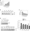
Detection of mutant neuroserpin polymers with an anti-polymer monoclonal antibody. (A) Binding of monoclonal antibodies 1A10 and 7C6 in a sandwich ELISA to recombinant monomeric wild-type neuroserpin (Wt) and monomeric (mon) or polymerized (pol) S49P neuroserpin as the antigens. (B) Binding of monoclonal antibodies 1A10 and 7C6 in a sandwich ELISA using different proportions of recombinant wild-type monomeric neuroserpin (mon) and polymerized S52R neuroserpin (pol) as the antigens. These are shown as the percentage of each species, for example mon75-pol25 is a mixture that is 75% monomer and 25% polymer. (C) Cell lysates from COS-7 cells transfected with wild-type neuroserpin or mutants of neuroserpin that cause FENIB (S49P, S52R, H338R, G392E) were analysed by sandwich ELISA using either 1A10 or 7C6 to detect neuroserpin. (D) Confocal microscopy analysis of COS-7 cells transiently transfected with each neuroserpin variant were immunostained for total neuroserpin with a rabbit anti-neuroserpin polyclonal antibody (red) and for neuroserpin polymers with monoclonal antibody 7C6 (green). The nucleus appears blue due to DNA staining with DAPI. Scale bar: 10 µm.
Figure 3.
Figure 3.
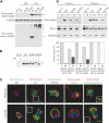
Increasing intracellular accumulation of mutant neuroserpin polymers correlates with increasing severity of FENIB. (A) Percentage of transfected COS-7 cells showing punctate neuroserpin accumulation 24 h after transfection. Neuroserpin accumulation was quantified by counting more than 100 transfected cells per experiment in three independent experiments for each neuroserpin variant. Each experiment was counted blind and cells were scored as containing punctate accumulation if there were at least 10 discrete protein ‘spots’ per cell. Percentages are averages ± SEM, and the differences were statistically significant when analysed by one-way ANOVA (P < 0.0001) followed by a post-test for linear trend (R2 = 0.93, P < 0.0001). (B) Cell lysates (C) and culture media (M) of COS-7 cells transfected with wild-type or each mutant neuroserpin or with a control plasmid expressing luciferase (Lucif.) were analysed 72 h after transfection by 8% w/v acrylamide SDS–PAGE and western-blot analysis with an anti-neuroserpin antibody. NS, control lane loaded with 20 ng of purified recombinant neuroserpin. Black arrow: fully glycosylated and secreted neuroserpin, 55 kDa; arrowhead: intracellular neuroserpin intermediate, 50 kDa; black and white arrow: non-glycosylated recombinant neuroserpin used as a control, 45 kDa. (C) The amount of total neuroserpin was determined by sandwich ELISA in cell lysates and culture media of COS-7 cells 72 h after transfection with wild-type or each mutant neuroserpin. The graph shows the proportion of neuroserpin that was present in the culture media. Values are averages ± SEM from three independent repeats, and the differences were statistically significant when analysed by one-way ANOVA (P < 0.0001) followed by a post-test for linear trend (R2 = 0.82, P < 0.0001). (D) Cells transfected with wild-type or each mutant neuroserpin were pulsed-labelled and chased for 6 h. Cells lysates (C) and culture media (M) were analysed by immunoprecipitation and 8% w/v SDS–PAGE. Arrow: fully glycosylated and secreted neuroserpin, 55 kDa; arrowhead: intracellular neuroserpin intermediate, 50 kDa; asterisk: a slower migrating second band in the culture medium of cells transfected with G392E neuroserpin. The graph shows the quantitation of the pulse-chase experiment using a phosphorimager. The amount of radioactivity in each sample is expressed as the percentage of the total radioactivity for each neuroserpin variant. Values are averages ± SEM from five independent repeats, and the differences were statistically significant when analysed by one-way ANOVA (P < 0.0001) followed by a post-test for linear trend (R2 = 0.59, P < 0.0001).
Figure 4.
Figure 4.
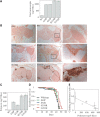
Trafficking of wild-type and mutant neuroserpin in stably transfected PC12 cell lines. (A) Lysates from PC12-Tet-On, PC12-Wt, PC12-S52R and PC12-G392E cells cultured with (on) or without (off) 10 µg/ml doxycycline to induce neuroserpin expression were analysed by SDS and non-denaturing PAGE followed by western-blot analysis with an anti-neuroserpin polyclonal antibody. The membrane from the SDS–PAGE was also analysed with an anti-GAPDH antibody as a loading control. (B) Neuroserpin from lysed PC12-Tet-On, PC12-Wt, PC12-S52R and PC12-G392E cells induced for 4 days was immunoprecipitated with the 1A10 anti-neuroserpin monoclonal antibody and treated (+) or not (−) with endoglycosidase H (eH) and analysed by SDS–PAGE and western blot analysis. S52R and G392E neuroserpin were sensitive to endoglycosidase H (arrow) whereas wild-type neuroserpin was resistant (arrowhead). (C) Immuno-co-localization of wild-type and G392E neuroserpin with resident proteins of the secretory pathway. PC12-wildtype and PC12-G392E cells were differentiated to a neuronal phenotype by plating in collagen and treating with NGF (150 ng/ml) for 7 days, and then induced to express neuroserpin with 10 µg/ml doxycycline for 3 days. Cells were co-stained with either a polyclonal anti-neuroserpin antibody or one of the anti-neuroserpin monoclonal antibodies (1A10 for total neuroserpin or 7C6 for neuroserpin polymers) and antibodies against calreticulin (ER), GM130 (Golgi), ERGIC-53/p58 (ERGIC) or chromogranin A (trafficked through the regulated secretory pathway). The colour corresponding to each antibody (red or green) is shown above the figure and only the merged images are presented. Yellow represents areas of overlapping red and green. The nucleus appears blue due to DNA staining with DAPI. Scale bar: 10 µm. (D) PC12-Wt, PC12-S52R and PC12-G392E cells were induced to express neuroserpin for 3 days with 10 µg/ml doxycycline and then incubated for 15 min with control or release buffer containing 5 or 55 mm KCl, respectively, to assess regulated secretion from dense core secretory granules. Neuroserpin was analysed in cell lysates and buffer solutions by SDS–PAGE and western blot and quantified by sandwich ELISA. The graph shows the averages for three independent experiments analysed by ELISA, expressed as percentages ± SEM. The amount of neuroserpin secreted from each cell line when treated with release buffer was statistically different when analysed by one-way ANOVA (P = 0.0003) followed by a post-test for linear trend (R2 = 0.93, P = 0.0001).
Figure 5.
Figure 5.
Polymers of mutant neuroserpin accumulate in the brain of transgenic flies and cause a locomotor phenotype. (A) Levels of total neuroserpin were determined in transgenic fly homogenates (see Materials and Methods) by sandwich ELISA. (B) Immunohistochemical detection of neuroserpin with the 1A10 monoclonal antibody in the brains of flies expressing the elav promotor (elav), wild-type (Wt), S49P (S49P), S52R (S52R), H338R (H338R) or G392E (G392E) neuroserpin at day 25 after eclosion. Intracellular accumulation of mutant neuroserpin (brown staining) was located within cortical neuronal cell bodies, adjacent to the mushroom bodies and lobula. Left and middle panels were taken with a 20× objective, enlarged details in right panels were obtained with a 100× oil immersion objective. (C) Levels of polymerized neuroserpin were determined in transgenic fly homogenates (see Materials and Methods) by sandwich ELISA with the 7C6 monoclonal antibody. (D) Cumulative survival plots for a hundred flies of each representative line chosen for each neuroserpin genotype. (E) Transgenic flies expressing all five variants of neuroserpin were subjected to climbing assays (see Materials and Methods) to assess their locomotor performance and the results were plotted against the levels of neuroserpin polymers detected by ELISA in (C). A negative correlation was found with an R2 = 0.84 that was statistically significant at P < 0.05.

References

    1. Carrell R.W., Lomas D.A. Conformational diseases. Lancet. 1997;350:134–138. - PubMed
    1. Lomas D.A., Mahadeva R. Alpha-1-antitrypsin polymerisation and the serpinopathies: pathobiology and prospects for therapy. J. Clin. Invest. 2002;110:1585–1590. - PMC - PubMed
    1. Kaiserman D., Whisstock J.C., Bird P.I. Mechanisms of serpin dysfunction in disease. Expert Rev. Mol. Med. 2006;8:1–19. - PubMed
    1. Huntington J.A., Pannu N.S., Hazes B., Read R., Lomas D.A., Carrell R.W. A 2.6Å structure of a serpin polymer and implications for conformational disease. J. Mol. Biol. 1999;293:449–455. - PubMed
    1. Dunstone M.A., Dai W., Whisstock J.C., Rossjohn J., Pike R.N., Feil S.C., Le Bonniec B.F., Parker M.W., Bottomley S.P. Cleaved antitrypsin polymers at atomic resolution. Protein Sci. 2000;9:417–420. - PMC - PubMed

Publication types

MeSH terms