. 2008 May 15;17(10):1446-56.
doi: 10.1093/hmg/ddn033. Epub 2008 Feb 11.

Dopamine determines the vulnerability of striatal neurons to the N-terminal fragment of mutant huntingtin through the regulation of mitochondrial complex II

Affiliations

Dopamine determines the vulnerability of striatal neurons to the N-terminal fragment of mutant huntingtin through the regulation of mitochondrial complex II

Alexandra Benchoua et al. Hum Mol Genet. .

Abstract

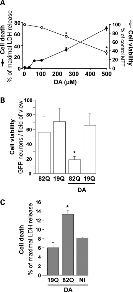
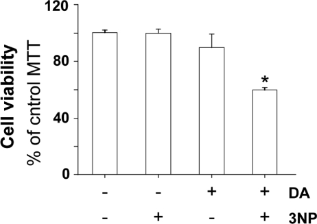
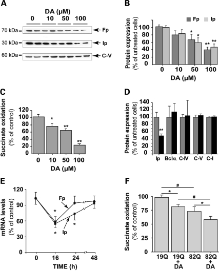
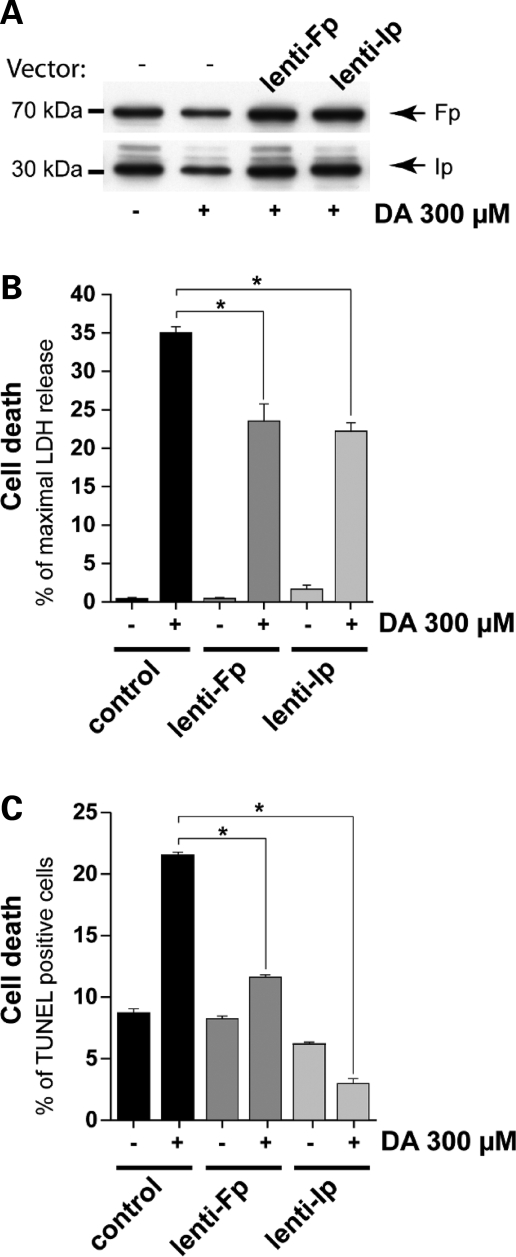
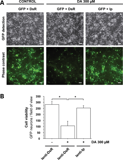
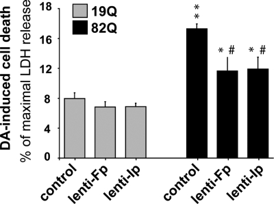
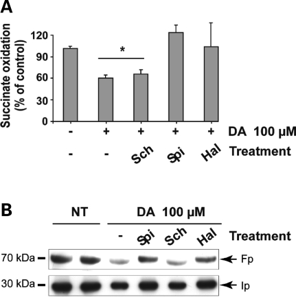
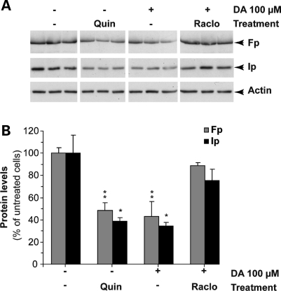
In neurodegenerative disorders associated with primary or secondary mitochondrial defects such as Huntington's disease (HD), cells of the striatum are particularly vulnerable to cell death, although the mechanisms by which this cell death is induced are unclear. Dopamine, found in high concentrations in the striatum, may play a role in striatal cell death. We show that in primary striatal cultures, dopamine increases the toxicity of an N-terminal fragment of mutated huntingtin (Htt-171-82Q). Mitochondrial complex II protein (mCII) levels are reduced in HD striatum, indicating that this protein may be important for dopamine-mediated striatal cell death. We found that dopamine enhances the toxicity of the selective mCII inhibitor, 3-nitropropionic acid. We also demonstrated that dopamine doses that are insufficient to produce cell loss regulate mCII expression at the mRNA, protein and catalytic activity level. We also show that dopamine-induced down-regulation of mCII levels can be blocked by several dopamine D2 receptor antagonists. Sustained overexpression of mCII subunits using lentiviral vectors abrogated the effects of dopamine, both by high dopamine concentrations alone and neuronal death induced by low dopamine concentrations together with Htt-171-82Q. This novel pathway links dopamine signaling and regulation of mCII activity and could play a key role in oxidative energy metabolism and explain the vulnerability of the striatum in neurodegenerative diseases.


Figures

Figure 1.
Figure 1.
Synergistic effects of mutated huntingtin and dopamine on striatal neuron degeneration. (A) Dose-dependent toxicity of DA (24 h treatment) assessed in striatal neurons (21 DIV) using the LDH and MTT cell death/viability assays. Note the clear-cut toxicity of DA above 250 µm. (B) Effect of 100 µm DA on the survival of striatal neurons 4 weeks after simultaneous transduction with a lentiviral vector encoding Htt171-82Q (mutated) or Htt171-19Q (wild-type), and a lentiviral vector encoding the reporter GFP. Neither DA alone nor Htt171-82Q alone is toxic to GFP-labeled neurons at this time point, but a combination of the two results in a 3-fold reduction in the number of neurons compared with Htt171-82Q controls. (C) Cell death assessed using the LDH assay in an experiment similar to that in (B), in the absence of lentiviral GFP. In the presence of DA, LDH release is significantly increased in cultures expressing Htt171-82Q. No LDH activity was observed without DA (data not shown). *P < 0.05; ANOVA and Fisher's post-hoc PLSD test.
Figure 2.
Figure 2.
Synergistic effects of mitochondrial complex II deficits and dopamine on striatal neuron degeneration. Cell viability assessed by the MTT assay after treatment for 24 h with 3-NP (75 µm), an irreversible inhibitor of mCII, and DA (100 µm). Note that when applied alone, 3-NP is non-toxic and DA is very mildly toxic. Treatment with both agents synergistically induces cell death. *P < 0.0001; ANOVA and Fisher's post-hoc PLSD test.
Figure 3.
Figure 3.
Cumulative effects of dopamine and mutated huntingtin on mCII expression and catalytic activity. Measurement of Ip and Fp protein expression levels as indicated by western blotting, and mCII activity in striatal cultures treated for 24 h with increasing concentrations of DA. (A) Representative western blot showing the reduction of Ip and Fp expression, while levels of the alpha-subunit of complex V (C-V) remain essentially unchanged. (B) Quantification of protein levels for Ip and Fp after western blotting. (C) Dose-dependent effect of a 24 h DA treatment on the catalytic activity (succinate dehydrogenase) of mCII. (D) Quantification of protein levels for BclXL, the alpha-subunit of complex V (C-V), subunit 4 of complex IV (C-IV) and the 39 kDa subunit of complex I (C-I) after 100 µm DA treatment. (E) Changes in Ip and Fp mRNA levels over time during a 100 µm DA treatment, using quantitative RT–PCR. Note the transient down-regulation of both transcripts. (F) mCII activity in striatal neurons transduced with lentiviral Htt171-82Q (mutant) or Htt171-19Q (wild-type), before treatment with 100 µm DA or vehicle. The effects of DA and mutated Htt on the reduction of mCII activity are cumulative, and correspond to the synergistic effects on neuronal degeneration seen in Figure 1. *P < 0.05; **P < 0.001; #P < 0.01; ANOVA and Fisher's post-hoc PLSD test.
Figure 4.
Figure 4.
Rescue of striatal neurons from DA toxicity by lentiviral mCII components, Fp and Ip. Effects of DA on mCII Ip and Fp subunit expression in neurons plated for 3 weeks in culture. Cultures were exposed to 300 µm DA for 24 h, 2 weeks after transduction with lentiviral vectors coding Ip (lenti-Ip) or Fp (lenti-Fp) proteins or vehicle alone. (A) Levels of Ip and Fp assessed by western blot analysis reveal that the overexpression of mCII subunits compensates for the loss induced by DA. (B and C) Evaluation of the protective effects of Ip and Fp overexpression using the LDH assay (B) and counts of TUNEL-positive cells by FACS (C). Note that the overexpression of Ip and Fp mCII subunits is neuroprotective. *P < 0.0001; ANOVA and Fisher's post-hoc PLSD test.
Figure 5.
Figure 5.
Microscopic analysis of the effects of Ip mCII subunit overexpression on DA-induced neuronal death. Cultures of striatal neurons expressing the reporter gene GFP, were transduced with a lentiviral vector coding the Ip subunit of mCII (lenti-Ip) or the DsRed reporter protein (lenti-DsR) as a control of transduction. (A) Representative field of view using phase contrast imaging (upper images) and fluorescence imaging of GFP (lower images) (Scale bar, 20 µm). (B) Histogram of GFP-positive cell counts in cultures transduced with lenti-Ds or lenti-Ip after a 24 h treatment with DA. Note the severity of neurodegeneration in cultures treated with DA, and the rescuing effect of Ip overexpression. *P < 0.005; ANOVA and Fisher's post-hoc PLSD test.
Figure 6.
Figure 6.
Rescue of striatal neurons by Ip and Fp overexpression from the synergistic degeneration induced by DA and mutated Htt. Striatal cell cultures transduced with either Ip or Fp subunits of mCII in addition to Htt171-82Q are less vulnerable than mock-infected controls to neurodegeneration induced by 100 µm DA treatment 6 weeks later (i.e. before the bulk of cell death produced by Htt171-82Q alone). Note that neurons expressing Htt171-82Q are more vulnerable to DA than those expressing Htt171-19Q. *P < 0.05 and **P < 0.0001 (Htt171-82Q versus htt171-19Q); #P < 0.005 (Fp or Ip transduction versus mock-infection, in htt171-82Q expressing cells). ANOVA and Fisher's post-hoc PLSD test.
Figure 7.
Figure 7.
D2 receptor inhibition reduces the DA-induced loss of mCII subunit expression and activity. Neurons were treated with 100 µm DA for 24 h in the presence or absence of the D2 receptor antagonists spiperone (SPI) and haloperidol (HAL), and the D1 receptor antagonist SCH23390 (SCH). (A) Quantification of the catalytic activity of mCII (succinate dehydrogenase). (B) Western blot analysis of Fp and Ip corresponding to the culture wells analyzed for mCII activity above. In both cases, the blockade of D2 receptors abolishes the loss of mCII triggered by DA, while D1 receptor blockade has no effect. *P < 0.05; ANOVA and Fisher's post-hoc PLSD test.
Figure 8.
Figure 8.
Activation of D2 receptors down-regulates the level of mCII subunits. Striatal cultures were treated for 24 h with either the D2 agonist quinpirole (QUIN) or with 100µm DA in the presence or absence of the D2 antagonist raclopride (RACLO). (A) Representative western blot showing the levels of Ip and Fp under the various experimental conditions (all images are from the same blot). (B) Quantification of western blots for Ip and Fp. Note that quinpirole mimics the effect of DA on Ip and Fp expression, while raclopride has a restorative effect similar to that of spiperone and haloperidol (see Fig. 7). *P < 0.05; **P < 0.01 (Untreated and raclopride+DA groups compared with quinpirole and DA only groups). ANOVA and Fisher's post-hoc PLSD test.

References

    1. Beal M.F. Does impairment of energy metabolism result in excitotoxic neuronal death in neurodegenerative illnesses? Ann. Neurol. 1992;31:119–130. - PubMed
    1. Harper P.S. Huntington's Disease. London: WB Saunders Company Ltd; 1991.
    1. Ferrante R.J., Kowall N.W., Beal M.F., Martin J.B., Bird E.D., Richardson E.P., Jr Morphologic and histochemical characteristics of a spared subset of striatal neurons in Huntington's disease. J. Neuropathol. Exp. Neurol. 1987;46:12–27. - PubMed
    1. The Huntington Disease Collaborative Research Group. A novel gene containing a trinucleotide repeat that is expanded and unstable on Huntington's disease chromosomes. The Huntington's Disease Collaborative Research Group. Cell. 1993;72:971–983. - PubMed
    1. Graham R.K., Deng Y., Slow E.J., Haigh B., Bissada N., Lu G., Pearson J., Shehadeh J., Bertram L., Murphy Z., et al. Cleavage at the caspase-6 site is required for neuronal dysfunction and degeneration due to mutant huntingtin. Cell. 2006;125:1179–1191. - PubMed

Publication types

MeSH terms