. 2008 Apr;28(8):2637-47.
doi: 10.1128/MCB.01601-07. Epub 2008 Feb 11.

Mice lacking Homer 1 exhibit a skeletal myopathy characterized by abnormal transient receptor potential channel activity

Affiliations

Mice lacking Homer 1 exhibit a skeletal myopathy characterized by abnormal transient receptor potential channel activity

Jonathan A Stiber et al. Mol Cell Biol. 2008 Apr.

Abstract

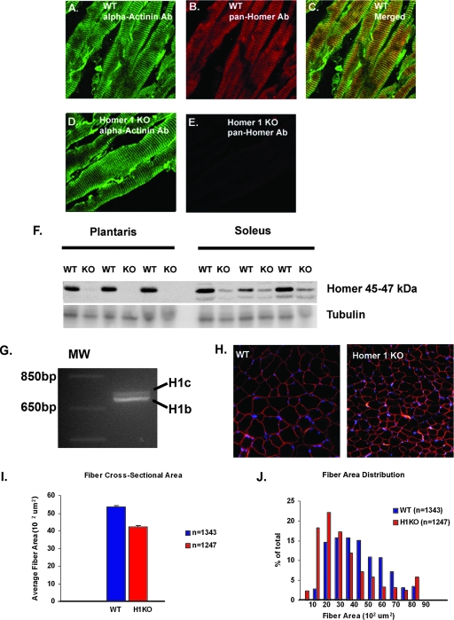
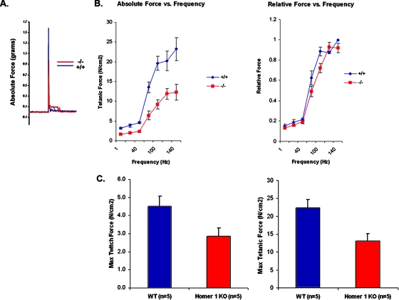
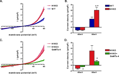
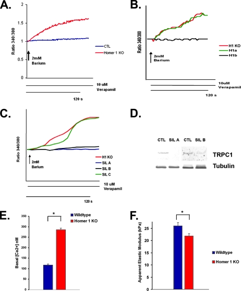
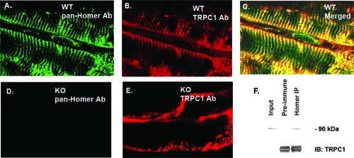
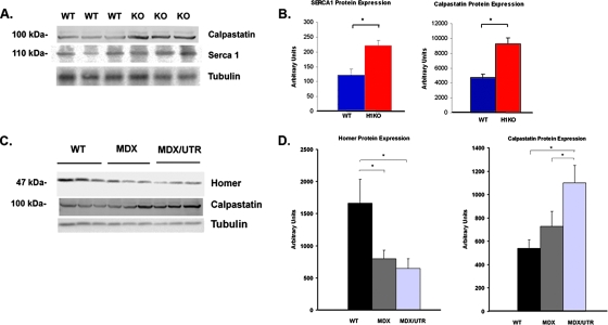
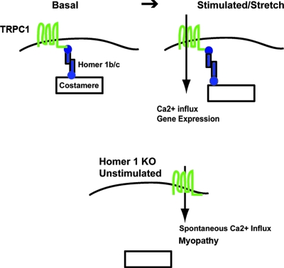
Transient receptor potential (TRP) channels are nonselective cation channels, several of which are expressed in striated muscle. Because the scaffolding protein Homer 1 has been implicated in TRP channel regulation, we hypothesized that Homer proteins play a significant role in skeletal muscle function. Mice lacking Homer 1 exhibited a myopathy characterized by decreased muscle fiber cross-sectional area and decreased skeletal muscle force generation. Homer 1 knockout myotubes displayed increased basal current density and spontaneous cation influx. This spontaneous cation influx in Homer 1 knockout myotubes was blocked by reexpression of Homer 1b, but not Homer 1a, and by gene silencing of TRPC1. Moreover, diminished Homer 1 expression in mouse models of Duchenne's muscular dystrophy suggests that loss of Homer 1 scaffolding of TRP channels may contribute to the increased stretch-activated channel activity observed in mdx myofibers. These findings provide direct evidence that Homer 1 functions as an important scaffold for TRP channels and regulates mechanotransduction in skeletal muscle.


Figures

FIG. 1.
FIG. 1.
Homer 1 colocalizes with alpha-actinin at the Z-disk. (A) Immunostaining of alpha-actinin in adult WT plantaris muscle. (B) Immunostaining of adult WT plantaris muscle using a pan-Homer Ab. (C) Merged image of panels A and B shows that Homer proteins and alpha-actinin colocalize. (D) Immunostaining of alpha-actinin in Homer 1 KO skeletal muscle. (E) Immunostaining of Homer 1 KO skeletal muscle using a pan-Homer Ab. (F) Western blotting using a pan-Homer Ab shows Homer expression in predominantly fast-twitch (plantaris) and predominantly slow-twitch (soleus) skeletal muscles from WT and Homer 1 KO mice. Non-Homer 1 isoforms are expressed in soleus muscle. (G) RT-PCR of cDNA generated from plantaris muscle shows the expression of coiled-coil Homer 1 isoforms, Homer 1b and Homer 1c, in adult mouse skeletal muscle. (H) DAPI (4′,6′-diamidino-2-phenylindole [blue]) and dystrophin (red) immunostaining of WT and Homer 1 KO mouse muscle. (I) Cross-sectional area of myofibers from muscles of Homer 1 KO (H1KO) mice (n = 1247) compared with WT littermates (n = 1343). Fiber cross-sectional area was 42.1 × 10² ± 80 μm2 in Homer 1 KO mice versus 53.6 × 10² ± 65 μm2 in WT littermate controls (mean ± SE; P < 0.05). (J) Distribution of muscle fiber cross-sectional areas from WT (blue) and Homer 1 KO mice (red).
FIG. 2.
FIG. 2.
Loss of Homer 1 results in decreased skeletal muscle contractility. (A) Representative tracings of twitch force generated by WT (blue) and Homer 1 KO (red) skeletal muscle display a delayed relaxation phase and a marked reduction in twitch force in Homer 1 KO muscle. (B) Force production was reduced over a range of stimulation frequencies (1 to 140 Hz), indicating a primary defect in contractility (n = 5, WT and KO). No significant difference was observed in the frequency at which maximum force was obtained (i.e., shift in the relative force-frequency curve) to suggest a defect in EC coupling in Homer 1 KO mice. (C) Homer 1 KO muscle exhibits significant decreases in both maximum twitch and tetanic force (P < 0.05). Force measurements shown are normalized to muscle cross-sectional area.
FIG. 3.
FIG. 3.
Membrane current of Homer 1 knockout (H1KO) versus WT day 5 myotubes. (A) Current density-voltage (I-V) relationships of whole-cell current of Homer 1 KO (H1KO; red) versus WT (blue) myotubes. Currents were induced by 200-ms voltage ramp protocols (1 mV/ms, from 100 mV to −100 mV) with a holding potential of −60 mV. Currents were normalized to membrane capacitance and shown in averaged traces. Vertical bars represent the standard error of the mean. (B) Group mean current amplitudes of WT (blue; n = 9) versus H1KO (red; n = 11) at −80 mV (downward) and +80 mV (upward) (**, P < 0.01). (C) Effect of tarantula toxin (5 μM GsMTx-4) on membrane current of KO myotubes. GsMTx-4 (green) inhibited membrane current at both positive and negative membrane potentials. (D) Group mean values of Homer 1 KO control (red) versus GsMTx-4 (green) at −80 mV (downward) and +80 mV (upward) (*, P < 0.05, n = 5).
FIG. 4.
FIG. 4.
Homer 1 KO myotubes exhibit abnormal TRP channel activation. (A) Homer 1 KO and WT myotubes were placed in a zero calcium solution in the presence of verapamil, followed by the readdition of 2 mM barium. Spontaneous barium influx was seen in Homer 1 KO myotubes (red) but not in controls (blue). (B) Homer 1 KO myotubes were transfected with empty vector expressing YFP alone (H1KO; n = 10) or along with an expression vector for either Homer 1a (H1a; n = 46) or Homer 1b (H1b; n = 44). Reexpression of Homer 1b (black trace) significantly inhibited the spontaneous cation influx observed in Homer 1 KO myotubes, while reexpression of Homer 1a (green trace) had no effect. (C) Homer 1 KO myotubes were transfected with either a control shRNA construct (H1KO; n = 22), an shRNA construct for silencing of TRPC1 (SIL A [n = 24] or SIL B [n = 28]), or a noneffective shRNA construct designed against TRPC1 (SIL C; n = 40). Silencing of TRPC1 (blue trace, SIL A; black trace, SIL B) significantly inhibited the spontaneous cation influx observed in Homer 1 KO myotubes. Marked spontaneous cation influx was still observed in Homer 1 KO myotubes transfected with a control shRNA construct (red trace) or a noneffective TRPC1 shRNA construct (green trace). (D) Knockdown of TRPC1 expression in myocytes with silencing constructs (SIL A and SIL B) was confirmed on a protein level by Western blotting after adenoviral infection. (E) Bar graph showing increased basal cytosolic calcium concentration in Homer 1 KO myotubes (n = 28) compared with controls (n = 16). (F) Primary skeletal myocytes were isolated from Homer 1 KO neonates and their WT littermate controls and allowed to differentiate into myotubes. After 5 days of differentiation, myotube stiffness was measured using AFM (n = 25 for WT and KO). Homer 1 KO myotubes exhibited a significant decrease in stiffness as expressed by the apparent elastic modulus. CTL, control.
FIG. 5.
FIG. 5.
Partial colocalization and coimmunoprecipitation of Homer and TRPC1 in adult skeletal muscle. (A) Immunostaining of adult WT plantaris muscle using a pan-Homer Ab. (B) Immunostaining of adult WT plantaris muscle for TRPC1. (C) Merged image of panels A and B shows that Homer 1 proteins and TRPC1 show significant colocalization at the costamere. (D) Immunostaining of Homer 1 KO plantaris muscle using a pan-Homer Ab. (E) Immunostaining of Homer 1 KO skeletal muscle using a TRPC1 Ab. (F) Coimmunoprecipitation of Homer and TRPC1 from a mouse gastrocnemius muscle protein lysate. Preimmune rat serum was used as a negative control. A pan-Homer Ab was used for immunoprecipitation (IP) of endogenous Homer protein, followed by SDS-PAGE and immunoblotting (IB) for TRPC1.
FIG. 6.
FIG. 6.
(A) Western blotting showing expression of calpastatin and SERCA1 in WT and Homer 1 KO gastrocnemius muscle. (B) Bar graphs summarizing expression data from Western blots in arbitrary units. (C) Western blots showing Homer protein and calpastatin expression in WT, MDX−/−, and MDX/UTR−/− double KO mice. (D) Bar graphs summarizing expression data from the above Western blots in arbitrary units.
FIG. 7.
FIG. 7.
Model of the role of Homer proteins in the regulation of mechanosensitive TRP channels. In the basal state, coiled-coil Homer proteins (i.e., Homer 1b and 1c) link mechanosensitive TRP channels to the Z-disk/costamere, where they are poised to respond to stretch. In response to stretch, mechanosensitive TRP channels are activated, resulting in activation of calcium-dependent signaling pathways. In the absence of Homer (bottom), dysregulation of mechanosensitive TRP channels results in spontaneous calcium influx, activation of calcium-dependent proteolysis via calpains, and degradation of cytoskeletal elements resulting in a myopathy.

References

    1. Ango, F., L. Prezeau, T. Muller, J. C. Tu, B. Xiao, P. F. Worley, J. P. Pin, J. Bockaert, and L. Fagni. 2001. Agonist-independent activation of metabotropic glutamate receptors by the intracellular protein Homer. Nature 411962-965. - PubMed
    1. Berthier, C., and S. Blaineau. 1997. Supramolecular organization of the subsarcolemmal cytoskeleton of adult skeletal muscle fibers. A review. Biol. Cell 89413-434. - PubMed
    1. Bloch, R. J., P. Reed, A. O'Neill, J. Strong, M. Williams, N. Porter, and H. Gonzalez-Serratos. 2004. Costameres mediate force transduction in healthy skeletal muscle and are altered in muscular dystrophies. J. Muscle Res. Cell Motil. 25590-592. - PubMed
    1. Bortoloso, E., N. Pilati, A. Megighian, E. Tibaldo, D. Sandona, and P. Volpe. 2006. Transition of Homer isoforms during skeletal muscle regeneration. Am. J. Physiol. 290C711—C718. - PubMed
    1. Bottai, D., J. F. Guzowski, M. K. Schwarz, S. H. Kang, B. Xiao, A. Lanahan, P. F. Worley, and P. H. Seeburg. 2002. Synaptic activity-induced conversion of intronic to exonic sequence in Homer 1 immediate early gene expression. J. Neurosci. 22167-175. - PMC - PubMed

Publication types