. 2008 Apr;28(8):2497-508.
doi: 10.1128/MCB.00405-07. Epub 2008 Feb 11.

Snu56p is required for Mer1p-activated meiotic splicing

Affiliations

Snu56p is required for Mer1p-activated meiotic splicing

Richard J Balzer et al. Mol Cell Biol. 2008 Apr.

Abstract

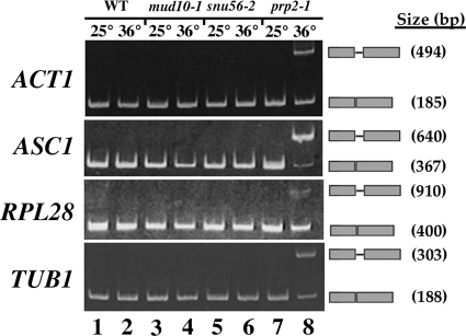
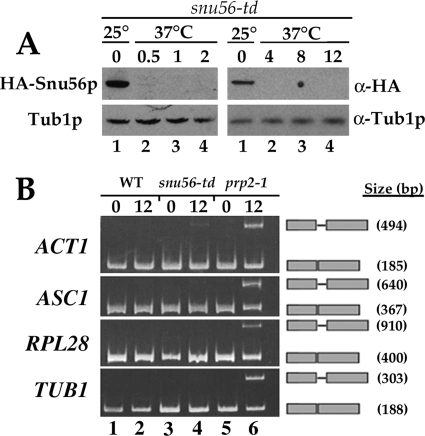
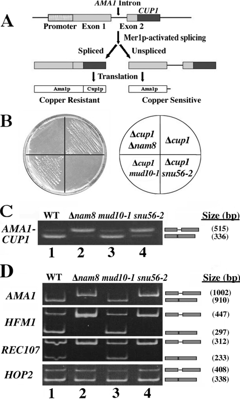
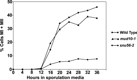
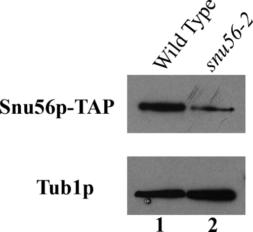
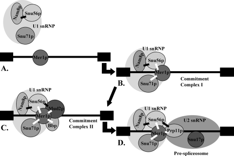
Alternative or regulated splicing can be applied to genes that are transcribed but whose products may be deleterious or unnecessary to the cell. In the yeast Saccharomyces cerevisiae, positive splicing regulation occurs during meiosis in which diploid cells divide to form haploid gametes. The Mer1 protein recruits the U1 snRNP to specific pre-mRNAs, permitting spliceosomal assembly and splicing. The mature transcripts are required for meiotic progression and, subsequently, sporulation. We have identified a novel allele (snu56-2) of the essential U1 snRNP protein Snu56p that exhibits a sporulation defect. Using a CUP1 reporter assay and reverse transcriptase PCR, we demonstrate that this allele specifically impairs Mer1p-activated splicing. This is not a reflection of a generally deficient spliceosome, as these cells splice vegetative transcripts efficiently. Furthermore, Snu56p depletion in vivo does not significantly impact mitotic splicing. Thus, its splicing function appears to be limited to Mer1p-activated meiosis-specific splicing. Two-hybrid studies indicate that Snu56p interacts with the other two U1 snRNP factors (Mer1p and Nam8p) required for this process. Interestingly, these two proteins do not interact, suggesting that Snu56p links pre-mRNA-bound Mer1p to Nam8p in the U1 snRNP. This work demonstrates that the Snu56 protein is required for splicing only during meiosis.


Figures

FIG. 1.
FIG. 1.
Vegetative mud10-1 and snu56-2 cells efficiently splice pre-mRNAs. Total RNA was prepared from exponential-phase wild-type (WT; lanes 1 and 2) and mud10-1 (lanes 3 and 4), snu56-2 (lanes 5 and 6), and prp2-1 (lanes 7 and 8) mutant cells grown at either 25°C or 36°C. ACT1, ASC1, RPL28, and TUB1 pre-mRNA splicing was analyzed by RT-PCR with the appropriate intron-flanking primer set. PCR products were resolved on 5% polyacrylamide gels and stained with ethidium bromide. The gene analyzed in each case is indicated to the left of the panel. Bands representing unspliced pre-mRNA and spliced mRNA are indicated to the right of each panel. PCR product sizes are also indicated on the right.
FIG. 2.
FIG. 2.
In vivo depletion of Snu56p does not affect vegetative splicing. (A) Snu56p-td is rapidly depleted at 37°C. Western blot analysis was performed with extracts from snu56-td mutant cells (MHY1481) grown at 25°C or shifted to 37°C for the times indicated above the lanes (hours). The antibodies used are indicated on the right. Tub1p was used as a loading control. HA, hemagglutinin. (B) RT-PCR analysis of ACT1, ASC1, RPL28, and TUB1 pre-mRNA splicing. Total RNA was prepared from exponential-phase wild-type (WT; MHY1482; lanes 1 and 2) and snu56-td (MHY1481; lanes 3 and 4) and prp2-1 (P30-6D; lanes 5 and 6) mutant cells grown at either 25°C or shifted to 37°C for 12 h. RNA was analyzed by RT-PCR with the appropriate intron-flanking primer set. PCR products were resolved on 5% polyacrylamide gels and stained with ethidium bromide. The gene analyzed in each case is indicated to the left of the panel. Bands representing unspliced pre-mRNA and spliced mRNA are indicated to the right of each panel. PCR product sizes are also indicated on the right.
FIG. 3.
FIG. 3.
Snu56p is required for Mer1p-activated splicing. (A) Schematic of the AMA1-CUP1 reporter assay used to monitor Mer1p-activated splicing in vegetative cells. The MER1 and AMA1-CUP1 fusion splicing reporter genes are constitutively expressed from separate plasmids. Mer1p-activated splicing results in the expression of an AMA1-CUP1 fusion protein conferring copper resistance. (B) Growth of cup1Δ mutant yeast on copper plates. Wild-type and snu56-2, mud10-1, and nam8Δ mutant cells that have had endogenous CUP1 knocked out and been transformed with MER1 and AMA1-CUP1 fusion splicing reporter plasmids are shown. Transformants were streaked onto synthetic complete medium containing 0.25 mM copper and lacking uracil and tryptophan (left). A plate schematic (right) indicates the mutant alleles of each strain. (C) RT-PCR analysis of AMA1-CUP1 pre-mRNA splicing. Total RNA was prepared from exponential-phase wild-type (WT; lane 1) and nam8Δ (lane 2), mud10-1 (lane 3), and snu56-2 (lane 4) mutant cells that have had endogenous CUP1 knocked out and been transformed with MER1 and AMA1-CUP1 fusion plasmids. RNA was analyzed by RT-PCR with primers complementary to the exons of the AMA1-CUP1 gene. PCR products were resolved on 1.75% agarose gels and stained with ethidium bromide. The gene analyzed in each case is indicated to the left of the panel. Bands representing unspliced pre-mRNA and spliced mRNA are indicated to the right of each panel. PCR product sizes are also indicated on the right. (D) AMA1, HFM1, and REC107 splicing during meiosis is Snu56p dependent. The RT-PCR products generated from total RNA samples were taken from wild-type (lane 1) and nam8Δ (lane 2), mud10-1 (lane 3), and snu56-2 (lane 4) mutant homozygous diploids 15 h after transfer to sporulation medium. PCR products were resolved on 5% acrylamide gels and stained with ethidium bromide. The gene analyzed in each case is indicated to the left of the panel. Bands representing unspliced pre-mRNA and spliced mRNA are indicated to the right of each panel. PCR product sizes are also indicated on the right.
FIG. 4.
FIG. 4.
Diploid snu56-2 mutant cells fail to execute meiosis. Meiotic nuclear divisions were examined by DAPI staining. Plotted is the percentage of cells that had undergone both nuclear divisions (MI and MII) at various times throughout sporulation. At least 200 cells from each strain were examined at each time point to determine the numbers of tri- and tetranucleate cells. Shown are averages of three independent experiments. All errors are within 5%.
FIG. 5.
FIG. 5.
Snu56 and snu56-2 protein expression levels. Western blot analysis was performed with exponential growth phase SNU56-TAP and snu56-2-TAP cell lysates. SNU56 alleles are indicated at the top of each lane, and the proteins visualized are indicated to the left. Tub1p was used as a loading control.
FIG. 6.
FIG. 6.
Synthetic lethality of snu56 alleles. Wild-type and mud10-1, snu56-2, nam8Δ, mud10-1 nam8Δ, and snu56-2 nam8Δ mutant cells bearing a CEN URA3 SNU56 plasmid are shown. Cells were streaked onto synthetic complete medium lacking uracil (left) or containing 5-FOA (center). The mutant allele(s) present in each strain is indicated by the plate schematic (right). Plates were incubated at 25°C for 2 days.
FIG. 7.
FIG. 7.
Snu56p protein-protein interactions during early spliceosome formation. Snu56p stabilizes Nam8p within the U1 snRNP (A) and associates with Mer1p to aid U1 snRNP recruitment to Mer1p-activated introns during commitment complex I formation (B). Later, interactions with Mud2p during commitment complex II assembly (C) and Prp11p during prespliceosome formation (D) may help stabilize these spliceosomal intermediates. Black boxes represent exons, and the black line represents a Mer1p-activated intron. Black bars between proteins represent two-hybrid interactions identified in this work, and white bars denote those previously identified (48).

References

    1. Ausubel, F. M., R. Brent, R. E. Kingston, D. D. Moore, J. G. seidman, J. A. Smith, and K. Struhl. 1994. Current protocols in molecular biology. John Wiley & Sons, Inc., New York, NY.
    1. Baudin-Baillieu, A., E. Guillemet, C. Cullin, and F. Lacroute. 1997. Construction of a yeast strain deleted for the TRP1 promoter and coding region that enhances the efficiency of the polymerase chain reaction-disruption method. Yeast 13353-356. - PubMed
    1. Bender, A., and J. R. Pringle. 1991. Use of a screen for synthetic lethal and multicopy suppressee mutants to identify two new genes involved in morphogenesis in Saccharomyces cerevisiae. Mol. Cell. Biol. 111295-1305. - PMC - PubMed
    1. Bossie, M. A., C. DeHoratius, G. Barcelo, and P. Silver. 1992. A mutant nuclear protein with similarity to RNA binding proteins interferes with nuclear import in yeast. Mol. Biol. Cell 3875-893. - PMC - PubMed
    1. Brow, D. A. 2002. Allosteric cascade of spliceosome activation. Annu. Rev. Genet. 36333-360. - PubMed

Publication types

MeSH terms

Substances

LinkOut - more resources