. 2008 Feb 18;205(2):339-46.
doi: 10.1084/jem.20071868. Epub 2008 Feb 11.

IL-33 reduces the development of atherosclerosis

Affiliations

IL-33 reduces the development of atherosclerosis

Ashley M Miller et al. J Exp Med. .

Erratum in

  • J Exp Med. 2012 Dec 17;209(13):2515

Abstract

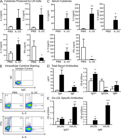
Atherosclerosis is a chronic inflammatory disease of the vasculature commonly leading to myocardial infarction and stroke. We show that IL-33, which is a novel IL-1-like cytokine that signals via ST2, can reduce atherosclerosis development in ApoE(-/-) mice on a high-fat diet. IL-33 and ST2 are present in the normal and atherosclerotic vasculature of mice and humans. Although control PBS-treated mice developed severe and inflamed atherosclerotic plaques in the aortic sinus, lesion development was profoundly reduced in IL-33-treated animals. IL-33 also markedly increased levels of IL-4, -5, and -13, but decreased levels of IFNgamma in serum and lymph node cells. IL-33 treatment also elevated levels of total serum IgA, IgE, and IgG(1), but decreased IgG(2a), which is consistent with a Th1-to-Th2 switch. IL-33-treated mice also produced significantly elevated antioxidized low-density lipoprotein (ox-LDL) antibodies. Conversely, mice treated with soluble ST2, a decoy receptor that neutralizes IL-33, developed significantly larger atherosclerotic plaques in the aortic sinus of the ApoE(-/-) mice compared with control IgG-treated mice. Furthermore, coadministration of an anti-IL-5 mAb with IL-33 prevented the reduction in plaque size and reduced the amount of ox-LDL antibodies induced by IL-33. In conclusion, IL-33 may play a protective role in the development of atherosclerosis via the induction of IL-5 and ox-LDL antibodies.


Figures

Figure 1.
Figure 1.
Expression of IL-33 and ST2 in vascular cells and tissues. The expression of mRNAs for IL-33, ST2, and β-actin were examined by RT-PCR in the thoracic aortas of 18-wk-old C57BL/6 control mice (normal diet) or ApoE−/− (normal diet or high-fat diet) mice (A; n = 3) and in primary cultured human umbilical vein ECs (HUVECs), human saphenous vein ECs (HSVECs), human saphenous vein SMCs (HSVSMCs), and human coronary artery SMCs (HCASMCs; B). (C) Quantitative PCR analysis of IL-33 gene expression in thoracic aortas of mice as described in A (expressed as a percentage of 18S endogenous control; n = 4–5). Data are the mean ± the SEM. Immunostaining and isotype controls in frozen vascular tissues of ApoE−/− mice for α-smooth muscle actin and IL-33 in the adventitia of the aorta (D), and for CD31 and IL-33 in ECs of small vessels of the heart (E). A, adventitia; M, media; L, lumen. Images shown are representative of seven sections. *, P < 0.05, Student's unpaired t test. Bars, 25 μm.
Figure 2.
Figure 2.
IL-33 treatment reduced atherosclerotic plaque size and macrophage and T cell accumulation in the aortic sinus of ApoE−/− mice. ApoE−/− mice were treated with PBS (open bars) or IL-33 (filled bars). (A) Plaque size (intimal area, mm2) in the aortic sinus (n = 12–15). (B) Representative photomicrographs of hematoxylin and eosin–stained aortic sections. (C) Quantification of plaque content: F4/80+ macrophages (percentage of total plaque area; n = 11–12), T cells (number of CD3+ T cells/millimeter2, n = 10–11), SMCs (percentage of total plaque area, n = 9–10), and collagen (percentage of total plaque area, n = 9–10). Data are the mean ± the SEM. (D) Representative photomicrographs of F4/80+ macrophages (brown), CD3+ T cells (brown), α-smooth muscle actin (brown), and collagen (blue) staining in plaques of ApoE−/− mice treated with PBS or IL-33. Data shown are from two independent experiments. *, P < 0.05; ***, P < 0.001, Student's unpaired t test. Bars: (B) 400 μm; (D; F4/80+ macrophages) 25 μm; (D; CD3+ T cells) 25 μm; (D; α-smooth muscle actin) 100 μm; (D; collagen) 100 μm.
Figure 3.
Figure 3.
IL-33 induced Th2 cytokines, Th2 antibodies, and ox-LDL–specific antibodies in serum and lymph node cells of ApoE−/− mice. ApoE−/− mice were treated with PBS (open bars) or IL-33 (filled bars). (A) Cytokine production (IL-4, -5, -13, and IFNγ) by lymph node cells restimulated with αCD3 and measured by ELISA. (B) Intracellular cytokine staining (IL-4, -5, and IFNγ) in CD4+ lymph node cells stimulated with PMA/ionomycin for 4 h. Numbers indicate the percentage of positive cells in each quadrant. (C) IL-4, -5, -6, -13 (all picogram/milliliter), and IFNγ (nanogram/milliliter) in serum measured by Luminex assay. (D) Total serum concentrations of Ig isotypes (IgA, IgE, IgG1, IgG2a, and IgM). (E) Ox-LDL–specific IgM and IgG1 antibodies in serum. Data shown are the mean ± the SEM. n = 10–13 mice/group from 2 independent experiments. *, P < 0.05; **, P < 0.01; ***, P < 0.001, Student's unpaired t test.
Figure 4.
Figure 4.
sST2 exacerbated atherosclerosis development in the aortic sinus and promoted Th1 response of ApoE−/− mice. ApoE−/− mice were treated with control IgG (open bars) or sST2 (filled bars). (A) Plaque size (intimal area, millimeter2) in the aortic sinus (n = 9–10). (B) Representative photomicrographs of hematoxylin and eosin–stained aortic sections. (C) Quantification of plaque content: F4/80+ macrophages (percentage of total plaque area, n = 9–10), T cells (number of CD3+ T cells/millimeter2, n = 9–10), SMCs (percentage of total plaque area, n = 9–10), and collagen (percentage of total plaque area, n = 9–10). (D) Representative photomicrographs of F4/80+ macrophages (brown), CD3+ T cells (brown), α-smooth muscle actin (brown), and collagen (blue) staining in plaques of ApoE−/− mice treated with IgG or sST2. (E) IFNγ production (nanogram/milliliter) by lymph node cells restimulated with αCD3 and measured by ELISA (n = 9–10). IL-5 (F) and IFNγ (G; picogram/milliliter) in serum determined by Luminex assay (n = 9–10). *, P < 0.05; ***, P < 0.001, Student's unpaired t test. Bars: (B) 400 μm; (D; F4/80+ macrophages) 25 μm; (D; CD3+ T cells) 25 μm; (D; α-smooth muscle actin) 100 μm; (D; collagen) 100 μm.
Figure 5.
Figure 5.
An anti–IL-5 mAb prevented the reduction in plaque size and the induction of ox-LDL antibodies in serum by IL-33. ApoE−/− mice were treated with IL-33 (filled bars) or IL-33/αIL-5 (striped bars). (A) Plaque size (intimal area, millimeter2) in the aortic sinus (n = 5). (B) Representative photomicrographs of hematoxylin and eosin–stained aortic sections. (C) Quantification of plaque content: F4/80+ macrophages (percentage of total plaque area, n = 5), T cells (number of CD3+ T cells/ millimeter2, n = 5), SMCs (percentage of total plaque area, n = 5), collagen (percentage of total plaque area, n = 5). (D) Representative photomicrographs of F4/80+ macrophages (brown), CD3+ T cells (brown), α-smooth muscle actin (brown), and collagen (blue) staining in plaques of ApoE−/− mice treated with IL-33 or IL-33/αIL-5. (E) Ox-LDL–specific IgM antibodies in serum (n = 5). (F) IL-5 and -12 (picogram/milliliter) in serum determined by ELISA (n = 5). *, P < 0.05; **, P < 0.01, Student's unpaired t test. Bars: (B) 400 μm; (D; F4/80+ macrophages) 25 μm; (D; CD3+ T cells) 25 μm; (D; α-smooth muscle actin) 100 μm; (D; collagen) 100 μm.

References

    1. Hansson, G.K., and P. Libby. 2006. The immune response in atherosclerosis: a double-edged sword. Nat. Rev. Immunol. 6:508–519. - PubMed
    1. Salonen, J.T., S. Yla-Herttuala, R. Yamamoto, S. Butler, H. Korpela, R. Salonen, K. Nyyssonen, W. Palinski, and J.L. Witztum. 1992. Autoantibody against oxidised LDL and progression of carotid atherosclerosis. Lancet. 339:883–887. - PubMed
    1. Major, A.S., S. Fazio, and M.F. Linton. 2002. B-lymphocyte deficiency increases atherosclerosis in LDL receptor-null mice. Arterioscler. Thromb. Vasc. Biol. 22:1892–1898. - PubMed
    1. Caligiuri, G., A. Nicoletti, B. Poirier, and G.K. Hansson. 2002. Protective immunity against atherosclerosis carried by B cells of hypercholesterolemic mice. J. Clin. Invest. 109:745–753. - PMC - PubMed
    1. de Boer, O.J., A.C. van der Wal, C.E. Verhagen, and A.E. Becker. 1999. Cytokine secretion profiles of cloned T cells from human aortic atherosclerotic plaques. J. Pathol. 188:174–179. - PubMed

Publication types

MeSH terms