. 2008 Feb 18;205(2):491-501.
doi: 10.1084/jem.20072041. Epub 2008 Feb 11.

VEGF and TGF-beta are required for the maintenance of the choroid plexus and ependyma

Affiliations

VEGF and TGF-beta are required for the maintenance of the choroid plexus and ependyma

Arindel S R Maharaj et al. J Exp Med. .

Abstract

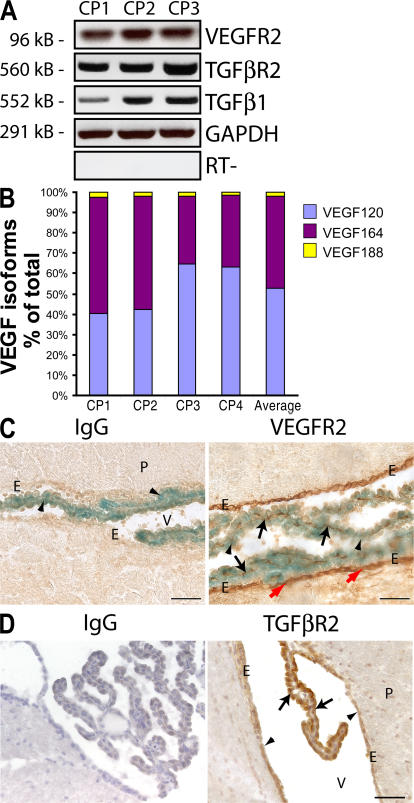
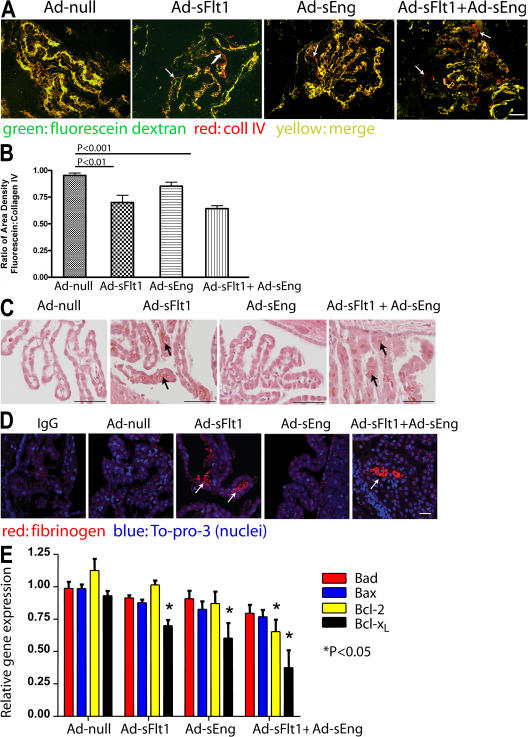
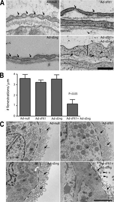
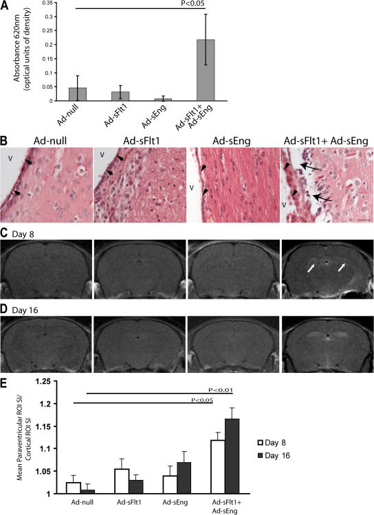
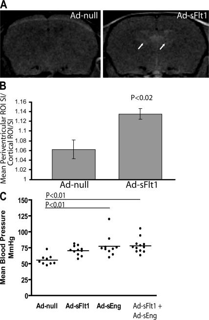
Although the role of vascular endothelial growth factor (VEGF) in developmental and pathological angiogenesis is well established, its function in the adult is less clear. Similarly, although transforming growth factor (TGF) beta is involved in angiogenesis, presumably by mediating capillary (endothelial cell [EC]) stability, its involvement in quiescent vasculature is virtually uninvestigated. Given the neurological findings in patients treated with VEGF-neutralizing therapy (bevacizumab) and in patients with severe preeclampsia, which is mediated by soluble VEGF receptor 1/soluble Fms-like tyrosine kinase receptor 1 and soluble endoglin, a TGF-beta signaling inhibitor, we investigated the roles of VEGF and TGF-beta in choroid plexus (CP) integrity and function in adult mice. Receptors for VEGF and TGF-beta were detected in adult CP, as well as on ependymal cells. Inhibition of VEGF led to decreased CP vascular perfusion, which was associated with fibrin deposition. Simultaneous blockade of VEGF and TGF-beta resulted in the loss of fenestrae on CP vasculature and thickening of the otherwise attenuated capillary endothelium, as well as the disappearance of ependymal cell microvilli and the development of periventricular edema. These results provide compelling evidence that both VEGF and TGF-beta are involved in the regulation of EC stability, ependymal cell function, and periventricular permeability.


Figures

Figure 1.
Figure 1.
Expression of VEGF, TGF-β1, VEGFR2, and TGF-βR2 in the CP and ependyma. (A) RT-PCR of complementary DNA from CP dissected from the lateral third and fourth ventricles of four adult mice (n = 3 pools) revealed expression of TGF-β1 and its receptor, TGF-βR2, as well as VEGFR2. (B) Real-time RT-PCR of CP complementary DNA (n = 4 pools) for VEGF isoforms (52.6% VEGF120, 45.5% VEGF164, and 1.8% VEGF 188). (C) Staining of VEGF-lacZ brain sections for lacZ (blue) and AEC immunostaining for VEGFR2 (red) revealed VEGFR2 in the CP vasculature (black arrows) adjacent to VEGF-expressing CP epithelial cells (arrowheads) and in the ependymal lining of the ventricle (red arrows). (D) DAB immunostaining followed by hematoxylin staining of adult brain sections for TGF-βR2 reveals TGF-βR2 immunoreactivity in the CP epithelia (arrows) and ependyma (arrowheads). E, ependyma; P, parenchyma; V, ventricle. Bars, 50 μm.
Figure 2.
Figure 2.
VEGF neutralization results in decreased CP vascular perfusion and fibrin deposition. (A) Mice perfused with high mol wt fluorescein dextran show a significant decrease in vessel perfusion in Ad-sFlt1 as well as in Ad-sFlt1 + Ad-sEng mice (arrows). Nonperfused vessels are identified as coll IV positive (red) and fluorescein negative. (B) Perfused blood vessels were quantified by measuring the proportion of sectional area occupied by fluorescein (green) and coll IV fluorescence (red; adapted from reference 63). Results represent the mean value of three sections 150 μm apart per mouse, and the mean of five to six animals. (C) H + E staining (black arrows) and (D) immunofluorescent staining (white arrows) for fibrinogen in CP show multiple fibrin clots in Ad-sFlt1 (C and D, arrows) and Ad-sFlt1 + Ad-sEng (C and D). (E) Relative messenger RNA expression of proapoptotic (bad and bax) and antiapoptotic (Bcl-2 and Bcl-xL) genes in CP were determined by real-time PCR analysis. Bars: (A and C): 50 μm; (D) 25 μm.
Figure 3.
Figure 3.
Effect of VEGF and TGF-β neutralization on the ultrastructure of CP vasculature and the ependyma. After 14 d of infection with the indicated Ad's, the CP and ependyma were examined by TEM. (A) In the CP vasculature, fenestrations (arrows) were unaffected in Ad-null, Ad-sFlt1, and Ad-sEng mice, whereas mice expressing Ad-sFlt1 + Ad-sEng exhibited loss of fenestrations, increased EC thickening (vertical bar), and the appearance of multiple caveolae (arrows). (B) Quantification of the number of fenestrations per 1 μm revealed no significant change between Ad-null (3.6 ± 0.37; n = 5), Ad-sFlt1 (3.22 ± 0.2; n = 3), and (Ad-sEng (3.53 ± 0.43; n = 5), whereas only 1.17 ± 0.41 (P < 0.05; n = 6) were present in Ad-sFlt1 + Ad-sEng–treated mice CP. (C) Electron microscopic visualization of the ependymal cells in all experimental groups revealed normal apical polarity of the mitochondria (labeled M). Dashed lines indicate the cell–microvilli border, arrows indicate microvilli, and arrowheads indicate cilia. Bars: (A) 1.25 μm; (C) 2.5 μm.
Figure 4.
Figure 4.
Effect of VEGF and TGF-β neutralization on brain permeability. (A) Experimental mice infected for 3 d were injected intravenously with 2% Evans blue in PBS. After 40 min, the brains were removed and quantified for Evans blue/albumin leakage. Simultaneous treatment with sFlt1 + sEng (n = 3) resulted in significantly increased Evans blue leakage into the extravascular space, whereas no changes could be detected with sFlt1 or sEng alone. Values represent means ± SD. (B) H + E staining of sections of the lateral ventricle of mice expressing sFlt1 + sEng for 14 d revealed marked periventricular edema (arrows) below the ependyma (arrowheads); no edema was observed in this region in other experimental or control mice. V, ventricle. Bar, 25 μm. (C and D) T1-weighted coronal MRI of experimental mice intravenously injected with Gd at 8 (C) and 16 (D) d after infection revealed focal lesions surrounding the lateral ventricles in the midcortex of mice expressing sFlt1 + sEng (white arrows) but not in the other experimental groups. (E) Quantification of the ratio of signal intensity in the periventricular zone versus the nonenhancing region in the cortex using ImageJ software (NIH). Values represent means ± SEM.
Figure 5.
Figure 5.
Effect of long-term VEGF neutralization on brain permeability, and effects of VEGF and TGF-β neutralization on blood pressure. (A and B) T1-weighted coronal MRI of Gd intravenously injected mice after 28 d of Ad-sFlt1 or Ad-null expression revealed focal lesions surrounding the lateral ventricles of significant intensity in the midcortex in Ad-sFlt1 mice. Areas in A represent the area of periventricular leakage observed on MRI. Values in B represent means ± SEM. (C) Noninvasive blood pressure measurement of experimental mice at day 7 Ad expression shows a small difference between Ad-null and Ad-sFlt1 mice but a significant difference between mice expressing Ad-null and Ad-sEng (P < 0.01) and Ad-sFlt1 + Ad-sEng (P < 0.01). Horizontal bars represent the average of the mean arterial pressure in the different groups.

References

    1. Coultas, L., K. Chawengsaksophak, and J. Rossant. 2005. Endothelial cells and VEGF in vascular development. Nature. 438:937–945. - PubMed
    1. Carmeliet, P., V. Ferreira, G. Breier, S. Pollefeyt, L. Kieckens, M. Gertsenstein, M. Fahrig, A. Vandenhoeck, K. Harpal, C. Eberhardt, et al. 1996. Abnormal blood vessel development and lethality in embryos lacking a single VEGF allele. Nature. 380:435–439. - PubMed
    1. Ferrara, N., K. Carver-Moore, H. Chen, M. Dowd, L. Lu, K.S. O'Shea, L. Powell-Braxton, K.J. Hillan, and M.W. Moore. 1996. Heterozygous embryonic lethality induced by targeted inactivation of the VEGF gene. Nature. 380:439–442. - PubMed
    1. Miquerol, L., B.L. Langille, and A. Nagy. 2000. Embryonic development is disrupted by modest increases in vascular endothelial growth factor gene expression. Development. 127:3941–3946. - PubMed
    1. Eming, S.A., and T. Krieg. 2006. Molecular mechanisms of VEGF-A action during tissue repair. J. Investig. Dermatol. Symp. Proc. 11:79–86. - PubMed

Publication types

MeSH terms

Substances