. 2008 Feb 11;180(3):537-48.
doi: 10.1083/jcb.200705085.

Akt regulates centrosome migration and spindle orientation in the early Drosophila melanogaster embryo

Affiliations

Akt regulates centrosome migration and spindle orientation in the early Drosophila melanogaster embryo

Graham J Buttrick et al. J Cell Biol. .

Abstract

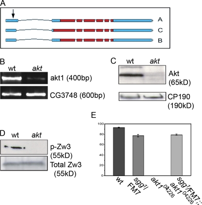
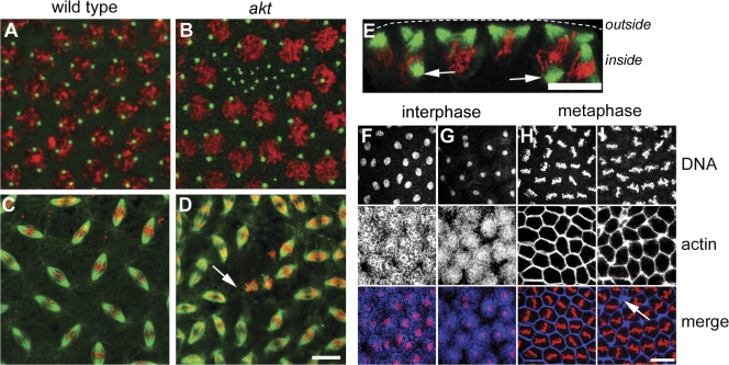
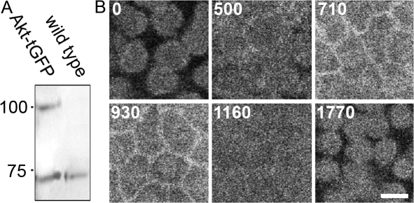
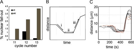
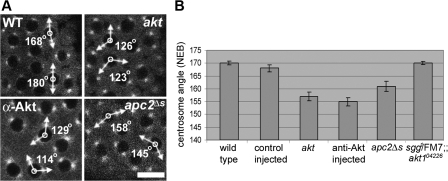
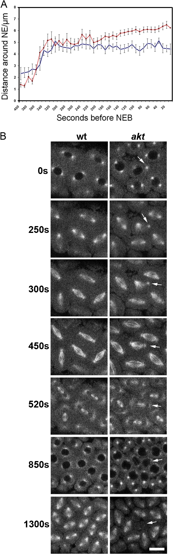
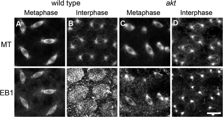
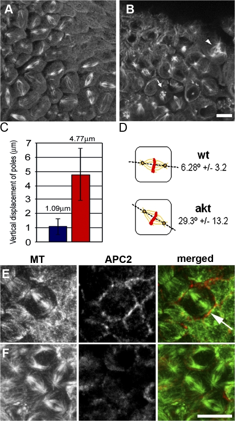
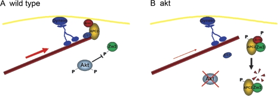
Correct positioning and morphology of the mitotic spindle is achieved through regulating the interaction between microtubules (MTs) and cortical actin. Here we find that, in the Drosophila melanogaster early embryo, reduced levels of the protein kinase Akt result in incomplete centrosome migration around cortical nuclei, bent mitotic spindles, and loss of nuclei into the interior of the embryo. We show that Akt is enriched at the embryonic cortex and is required for phosphorylation of the glycogen synthase kinase-3beta homologue Zeste-white 3 kinase (Zw3) and for the cortical localizations of the adenomatosis polyposis coli (APC)-related protein APC2/E-APC and the MT + Tip protein EB1. We also show that reduced levels of Akt result in mislocalization of APC2 in postcellularized embryonic mitoses and misorientation of epithelial mitotic spindles. Together, our results suggest that Akt regulates a complex containing Zw3, Armadillo, APC2, and EB1 and that this complex has a role in stabilizing MT-cortex interactions, facilitating both centrosome separation and mitotic spindle orientation.


Figures

Figure 1.
Figure 1.
D. melanogaster Akt regulates Zw3 during early embryonic development. (A) Representation of the akt locus showing differential splicing of the three proposed isoforms (A, B, and C) and the site of insertion of the PZ P element (arrow). Coding regions are shown in red and untranslated regions are shown in blue (adapted from Flybase; http://www.flybase.indiana.org). (B) RT-PCR of a 400-bp region of the akt coding region from cDNA extracted from wild-type and akt mutant flies. (C) Western blot of 0–3-h embryo extracts probed with antibodies specific for Akt. CP190 is shown as a loading control. (D) Western blot of embryo extracts probed with an antibody specific for Zw3 phosphorylated on the consensus Akt phosphorylation site (Ser12). (E) Histogram demonstrating the rescue of akt104426 female sterility through the introduction of one copy of sgg1. Error bars indicate the SEM.
Figure 2.
Figure 2.
Reduced Akt levels lead to defects in syncytial mitoses. (A–D) Syncytial stage embryos fixed with methanol to reveal DNA (red), centrosomes (A and B, green), or MTs (C and D, green). (A and C) Wild-type embryos. (B and D) akt embryos show gaps in their cortex, which is devoid of nuclei but contains many centrosomes. Although many spindles look normal, regions of akt embryos show poorly formed spindles that are irregularly spaced (D, arrow). (E) A cross-sectional view of an akt embryo in prometaphase. Arrows indicate spindle poles not connected to the embryonic cortex. (F–I) Gross actin morphology is not perturbed in akt embryos. During interphase in wild-type (F) or akt embryos (G), actin is present in cortical caps. During metaphase in wild-type (H) and akt (I) embryos, actin is present in pseudocleavage furrows. Bars, 10 μm.
Figure 3.
Figure 3.
The regulation of APC2 and Arm is altered in akt embryos. (A) Immunoprecipiation of Arm from 0–3-h wild-type and akt104226 embryo extracts. Pros-35 is shown as a control. T, total extract; S, supernatant; P, pellet. (B) The mobility of APC2 in akt104226 embryos can be altered by treatment with λ phosphatase. (C) Wild-type and akt104226 embryos fixed and stained to visualize DNA and APC2. In akt embryos, APC2 fails to localize. Bar, 10 μm.
Figure 4.
Figure 4.
Akt localizes to the embryonic cortex in a cell cycle–dependent manner. (A) Western blot of 0–3-h wild-type embryos and embryos containing UAS-Akt-turbo-GFP driven by Nanos Gal4. Akt-tGFP is expressed at similar levels to endogenous Akt. Molecular mass (kD) is indicated on the left. (B) Live confocal analysis of embryos expressing Akt-tGFP under the UAS-Gal4 system. Akt-tGFP appears to cycle between cortical caps (0 and 1,770 s) and pseudocleavage furrows (710 and 930 s). Time is indicated in seconds. See Video 1 (available at http://www.jcb.org/cgi/content/full/jcb.200705085/DC1). Bar, 10 μm.
Figure 5.
Figure 5.
The timing of syncytial mitoses is not affected in akt embryos. Chromosome dynamics in wild-type and akt1q/akt104226 embryos expressing GFP-histone. (A) Histogram representing the percentage of nuclei that move into the interior of the embryo (nuclear fallout) during cycles 11, 12, and 13. Data were obtained from 20 wild-type and 20 akt1q/akt104226 embryos. (B) Graphical representation of an idealized graph applied to chromosomes throughout mitosis (Yau and Wakefield, 2007). Parameters of interest are automatically obtained using this software, such as (i) rate of chromosome condensation, (ii) length of metaphase, and (iii) rate of chromosome segregation. (C) Chromosome dynamics during cycle 12 in wild-type (black) and akt1q/akt104226 (red) embryos. Each line is the mean of 10 nuclei from a single embryo. Data were obtained from seven different wild-type or akt1q/akt104226 embryos. Similar results were found for embryos in cycles 11 and 13 (unpublished data). See Videos 2 and 3 (available at http://www.jcb.org/cgi/content/full/jcb.200705085/DC1).
Figure 6.
Figure 6.
Centrosome separation is inhibited after inactivation of Akt or APC2. (A) Live confocal analysis of embryos expressing α-tubulin–GFP. Examples of the angle of centrosome separation 20 s before NEB in wild-type (WT) embryos, akt1q/akt104226 embryos (akt), embryos injected with anti-Akt antibodies (α-Akt), and embryos expressing an allele of APC2 unable to bind to Arm (apc2Δs). Bar, 10 μm. (B) Histogram representing the mean angle of centrosome separation before NEB in embryos. Data for each class was obtained from 100 individual nuclei. Errors bars indicate the SEM. See Videos 4–8 (available at http://www.jcb.org/cgi/content/full/jcb.200705085/DC1).
Figure 7.
Figure 7.
The dynamics of centrosome migration in akt embryos. (A) Purpose designed automated tracking software was used to identify and follow centrosome movement in wild-type or akt embryos. Data were obtained from 26 wild-type and 16 akt1q/akt104226 centrosomal pairs. Error bars show the SEM based on one standard deviation. (B) Stills from time-lapse videos of cycle 11 wild-type (wt) and akt1q/akt104226 (akt) embryos. In the akt embryo, a centrosome that appears to have detached from the nuclear envelope does not initially contribute to spindle formation (arrows). The bent, short spindle formed recaptures the centrosome and is capable of chromosome segregation. Nonetheless, the resulting nuclei move into the interior of the embryo during the following interphase. See Videos 9 and 10 (available at http://www.jcb.org/cgi/content/full/jcb.200705085/DC1). Bar, 10 μm.
Figure 8.
Figure 8.
The localization of EB1 is altered in akt embryos. Wild-type (A and B) and akt104226/akt104226 (C and D) embryos fixed and stained with antibodies to α-tubulin and EB1. The ability of EB1 to bind centrosomes and spindles during metaphase is similar in wild-type and akt embryos. However, although wild-type embryos accumulate EB1 at the embryonic cortex and at centrosomes during interphase, akt embryos only accumulate EB1 on centrosomes. Bar, 10 μm.
Figure 9.
Figure 9.
The axis of spindle formation is perturbed in cellularized akt embryos. (A and B) Single plane confocal analysis of 2–4-h wild-type and akt104226 embryos fixed and stained with anti-tubulin antibodies. (A) Mitotic spindles in wild-type embryos appear parallel to the embryonic cortex. (B) In akt embryos, mitotic spindles are positioned in many different orientations. (C) Quantitative analysis of spindle orientation in wild-type and akt embryos. Data were obtained from 100 spindles from four wild-type and akt embryos. Error bars indicate the SEM. Blue, wild-type embryos; red, akt embryos. (D) Diagram of the results shown in C. (E and F) 2–4-h wild-type (E) and akt104226 (F) embryos fixed and stained with antibodies to α-tubulin and APC2. Arrows indicate mitotic spindles oriented perpendicular to the most embryonic cortex. Bar, 10 μm.
Figure 10.
Figure 10.
A model of how Akt contributes to centrosome separation in the syncytial blastoderm. (A) In wild-type embryos, cortical Akt phosphorylates Zw3, ensuring it remains inactive. A complex of Arm and APC2 is able to interact with the actin cortex and with the MT + Tip protein EB1. The stable interaction between the cortex and MTs allows cortical dynein to generate the force required for full centrosome separation. (B) In embryos in which akt levels are severely reduced, Zw3 is no longer phosphorylated. The active Zw3 kinase can now phosphorylate both APC2 and Arm. Phosphorylated Arm is targeted for degradation, disrupting the cortical complex. EB1, although able to bind MTs, cannot stably associate with the actin cortex in the absence of cortical Arm–APC2. Cortical dynein is able to transiently interact with MTs and generate force. However, the force required for full centrosome separation is now greater than can be generated in the absence of Arm–APC2. Consequently, centrosome separation stops before completion.

References

    1. Allan, V., and I.S. Nathke. 2001. Catch and pull a microtubule: getting a grasp on the cortex. Nat. Cell Biol. 3:E226–E228. - PubMed
    1. Andjelkovic, M., D.R. Alessi, R. Meier, A. Fernandez, N.J. Lamb, M. Frech, P. Cron, P. Cohen, J.M. Lucocq, and B.A. Hemmings. 1997. Role of translocation in the activation and function of protein kinase B. J. Biol. Chem. 272:31515–31524. - PubMed
    1. Berrueta, L., J.S. Tirnauer, S.C. Schuyler, D. Pellman, and B.E. Bierer. 1999. The APC-associated protein EB1 associates with components of the dynactin complex and cytoplasmic dynein intermediate chain. Curr. Biol. 9:425–428. - PubMed
    1. Brunet, A., A. Bonni, M.J. Zigmond, M.Z. Lin, P. Juo, L.S. Hu, M.J. Anderson, K.C. Arden, J. Blenis, and M.E. Greenberg. 1999. Akt promotes cell survival by phosphorylating and inhibiting a Forkhead transcription factor. Cell. 96:857–868. - PubMed
    1. Cliffe, A., J. Mieszczanek, and M. Bienz. 2004. Intracellular shuttling of a Drosophila APC tumour suppressor homolog. BMC Cell Biol. 5:37. - PMC - PubMed

Publication types

MeSH terms