. 2008 Feb 12;105(6):2198-202.
doi: 10.1073/pnas.0711074105. Epub 2008 Feb 11.

Remodeling of ryanodine receptor complex causes "leaky" channels: a molecular mechanism for decreased exercise capacity

Affiliations

Remodeling of ryanodine receptor complex causes "leaky" channels: a molecular mechanism for decreased exercise capacity

Andrew M Bellinger et al. Proc Natl Acad Sci U S A. .

Abstract

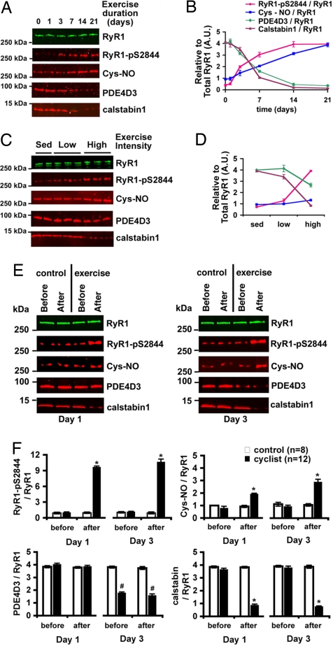
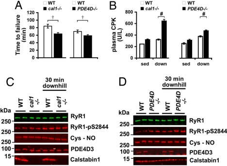
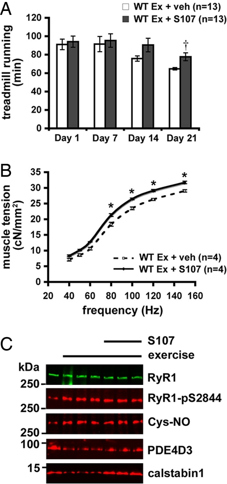
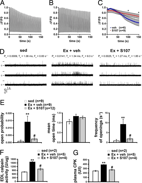
During exercise, defects in calcium (Ca2+) release have been proposed to impair muscle function. Here, we show that during exercise in mice and humans, the major Ca2+ release channel required for excitation-contraction coupling (ECC) in skeletal muscle, the ryanodine receptor (RyR1), is progressively PKA-hyperphosphorylated, S-nitrosylated, and depleted of the phosphodiesterase PDE4D3 and the RyR1 stabilizing subunit calstabin1 (FKBP12), resulting in "leaky" channels that cause decreased exercise tolerance in mice. Mice with skeletal muscle-specific calstabin1 deletion or PDE4D deficiency exhibited significantly impaired exercise capacity. A small molecule (S107) that prevents depletion of calstabin1 from the RyR1 complex improved force generation and exercise capacity, reduced Ca2+-dependent neutral protease calpain activity and plasma creatine kinase levels. Taken together, these data suggest a possible mechanism by which Ca2+ leak via calstabin1-depleted RyR1 channels leads to defective Ca2+ signaling, muscle damage, and impaired exercise capacity.


Conflict of interest statement

Conflict of interest statement: A.R.M. and D.W.L. are on the scientific advisory board and own shares in ARMGO Pharma, Inc., a start-up company that is developing RyR targeted drugs for clinical use in the treatment of heart failure and sudden death. S.R. is a consultant for ARMGO Pharma, Inc.

Figures

Fig. 1.
Fig. 1.
The RyR1 macromolecular complex undergoes remodeling during repeated exercise. (A) RyR1 complex in EDL muscle after exercise (twice daily swimming) analyzed by immunoprecipitation and immunoblotting for RyR1, RyR1-pS2844, cysteine S-nitrosylation of RyR1 (Cys-NO), and PDE4D3 and calstabin1 bound to the receptor. (B) Densitometric quantification of A, where each value is relative to the total RyR1 immunoprecipitated. RyR1 levels did not significantly change under any exercise condition. (C) Composition of the RyR1 complex in EDL muscle after low-intensity (swimming 15 min twice daily) and high-intensity (90 min twice daily) exercise for 5 days. (D) Densitometric quantification of C. (E) Immunoblot of the RyR1 complex immunoprecipitated from 100 μg of muscle homogenate from individual human thigh biopsies before and after exercise on days 1 and 3 of a cycling protocol (3 h at 70% VO2 max). Control cyclists sat in the exercise room but did not exercise. (F) Quantification by densitometry of E. Bar graphs depict PKA phosphorylation, S-nitrosylation, and PDE4D3, and calstabin1 levels in the RyR1 complex normalized to total RyR1 from control (n = 6) and exercise (n = 12) biopsies on each day. All data are mean ± SEM; *, P < 0.01 after exercise versus before; #, P < 0.01 exercise versus control. In all cases, the product of a single RyR1 immunoprecipitation was separated on a 4–20% gradient polyacrylamide gel, transferred, and probed for both total RyR1 and one or more of the modifications noted. The blots shown are representative of three or more independent experiments.
Fig. 2.
Fig. 2.
Muscle-specific cal1−/− and PDE4D−/− mice have impaired exercise capacity. (A) Treadmill running times of 2-month-old cal1−/− (n = 17) mice and WT (n = 12) littermates and PDE4D−/− (n = 6) mice and WT (n = 6) littermates. (B) Plasma creatine kinase (CPK) levels at rest and after a single downhill eccentric treadmill run (n = 4 mice, in triplicate, at each condition). (C and D) RyR1 immunoprecipitated from EDL muscle from cal1−/− (C) and PDE4D−/− (D) mice and immunoblotted for RyR, RyR1-pS2844, Cys-NO, PDE4D3, and calstabin1. Data presented as mean ± SEM; †, P < 0.05, Wilcoxon rank-sum test; #, P < 0.01, unpaired t test. sed, sedentary; down, downhill.
Fig. 3.
Fig. 3.
Pharmacologic prevention of calstabin1 depletion from the RyR1 complex improves in vivo exercise capacity. (A) Time to failure during treadmill assays on indicated days of a 28-day treatment trial with S107. (B) Force–frequency curves of EDL muscle isolated immediately after the 21st day of exercise and isometrically stimulated in an oxygenated muscle bath. Forces (cN) are normalized to muscle cross-sectional area. (C) RyR1 immunoprecipitated from EDL and immunoblotted for RyR, RyR1-pS2844, Cys-NO, PDE4D3, and calstabin1. Data are presented as mean ± SEM; †, P < 0.05, Wilcoxon rank-sum test WT + S107 vs. WT + vehicle (veh); *, P < 0.05, unpaired t test. ex, exercise.
Fig. 4.
Fig. 4.
Stabilization of RyR1 channels slows muscle fatigue and reduces damage. (A) Representative trace of fluorescence (ΔF/F0) from a vehicle-treated FDB fiber loaded with fluo-4 normalized to the peak during repeated 300-ms, 120-Hz field-stimulated tetani at 0.5 Hz. Isolated cells were continuously perfused with Hepes-buffered Tyrodes solution at room temperature. (B) Representative (ΔF/F0) Ca2+ tetanic trace from an S107-treated FDB fiber. (C) Mean peak tetanic Ca2+ normalized to the peak during fatiguing stimulation (n = 6, vehicle; n = 9, S107). *, P < 0.05 unpaired t test. (D) Representative traces of RyR1 channel activity at 90 nM [Ca2+]cis from sedentary mice (sed, Left), mice exercised and treated with vehicle (Ex + veh, Center), and mice exercised and treated with S107 (Ex + S107, Right). Single channel openings are plotted as upward deflections; the open and closed (c) states of the channel are indicated by horizontal bars at the beginning of the traces. Channel open probability (Po), mean open time (To) and frequency of openings (Fo) are shown above each group of traces and represent average values from all experiments. (E) Average values of Po (Left), To (Center), and Fo (Right) of RyR1 from sedentary mice (sed, n = 9) and exercised mice treated either with vehicle (Ex + veh, n = 9) or S107 (Ex + S107, n = 12). (F) Calpain activity levels in EDL homogenates. (G) Plasma creatine kinase (CPK) activity levels in sedentary and exercised mice with, and without, calstabin1 rebinding with S107. Data are presented as mean ± SEM; **, P < 0.01 compared with sed; #, P < 0.01 compared with Ex + veh.

References

    1. Brillantes AB, et al. Stabilization of calcium release channel (ryanodine receptor) function by FK506-binding protein. Cell. 1994;77:513–523. - PubMed
    1. Marx SO, et al. Phosphorylation-dependent regulation of ryanodine receptors: A novel role for leucine/isoleucine zippers. J Cell Biol. 2001;153:699–708. - PMC - PubMed
    1. Marx SO, Ondrias K, Marks AR. Coupled gating between individual skeletal muscle Ca2+ release channels (ryanodine receptors). Science. 1998;281:818–821. - PubMed
    1. Lamb GD, Stephenson DG. Effects of FK506 and rapamycin on excitation–contraction coupling in skeletal muscle fibres of the rat. J Physiol. 1996;494:569–576. - PMC - PubMed
    1. Avila G, Lee EH, Perez CF, Allen PD, Dirksen RT. FKBP12 binding to RyR1 modulates excitation–contraction coupling in mouse skeletal myotubes. J Biol Chem. 2003;278:22600–22608. - PubMed

Publication types

Substances