. 2008 Feb 19;105(7):2274-9.
doi: 10.1073/pnas.0708088105. Epub 2008 Feb 11.

Hydrophobicity of protein surfaces: Separating geometry from chemistry

Affiliations

Hydrophobicity of protein surfaces: Separating geometry from chemistry

Nicolas Giovambattista et al. Proc Natl Acad Sci U S A. .

Abstract

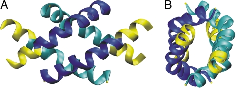
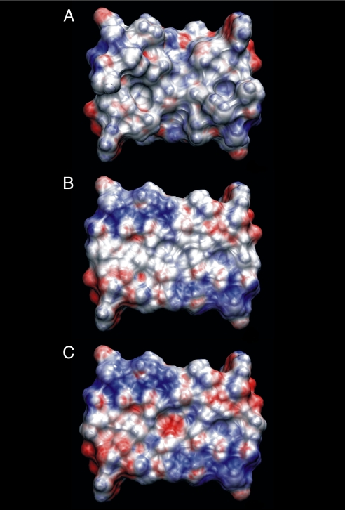
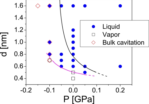
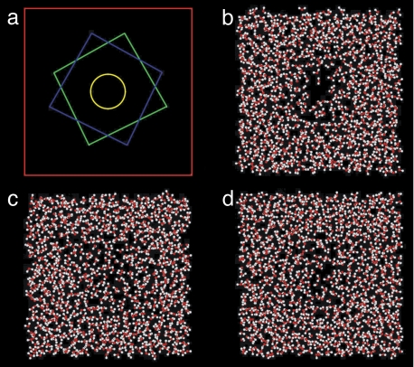
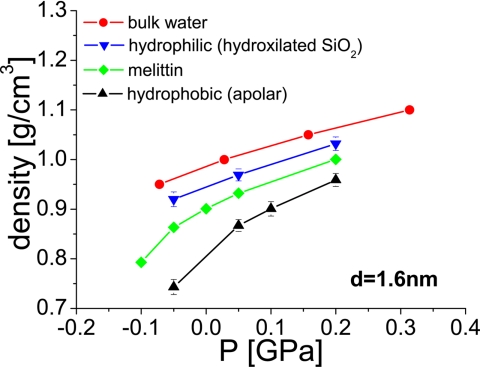
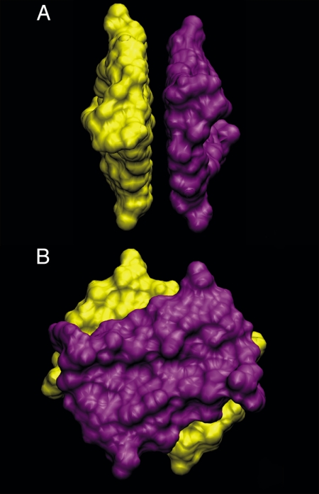
To better understand the role of surface chemical heterogeneity in natural nanoscale hydration, we study via molecular dynamics simulation the structure and thermodynamics of water confined between two protein-like surfaces. Each surface is constructed to have interactions with water corresponding to those of the putative hydrophobic surface of a melittin dimer, but is flattened rather than having its native "cupped" configuration. Furthermore, peripheral charged groups are removed. Thus, the role of a rough surface topography is removed, and results can be productively compared with those previously observed for idealized, atomically smooth hydrophilic and hydrophobic flat surfaces. The results indicate that the protein surface is less hydrophobic than the idealized counterpart. The density and compressibility of water adjacent to a melittin dimer is intermediate between that observed adjacent to idealized hydrophobic or hydrophilic surfaces. We find that solvent evacuation of the hydrophobic gap (cavitation) between dimers is observed when the gap has closed to sterically permit a single water layer. This cavitation occurs at smaller pressures and separations than in the case of idealized hydrophobic flat surfaces. The vapor phase between the melittin dimers occupies a much smaller lateral region than in the case of the idealized surfaces; cavitation is localized in a narrow central region between the dimers, where an apolar amino acid is located. When that amino acid is replaced by a polar residue, cavitation is no longer observed.


Conflict of interest statement

The authors declare no conflict of interest.

Figures

Fig. 1.
Fig. 1.
Front (A) and side (B) view of the melittin tetramer from the x-ray crystal structure (30). The residues that are truncated for our simulation purposes are shown in yellow. Each dimer pair is shown in light (dark) blue.
Fig. 2.
Fig. 2.
Solvent accessible surface area colored by electrostatic potential for the truncated melittin dimer (A), flattened, truncated melittin dimer (B), and mutated, flattened, and truncated melittin dimer (C). The coloring gradient ranges from −5kBT/e (red) to 5kBT/e (blue)
Fig. 3.
Fig. 3.
Phase diagram. Magenta line separates approximately the state points at which water confined by the melittin dimers remains in the liquid (blue points) or vapor (open squares) phase. The black line, included for comparison, is from ref. , for water confined between hydrophobic atomically flat nanoscale walls.
Fig. 4.
Fig. 4.
Snapshots of confined water. (a) Scheme showing the simulation box cross section (red square, side length, L ≈ 4.75 nm), and the two melittin dimers (green and blue squares), one on top of the other. The yellow circle (with a radius of 0.5 nm), centered along the axis connecting both dimers, corresponds to the base area of the cylindrical volume used for the calculation in Fig. 5. Snapshots obtained from simulations at: P = 0, d = 0.5 nm, 500 ps (b); P = 0.05GPa, d = 0.5 nm, 900 ps (c); P = 0, d = 0.6 nm, 500 ps (d). Only b shows clear cavitation which is reversed by small increases in pressure in c. Increasing the separation between the melittin dimers results in a thicker water layer between the surfaces (d).
Fig. 5.
Fig. 5.
Density of water in the confined volume as a function of the applied pressure for different surfaces. Data for bulk water are from ref. . Data for water confined by hydrophobic and hydrophilic surfaces is from ref. . In the melittin case (green; this work), the density was computed in a cylindrical region, as shown in Fig. 4a. The density calculated at P = −0.01 GPa with the melittin dimers is obtained before bulk cavitation occurs.
Fig. 6.
Fig. 6.
Side (A) and (B) top view of the melittin dimers in the configuration implemented in our simulations (see also Fig. 1).

References

    1. Hummer G, Garde S, Garcia AE, Pratt LR. Chem Phys. 2000;258:349–370.
    1. Dill KA. Biochemistry. 1990;29:7133–7155. - PubMed
    1. Guo WH, Lampoudi S, Shea JE. Biophys J. 2003;85:61–69. - PMC - PubMed
    1. Sharp KA, Nicholls A, Fine RF, Honig B. Science. 1991;252:106–109. - PubMed
    1. Harano Y, Kinoshita M. Chem Phys Lett. 2004;399:342–348.

Publication types