. 2008 Feb 19;105(7):2538-43.
doi: 10.1073/pnas.0707730105. Epub 2008 Feb 11.

Mutations in FN1 cause glomerulopathy with fibronectin deposits

Affiliations

Mutations in FN1 cause glomerulopathy with fibronectin deposits

Federica Castelletti et al. Proc Natl Acad Sci U S A. .

Abstract

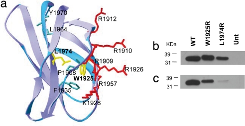
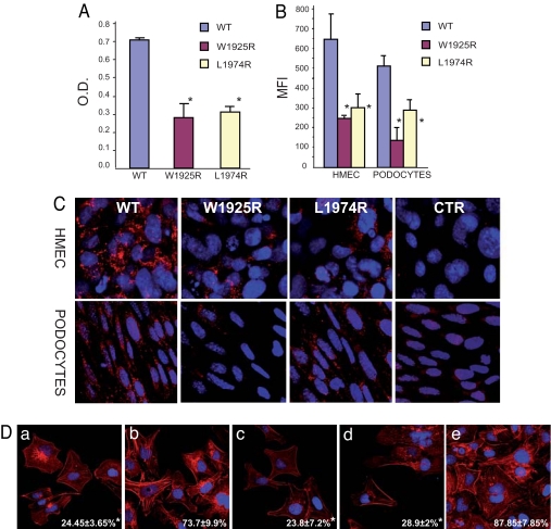
Glomerulopathy with fibronectin (FN) deposits (GFND) is an autosomal dominant disease with age-related penetrance, characterized by proteinuria, microscopic hematuria, hypertension, and massive glomerular deposits of FN that lead to end-stage renal failure. The genetic abnormality underlying GFND was still unknown. We hypothesized that mutations in FN1, which encodes FN, were the cause of GFND. In a large Italian pedigree with eight affected subjects, we found linkage with GFND at the FN1 locus at 2q32. We sequenced the FN1 in 15 unrelated pedigrees and found three heterozygous missense mutations, the W1925R, L1974R, and Y973C, that cosegregated with the disease in six pedigrees. The mutations affected two domains of FN (Hep-II domain for the W1925R and the L1974R, and Hep-III domain for the Y973C) that play key roles in FN-cell interaction and in FN fibrillogenesis. Mutant recombinant Hep-II fragments were expressed, and functional studies revealed a lower binding to heparin and to endothelial cells and podocytes compared with wild-type Hep-II and an impaired capability to induce endothelial cell spreading and cytoskeletal reorganization. Overall dominant mutations in FN1 accounted for 40% of cases of GFND in our study group. These findings may help understanding the pathogenesis of proteinuria and glomerular FN deposits in GFND and possibly in more common renal diseases such as diabetic nephropathy, IgA nephropathy, and lupus nephritis. To our knowledge no FN1 mutation causing a human disease was previously reported.


Conflict of interest statement

The authors declare no conflict of interest.

Figures

Fig. 1.
Fig. 1.
FN1 mutations in GFND. (a) (Upper) Haplotype analysis at FN1 locus in pedigree F233. Microsatellite loci are on the left. (Lower) Multipoint linkage analysis by GENEHUNTER (model: autosomal dominant transmission with age-related penetrance, analysis with liability classes). For markers D1S128 and D1S2361, the maximum lod score was Zmax = 3.084, as indicated. *, subjects previously published. ‡, biopsy-proven GFND. Solid symbols, affected individuals; crossed symbols, deceased; violet arrow, proband: the whole FN1 was sequenced; red dots, FN1 mutation carriers. ¶, subjects screened for the FN1 mutation and for SNP segregation; un, unavailable. (b) Schematic diagram of fibronectin. Fibronectin monomer consists of type I (blue), II (green), and III (orange) repeats and the alternatively spliced sites EDI, EDII, and IIICS. The three main heparin-binding domains and the binding sites for integrins are shown. Positions of the GFND-associated mutations are indicated by arrows. (c Upper) Pedigrees of the other five families with FN1 mutations. (Lower) The number of affected subjects, the mutation, and the origin of all of the six mutated families, are reported.
Fig. 2.
Fig. 2.
Expression of recombinant wild-type and mutant Hep-II domains of fibronectin. (a) Structure of FN III13. Amino acid residues are color marked for positively charged (red), hydrophobic core (green), and residues W1925 and L1974 (yellow). (b and c) WT and mutant purified recombinant proteins were analyzed by SDS/PAGE on 12% gels and visualized by Western blotting with either an antibody anti-His (C-term) (b) or an antifibronectin mAb against the Hep-II domain. (c) Position of standards (kDa) are shown. Equal amounts (5 μg each) of WT and mutant proteins were loaded. Separate lanes were labeled with Coomassie blue as control for loading. Unt, untransfected.
Fig. 3.
Fig. 3.
Mutations in FN Hep-II domain cause reduced binding to heparin, endothelial cells, and podocytes and impair stress fiber formation. (A) Binding of III12–14wt, III12–14W1925R, and III12–14L1974R to heparin by ELISA. O.D., optical density. (B and C) Binding of WT and mutant poli-His-tagged III12–14W1925R and III12–14L1974R recombinants added to human endothelial cells (HMEC) and mouse podocytes (b) FACS analysis. (C) Confocal microscopy; original magnification, ×600). Staining was done with an anti-His antibody plus FITC-conjugated (FACS) or Cy3-conjugated (confocal, red) secondary antibodies. MFI, median fluorescence intensity. (D) HMEC were plated on a 120-kDa N-terminal FN fragment in the absence (a) or presence of III12–14wt (b), III12–14W1925R (c), III12–141974R (d), or full length FN (e), in serum-free medium for 3 h and then labeled with rhodamine-phalloidin to visualize stress fibers. The percent of stress-positive cells (mean ± SD) is shown in the bottom (white numbers). Data are mean ± SD of three independent experiments. *, P < 0.01 vs. WT. O.D. and MFI values were calculated after subtracting values recorded with addition of buffer alone (blanks).

References

    1. Strom EH, Banfi G, Krapf R, Abt AB, Mazzucco G, Monga G, Gloor F, Neuweiler J, Riess R, Stosiek P, et al. Kidney Int. 1995;48:163–170. - PubMed
    1. Kornblihtt AR, Pesce CG, Alonso CR, Cramer P, Srebrow A, Werbajh S, Muro AF. FASEB J. 1996;10:248–257. - PubMed
    1. Hildebrandt F, Strahm B, Prochoroff A, Cybulla M, Gemperle O, Krapf R, Brandis M. Am J Med Genet. 1996;63:323–327. - PubMed
    1. Zhang Z, Kundu GC, Yuan CJ, Ward JM, Lee EJ, DeMayo F, Westphal H, Mukherjee AB. Science. 1997;276:1408–1412. - PubMed
    1. Vollmer M, Krapf R, Hildebrandt F. Nephrol Dial Transplant. 1998;13:2417–2418. - PubMed

Publication types

MeSH terms