. 2008 Sep 6;5(26):1023-9.
doi: 10.1098/rsif.2007.1351.

Cubic membranes: a structure-based design for DNA uptake

Affiliations

Cubic membranes: a structure-based design for DNA uptake

Zakaria Almsherqi et al. J R Soc Interface. .

Abstract

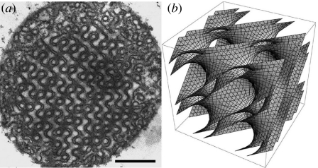
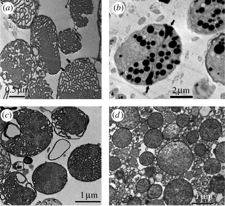
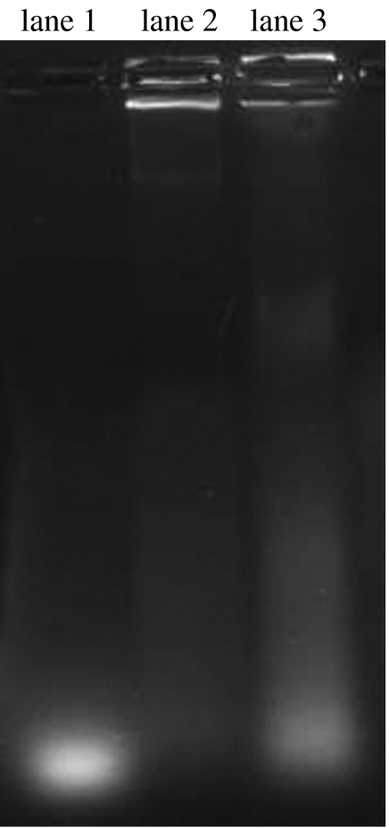
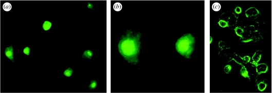
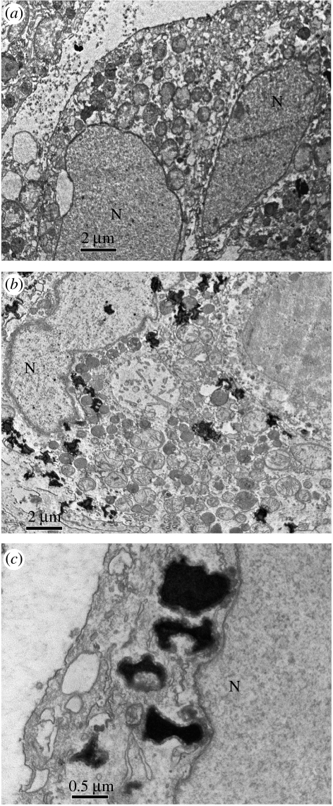
Cubic membranes are soft three-dimensional crystals found within cell organelles in a variety of living systems, despite the aphorism of Fedorov: 'crystallization is death'. They consist of multi-bilayer lipid-protein stacks, folded onto anticlastic surfaces that resemble triply periodic minimal surfaces, forming highly swollen crystalline sponges. Although cubic membranes have been observed in numerous cell types and under different pathophysiological conditions, knowledge about the formation and potential function(s) of non-lamellar, cubic structures in biological systems is scarce. We report that mitochondria with this cubic membrane organization isolated from starved amoeba Chaos carolinense interact sufficiently with short segments of phosphorothioate oligonucleotides (PS-ODNs) to give significant ODNs uptake. ODNs condensed within the convoluted channels of cubic membrane by an unknown passive targeting mechanism. Moreover, the interaction between ODNs and cubic membrane is sufficient to retard electrophoretic mobility of the ODN component in the gel matrix. These ODN-cubic membrane complexes are readily internalized within the cytoplasm of cultured mammalian cells. Transmission electron microscopic analysis confirms ODNs uptake by cubic membranes and internalization of ODN-cubic membrane complexes into the culture cells. Cubic membranes thus may offer a new, potentially benign medium for gene transfection.


Figures

Figure 1
Figure 1
(a) Two-dimensional transmission electron microscopic image and (b) three-dimensional mathematical model of the same cubic membrane organization observed in 10-day starved amoeba Chaos mitochondrion. Scale bar, 500 nm.
Figure 2
Figure 2
Two-colour fluorescence images analysis. (a) Green fluorescent-tagged ODNs were incubated with (b) amoeba Chaos cubic mitochondria labelled with MitoTracker Red for an hour at room temperature. (c) Overlay of the two images shows co-localization of the fluorescent-tagged ODNs and Chaos cubic mitochondria, suggesting mitochondrial uptake of the ODNs. The results illustrate a representative experiment of at least three other times with similar findings.
Figure 3
Figure 3
TEM images of mitochondria containing cubic membrane structure isolated from 10-day starved Chaos cells (a) before and (b) after incubation with ODNs. ODN molecules interact significantly with the mitochondria exhibiting cubic membrane organization (b) but not with the same amount of ‘non-cubic’ mitochondria isolated from (c) well-fed amoeba or (d) mice liver. Multiple electron-dense intra-mitochondrial inclusions may represent cubic membrane-mediated ODN interactions. The electron-dense core may consist of ODNs–membrane lipid complexes encapsulated within distinct vesicles. A significant increase in the mitochondrial size and volume has been observed post ODN treatment, which could be attributed to the (b) ‘adsorption’ of substantial amount of ODNs. The multiple pores (arrows in (a,b)) at the surface of cubic mitochondria may play an important role in facilitating passive uptake of ODNs. Data are from a representative independent experiment performed at least five other times with similar findings.
Figure 4
Figure 4
Gel retardation study. The same amounts of mitochondrial proteins (26 μg μl−1) from amoeba cubic mitochondria and mice liver mitochondria were incubated with the same amount of ODN (0.1 μg μl−1) molecules. The mitochondria with cubic membrane organization are able to retard ODN mobilization (lane 2) towards the positive pole when compared with the same amount of pure ODNs (lane 1) and ODNs incubated with isolated mice liver mitochondria (lane 3). The results illustrate a representative experiment of at least three other times with similar findings, showing one of the duplicate samples.
Figure 5
Figure 5
In vitro study of ODN–cubic membrane complex internalization into MCF-7 cells. The MCF-7 cells were incubated with stable submicrometre-sized particles of cubic membranes containing ODNs. After 5 hours of treatment, highly fluorescent MCF-7 cells were observed under (a) low, (b) high magnification of fluorescence and (c) phase-contrast microscope. The green fluorescent-tagged ODNs are observed in the cytoplasm and the nucleus ((a,c) 64×, (b) 200×). The results illustrate a representative experiment of at least three to five other times with similar findings.
Figure 6
Figure 6
TEM images of the MCF-7 cells, (a) before and 5 hours post treatment with (b) ODNs–membranes complexes. Multiple electron-dense inclusion bodies (ODN–cubic membrane complexes) are frequently observed within the cytoplasm and some targeted the nucleus (N) of the treated MCF-7 cells (b) while there are no such inclusion bodies observed in MCF-7 cells before ODN treatment (a). At higher magnification, membrane association was frequently observed between electron-dense inclusion bodies and nuclear membranes (c). Data are from a representative experiment performed at least three to five other times with similar findings.

References

    1. Almsherqi Z.A, Kohlwein S.D, Deng Y. Cubic membranes: a legend beyond the Flatland* of cell membrane organization. J. Cell Biol. 2006;173:839–844. doi: 10.1083/jcb.200603055. - DOI - PMC - PubMed
    1. Angelov B, Angelova A, Ollivon M, Bourgaux C, Campitelli A. Diamond-type lipid cubic phase with large water channels. J. Am. Chem. Soc. 2003;125:7188–7189. doi: 10.1021/ja034578v. - DOI - PubMed
    1. Bradford M.M. A rapid and sensitive method for the quantitation of microgram quantities of protein utilizing the principle of protein–dye binding. Anal. Biochem. 1976;72:248–254. doi: 10.1016/0003-2697(76)90527-3. - DOI - PubMed
    1. Conner S.D, Schmid S.L. Regulated portals of entry into the cell. Nature. 2003;422:37–44. doi: 10.1038/nature01451. - DOI - PubMed
    1. Conwell C.C, Huang L. Recent advances in non-viral gene delivery. Adv. Genet. 2005;53:1–18. - PubMed

Publication types

LinkOut - more resources