. 2008 Apr 18;283(16):11038-49.
doi: 10.1074/jbc.M704398200. Epub 2008 Feb 12.

Structural basis for ubiquitin recognition by the Otu1 ovarian tumor domain protein

Affiliations

Structural basis for ubiquitin recognition by the Otu1 ovarian tumor domain protein

Troy Eugene Messick et al. J Biol Chem. .

Abstract

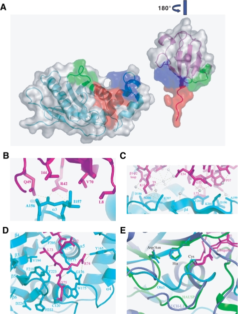
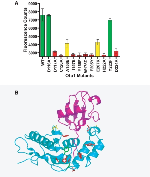
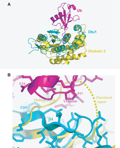
Ubiquitination of proteins modifies protein function by either altering their activities, promoting their degradation, or altering their subcellular localization. Deubiquitinating enzymes are proteases that reverse this ubiquitination. Previous studies demonstrate that proteins that contain an ovarian tumor (OTU) domain possess deubiquitinating activity. This domain of approximately 130 amino acids is weakly similar to the papain family of proteases and is highly conserved from yeast to mammals. Here we report structural and functional studies on the OTU domain-containing protein from yeast, Otu1. We show that Otu1 binds polyubiquitin chain analogs more tightly than monoubiquitin and preferentially hydrolyzes longer polyubiquitin chains with Lys(48) linkages, having little or no activity on Lys(63)- and Lys(29)-linked chains. We also show that Otu1 interacts with Cdc48, a regulator of the ER-associated degradation pathway. We also report the x-ray crystal structure of the OTU domain of Otu1 covalently complexed with ubiquitin and carry out structure-guided mutagenesis revealing a novel mode of ubiquitin recognition and a variation on the papain protease catalytic site configuration that appears to be conserved within the OTU family of ubiquitin hydrolases. Together, these studies provide new insights into ubiquitin binding and hydrolysis by yeast Otu1 and other OTU domain-containing proteins.


Figures

FIGURE 1.
FIGURE 1.
Kinetic analysis of Otu1. A, Otu1 labeling by ubiquitin vinyl sulfone. The quantitative labeling of Otu1 by ubiquitin vinyl sulfone was analyzed by SDS-polyacrylamide gel and stained with Sypro Ruby Red. B, deubiquitinase activity of Otu1. Lineweaver-Burk plot of activity data obtained for recombinant full-length Otu1 (blue diamonds) and Otu1ΔUBX (magenta boxes) using a ubiquitin-AMC substrate. The release of AMC as a function of added protein is monitored by fluorescence. C, Otu1 catalytic activity in the presence of Cdc48. Activity of Otu1 is measured in the presence or absence of a 2-fold molar excess of the Cdc48 hexamer. D, Otu1 cleavage of native polyubiquitin chains. Otu1-mediated cleavage of Lys29-, Lys48-, or Lys63-linked Ub4 chains is analyzed by SDS-polyacrylamide gel and Western blotting with a monoclonal ubiquitin antibody and visualized by ECL.
FIGURE 2.
FIGURE 2.
Protein-protein interactions of Otu1. A, ubiquitin binding activity of Otu1. Otu1 binding to ubiquitin and Ub4 resins in the presence or absence of ΔOtu1 yeast lysate is analyzed by SDS-polyacrylamide gel stained with Sypro Ruby Red. The properties of the resin are indicated above the gel, and the presence or absence of Otu1 protein and ΔOtu1 yeast lysate is indicated below the gel. B, requirement of the Otu1 NH2 terminus for Cdc48 association. Cdc48 association with resins containing monoubiquitin or Lys48-linked Ub4 in the presence or absence of Otu1 and Otu1ΔUBX is analyzed by a SDS-polyacrylamide gel and stained with Sypro Ruby Red. The Otu1 load lanes are 4% of total Otu1 input, and the remaining lanes are all elution fractions from the resins. The arrows indicate the Otu1 interactor, Cdc48. C, requirement of the NH2 terminus of Otu1 for Cdc48 association. His6Cdc48 was incubated with control or OTU1 containing resins in the presence or absence of 1 mm ATP. Cdc48 in the flow-through and elution fractions was detected with anti-His6 antibody.
FIGURE 3.
FIGURE 3.
Overall structure of the Otu1-ubiquitin complex. A, overall structure of the Otu1-Ub complex. A schematic representation of the complex is shown with Otu1 and ubiquitin color-coded cyan and purple, respectively. Secondary structure elements are as labeled. B, the ubiquitin binding pocket on Otu1 (a view orthogonal to A looking down on the ubiquitin binding site Otu1). Otu1 is shown in a surface representation with a mapping of OTU domain-conserved residues mapped onto the protein and ubiquitin shown in a schematic representation. The color-coding for conservation is as follows: yellow, 50% conserved; orange, 75% conserved; red, 95% conserved. C, sequence alignment of OTU domains. A subset of OTU domains from different organisms is shown. The degree of conservation is color-coded according to B. The secondary structure of the Otu1 catalytic domain is shown above the sequence alignment, and residues that participate in ubiquitin contact are indicated with a star above the sequence alignment. Mutated residues (Fig. 5A) are underlined.
FIGURE 4.
FIGURE 4.
The Otu1-ubiquitin interface. A, three regions of interaction between Otu1 and ubiquitin. The figure is rendered in a surface representation and shown in “open book” format highlighting contact regions 1 (blue), 2 (green), and 3 (red). B, close-up of Otu1-ubiquitin interactions in region 1. Side chains that mediate hydrogen bonds (dotted line) and van der Waals interactions are shown. C, close-up of Otu1-ubiquitin interactions in region 2. Water molecules are indicated as white spheres. D, close-up of Otu1-ubiquitin interactions in region 3. E, superimposed active sites of deubiquitinating enzymes. The catalytic triad cysteine, histidine, and aspartate as well as ubiquitin (magenta) are shown as a stick models. Otu1 is shown as cyan, UCH-L3 (Protein Data Bank code 1xd3) is shown in violet, and HAUSP/USP7 (Protein Data Bank code 1nbf) is shown in green.
FIGURE 5.
FIGURE 5.
Mutational analysis of the Otu1-ubiquitin interface. A, deubiquitinase activity of recombinant Otu1 and single site mutants. Each of the reactions was performed in triplicate and in the linear range of time and enzyme and substrate concentration. Activities are color-coded according to severity (green, wild type activity; yellow, about 50% of wild-type activity; red, less than 30% of wild-type activity). B, mapping of mutational studies onto the Otu1-ubiquitin complex. Side chains that were mutated in A are color-coded according to their severity on enzymatic activity as described in A.
FIGURE 6.
FIGURE 6.
Comparison of the Otu1-ubiquitin complex with otubain 2. A, superposition of the Otu1-ubiquitin complex (cyan and magenta, respectively) with nascent otubain 2 (yellow). B, close-up view of the β4-loop region of Otu1 (cyan) with bound ubiquitin (magenta) with the corresponding region of nascent otubain 2 (yellow).

References

    1. Haglund, K., and Dikic, I. (2005) EMBO J. 24 3353-3359 - PMC - PubMed
    1. Reinstein, E., and Ciechanover, A. (2006) Ann. Intern. Med. 145 676-684 - PubMed
    1. Hochstrasser, M. (2000) Nat. Cell Biol. 2 153-157 - PubMed
    1. Pickart, C. M. (2001) Annu. Rev. Biochem. 70 503-533 - PubMed
    1. Fang, S., and Weissman, A. M. (2004) Cell Mol. Life Sci. 61 1546-1561 - PMC - PubMed

Publication types

MeSH terms

Associated data