

Cerebral microbleeds in the population based AGES-Reykjavik study: prevalence and location
- PMID: 18270235
- PMCID: PMC11090473
- DOI: 10.1136/jnnp.2007.121913
Cerebral microbleeds in the population based AGES-Reykjavik study: prevalence and location
Abstract
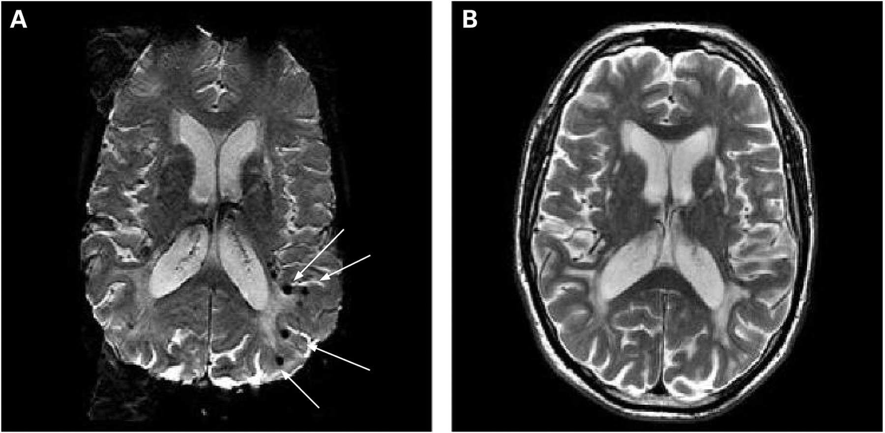
Background and purpose: Incidental foci of signal loss suggestive of cerebral microbleeds (CMBs) are frequent findings on gradient echo T2* weighted MRI (T2* MRI) of patients with haemorrhagic or ischaemic stroke. There are few prevalence data on older populations. This paper reports on the prevalence and location of CMBs in a community based cohort of older men and women (born 1907-1935) who participated in the Age Gene/Environment Susceptibility (AGES)-Reykjavik Study, a population based cohort study that followed the Reykjavik Study
Methods: As part of the examination, all eligible and consenting cohort members underwent a full brain MRI, and blood was drawn for genotyping. Results are based on the first 1962 men (n = 820) and women (n = 1142), mean age 76 years, with complete MRI and demographic information available.
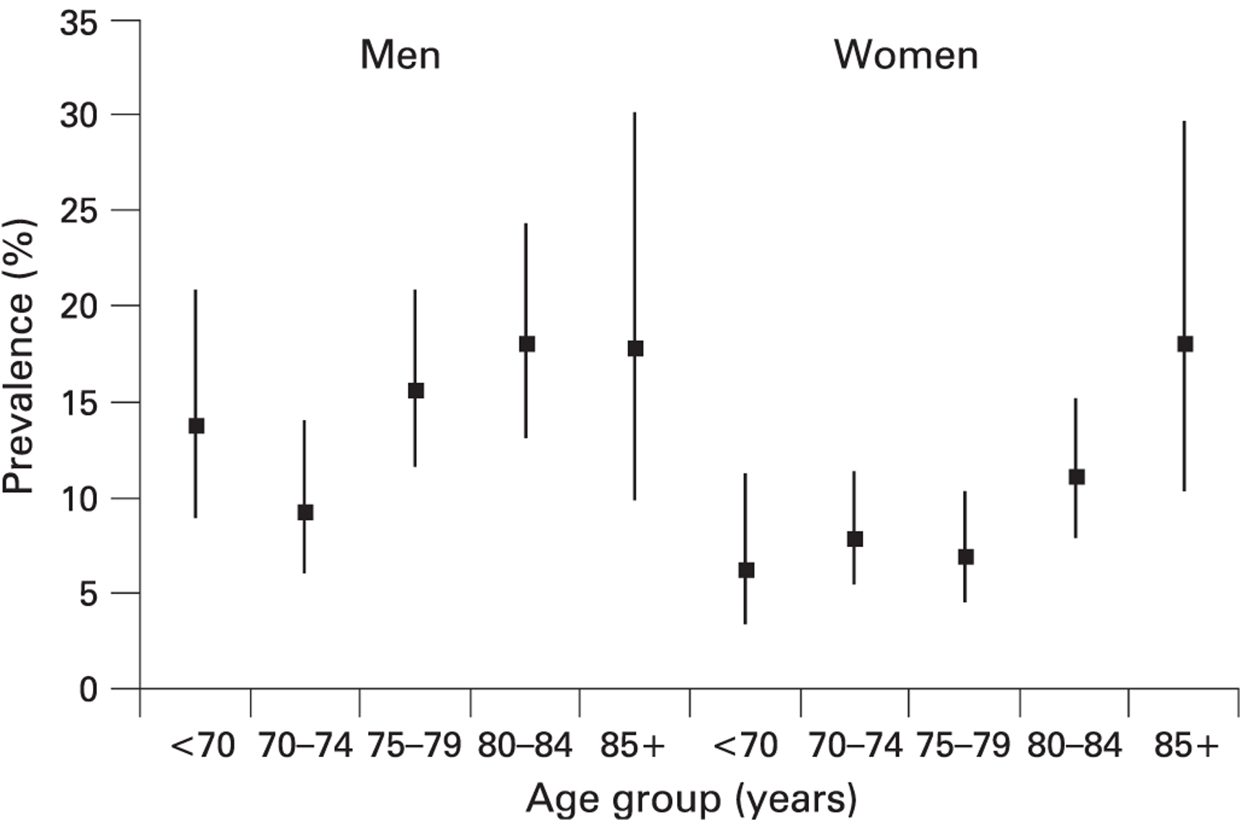
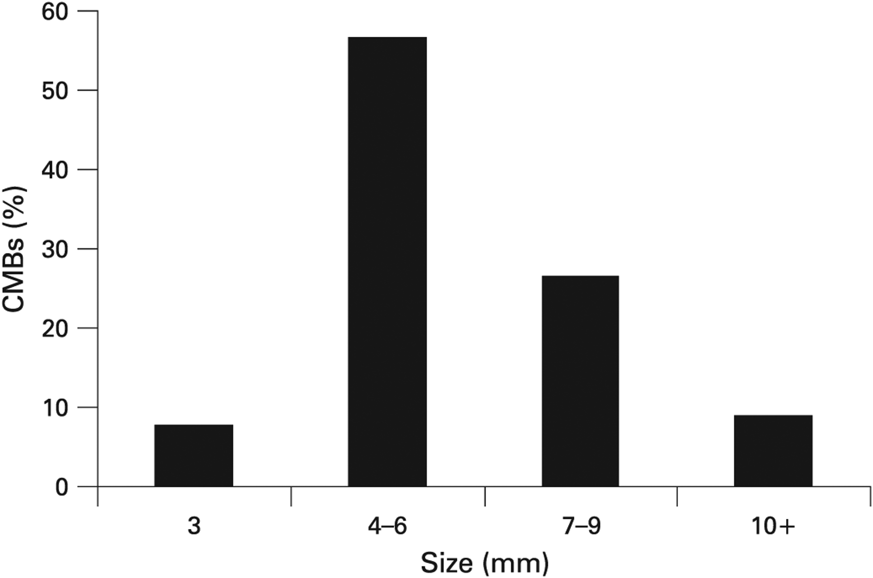
Results: Evidence of CMBs was found in 218 participants (11.1% (95% CI 9.8% to 12.6%)); men had significantly more CMBs than women (14.4% vs 8.8%; p = 0.0002, age adjusted). The prevalence of CMBs increased with age (p = 0.0001) in both men (p = 0.006) and women (p = 0.007). CMBs were located in the cerebral lobes (70%), the basal ganglia region (10.5%) and infratentorium (18.6%). Having a CMB was significantly associated with a homozygote Apo E epsilon4epsilon4 genotype (p = 0.01).
Conclusion: Cerebral microbleeds are common in older persons. The association with homozygote Apo E epsilon4 genotype and finding a relative predominance in the parietal lobes might indicate an association with amyloid angiopathy.
Conflict of interest statement
Competing interests: None.
Figures

References
-
- Viswanathan A, Chabriat H. Cerebral microhemorrhage. Stroke 2006;37:550–5. - PubMed
-
- Cordonnier C, Al-Shahi Salman R, Wardlaw J. Spontaneous brain microbleeds: systematic review, subgroup analyses and standards for study design and reporting. Brain 2007;130:1988–2003. - PubMed
-
- Roob G, Lechner A, Schmidt AR, et al. Frequency and location of microbleeds in patients with primary intracerebral hemorrhage. Stroke 2000;31:2556–69. - PubMed
-
- Tanaka A, Ueno Y, Nakayama Y, et al. Small chronic hemorrhages and ischemic lesions in association with spontaneous intracerebral hematomas. Stroke 1999;30:1637–42. - PubMed
Publication types
MeSH terms
Substances
Grants and funding
LinkOut - more resources
Full Text Sources
Other Literature Sources
Research Materials