. 2008 Mar 1;121(Pt 5):685-94.
doi: 10.1242/jcs.020347. Epub 2008 Feb 12.

The tumour-associated antigen L6 (L6-Ag) is recruited to the tetraspanin-enriched microdomains: implication for tumour cell motility

Affiliations

The tumour-associated antigen L6 (L6-Ag) is recruited to the tetraspanin-enriched microdomains: implication for tumour cell motility

Tamara Lekishvili et al. J Cell Sci. .

Abstract

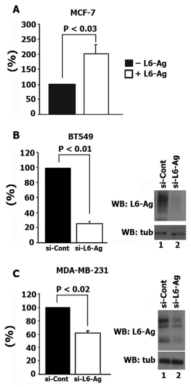
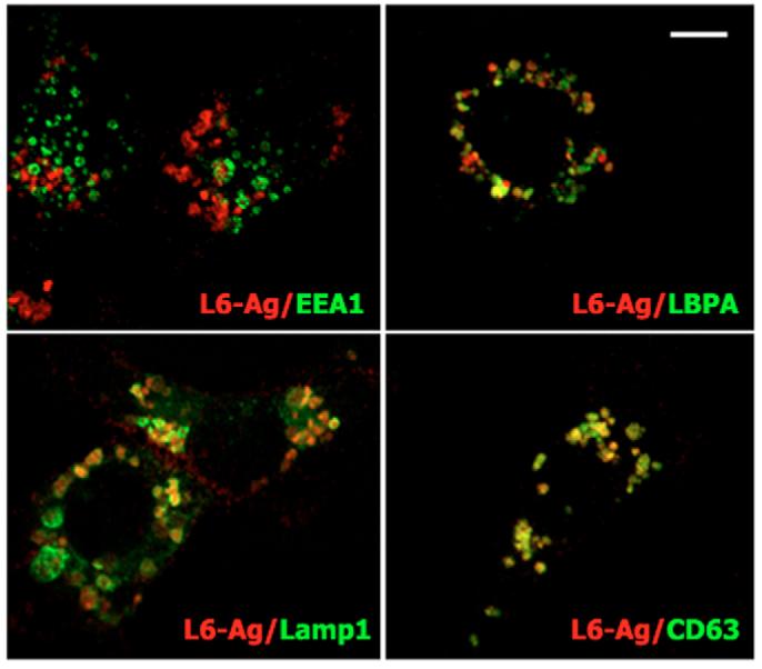
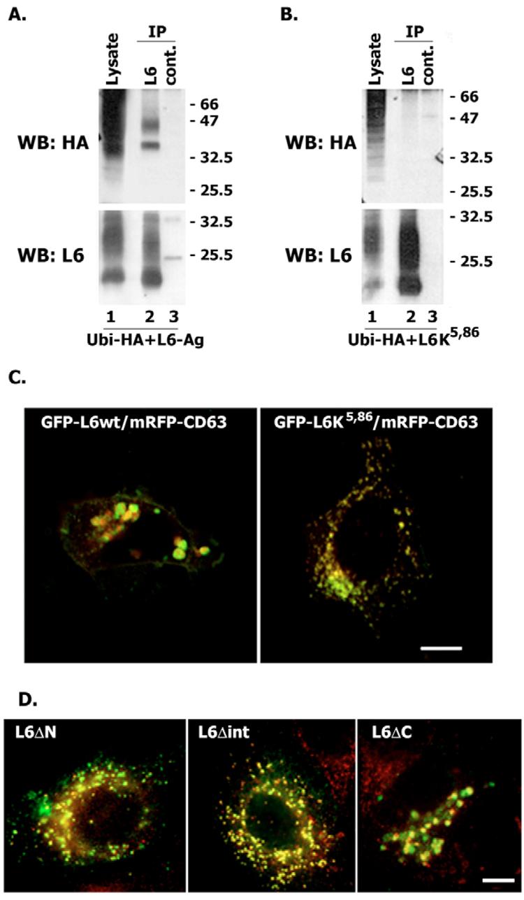
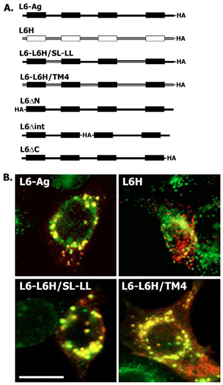
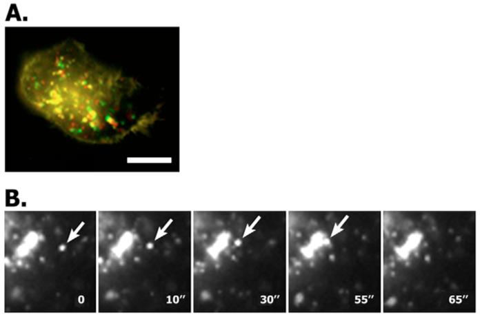
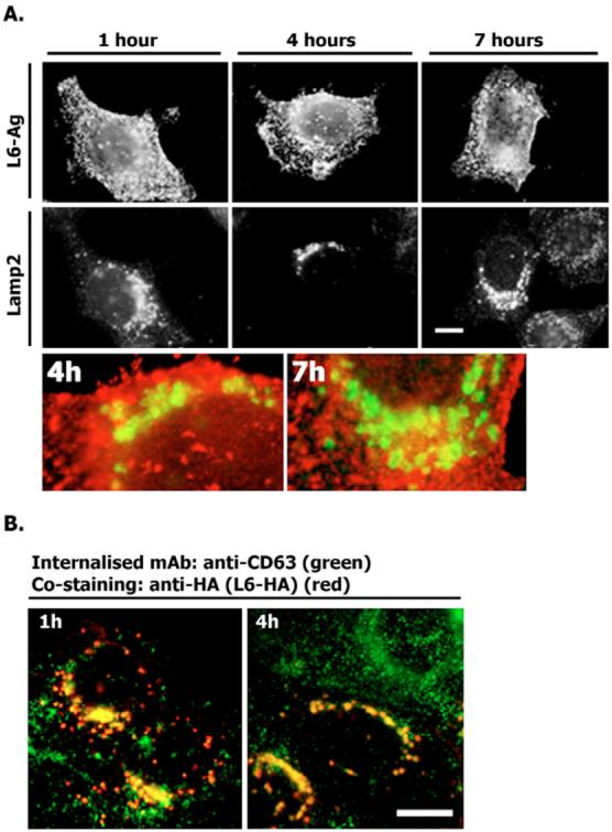
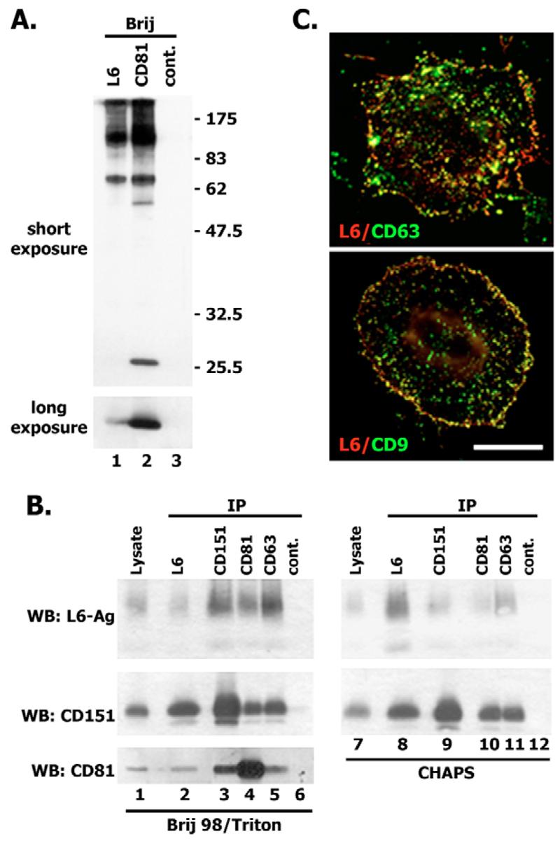
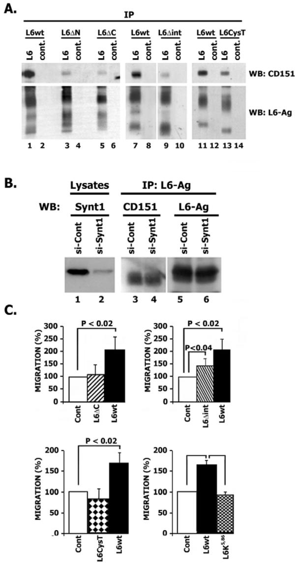
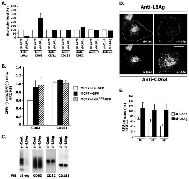
Tumour-associated antigen L6 (L6-Ag, also known as TM4SF1) regulates tumour cell motility and invasiveness. We found that L6-Ag is abundant on the plasma membrane and on intracellular vesicles, on which it is co-localised with the markers for late endosomal/lysosomal compartments, including Lamp1/Lamp2 proteins and LBPA. Antibody internalisation and live-imaging experiments suggested that L6-Ag is targeted to late endocytic organelles (LEO) predominantly via a biosynthetic pathway. Mapping experiments showed that the presence of transmembrane regions is sufficient for directing L6-Ag to LEO. On the plasma membrane, L6-Ag is associated with tetraspanin-enriched microdomains (TERM). All three predicted cytoplasmic regions of L6-Ag are crucial for the effective recruitment of the protein to TERM. Recruitment to TERM correlated with the pro-migratory activity of L6-Ag. Depletion of L6-Ag with siRNA has a selective effect on the surface expression of tetraspanins CD63 and CD82. By contrast, the expression levels of other tetraspanins and beta1 integrins was not affected. We found that L6-Ag is ubiquitylated and that ubiquitylation is essential for its function in cell migration. These data suggest that L6-Ag influences cell motility via TERM by regulating the surface presentation and endocytosis of some of their components.


Figures

Fig. 1
Fig. 1
L6-Ag regulates motility of breast cancer cells. (A) MCF-7 cells were co-transfected with plasmids encoding L6-Ag and GFP. Migration experiments towards fibronectin were performed 48 hours after transfection. (B,C) Cells were electroporated with control siRNA (si-Cont) or siRNA that targets L6-Ag (si-L6-Ag). Migration experiments were performed 72 hours after transfection. Migration was quantified by counting cells in seven random fields per membrane (∼10-30 cells/field) as described in detail in Materials and Methods. Data are reported as fold increases/decreases over migration of cells transfected with either control plasmid (GFP, A) or control siRNA (B,C). Data (all graph panels) are shown as mean ± standard deviation (s.d.) calculated from at least three separate experiments each performed in triplicate. P values were calculated using the two-tailed t-test. Right panels show representative western blots (WB) using lysates prepared 72 hours after transfection.
Fig. 2
Fig. 2
L6-Ag is found in late endocytic organelles. MDA-MB-231 cells were grown on glass coverslips, fixed with 2% paraformaldehyde and permeabilised with 0.1% Triton X-100. Immunofluorescence staining was carried out using mouse mAb to human L6-Ag (IgG2a) in combination with mouse mAbs to proteins localised to various intracellular compartments (all IgG1): EEA1, early endosomes; LBPA, late endosomes; Lamp1, late endosomes/lysosomes. Staining was visualised using Alexa-Fluor-594-conjugated goat anti-mouse IgG2a Ab (red) and Alexa-Fluor-488-conjugated goat anti-mouse IgG1 Ab (green). Shown are representative images acquired using a LSM510 microscope. Note that, after permeabilisation, surface pools of L6-Ag and CD63 are almost completely lost. Scale bar: 10 μm.
Fig. 3
Fig. 3
L6-Ag is ubiquitylated. Cellular distribution of ubiquitylation-deficient L6-Ag. (A,B) 293T cells were transiently transfected with the plasmids encoding wild-type L6-Ag (A) or L6K5,86 (B) and HA-tagged ubiquitin. 48 hours later, cells were lysed in 1% Triton X-100 and the immunoprecipitation (IP) was carried out using the anti-L6-Ag mAb (lanes 2) or a negative control mAb (lanes 3). The protein lysates were used as a positive control for transfection (lanes 1). Proteins were resolved in 11% SDS-PAGE and transferred to nitrocellulose membranes. The membranes were probed with either anti-HA polyclonal Ab or the anti-L6-Ag mAb (L6pke). (C) MCF-7 cells were co-transfected with the plasmid encoding GFP-L6-Ag [wild type (wt), or GFP-L6K5,86] and mRFP-CD63. Co-distribution of tagged proteins was analysed 48 hours after transfection. Shown are representative images acquired using a LSM510 microscope. (D) MCF-7 cells were transfected with the plasmid encoding L6-Ag mutants and 48 hours later the cells were processed for double-immunofluorescence staining using specific Abs as described in the legend to Fig. 2. Staining was visualised using Alexa-Fluor-488-conjugated goat anti-mouse IgG2a Ab (green, L6-Ag) and Alexa-Fluor-594-conjugated goat anti-mouse IgG1 Ab (red). Shown is the co-distribution of L6-Ag mutants and Lamp2. Scale bars: 10 μm.
Fig. 4
Fig. 4
The presence of transmembrane domains is sufficient to direct trafficking of L6 to late endocytic organelles. (A) Schematic diagram shows the constructs used for transfection of MCF-7 cells. (B) MCF-7 cells were transfected with the plasmid encoding the L6H-HA, L6-Ag-HA and L6-L6H chimeras, and 48 hours later the cells were processed for double-immunofluorescence staining using specific Abs as described in the legend to Fig. 2. Co-distribution of the tagged proteins with Lamp2 was analysed using anti-HA mAb F7 (IgG2a) and anti-Lamp2 mAb H4B4 (IgG1). Staining was visualised using Alexa-Fluor-594-conjugated goat anti-mouse IgG2a Ab (red) and Alexa-Fluor-488-conjugated goat anti-mouse IgG1 (green). Scale bar: 20 μm.
Fig. 5
Fig. 5
Motility of L6-Ag-positive vesicles. MCF-7 cells were transiently transfected with the plasmid encoding GFP–L6-Ag and 48 hours later motility of the L6-Ag-positive endosomes was analysed by time-lapse video microscopy. Images were collected every 3 seconds for 1 minute (A) or every 3-5 seconds for 2 minutes (B). (A) Represents a superposition of the first and the last images artificially coloured in red and green, respectively. (B) Arrows point to a vesicle that fused with the enlarged L6-Ag-positive endosomes (video sequence). Scale bar: 10 μm.
Fig. 6
Fig. 6
Internalisation of surface-labelled L6-Ag and CD63. (A) MCF-7 cells were transiently transfected with the plasmid encoding HA–L6-Ag. 48 hours later, cells were surface labelled with the anti-L6-Ag mAb (L6, IgG2a) for 1 hour at 4°C and then placed to 37°C for the indicated durations. Cells were subsequently fixed and permeabilised as described in the legend to Fig. 2. The internalised mAbs were visualised with Alexa-Fluor-594-conjugated goat anti-mouse IgG2a (red). Late endocytic organelles were visualised with anti-Lamp2 mAb and Alexa-Fluor-488-conjugated goat anti-mouse IgG1 Ab (green). (B) MCF-7 cells were prepared for the experiments as described in A except that they were incubated with the anti-CD63 mAb (6H1, IgG1) instead of the anti-L6-Ag mAb. The internalised mAbs were visualised with Alexa-Fluor-488-conjugated goat anti-mouse IgG1 Ab (green). Late endocytic organelles were visualised with the anti-HA mAb (F7, IgG2a) and Alexa-Fluor-594-conjugated goat anti-mouse IgG2a Ab (red). Scale bars: 10 μm.
Fig. 7
Fig. 7
L6-Ag is associated with tetraspanins. (A) HT1080 cells were surface labelled with EZ-link Sulfo-NHS-LC-biotin and lysed in 0.8% Brij98/0.2% Triton X-100. Protein complexes were immunoprecipitated with anti-L6-Ag mAb (lane 1), anti-CD81 and mAb M38 (lane 2) or control mAb (lane 3). Immunocomplexes were separated in 11% SDS-PAGE and transferred onto nitrocellulose membranes. Membranes were developed with streptavidin conjugated to horse radish peroxidase (HRPO). (B) HT1080 cells were lysed in either 0.8% Brij98/0.2% Triton X-100 or 0.5% CHAPS. Protein complexes were immunoprecipitated with anti-L6-Ag mAb (lane 2), anti-CD151 mAb 5C11 (lane 3), anti-CD81 mAb M38 (lane 4) or anti-CD63 mAb 6H1 (lane 5). Irrelevant mAb (187.1) was used as a negative control (lane 6). The protein lysate (lane 1) was used as a positive control. Immunocomplexes were separated in 11% SDS-PAGE and transferred onto nitrocellulose membranes. Membranes were developed with the mAbs to L6-Ag (L6pke) and with polyclonal anti-CD151 Ab. (C) Co-localisation of L6-Ag with tetraspanins on the surface of BT549 cells. Cells grown on glass coverslips were fixed with 2% paraformaldehyde and subsequently stained with combinations of anti-L6-Ag plus anti-CD63 mAbs (or anti-L6-Ag plus anti-CD9 mAb). Staining was visualised using Alexa-Fluor-594-conjugated goat anti-mouse IgG2a Ab (red) and Alexa-Fluor-488-conjugated goat anti-mouse IgG1 Ab (green). Scale bar: 15 μm.
Fig. 8
Fig. 8
Association with TERM is essential for the pro-migratory activity of L6-Ag. (A) Predicted cytoplasmic regions are involved in recruitment of L6-Ag to TERM. 293T cells were transiently transfected with the plasmids encoding wild-type L6-Ag or various mutants of L6-Ag. After 48 hours cells were lysed in 0.8% Brij 98/0.2% Triton X-100 and immunoprecipitation was carried out using the anti-L6-Ag mAb (odd lanes) or a negative control mAb (even lanes). Proteins were resolved in 11% SDS-PAGE and transferred to nitrocellulose membranes. The membranes were probed with either anti-CD151 polyclonal Ab or the anti-L6-Ag mAb (L6pke). (B) Syntenin-1 is not involved in the recruitment of L6-Ag to TERM. MDA-MB-231 cells were transiently transfected with either control siRNA (si-Cont) or siRNA that targets syntenin-1 (si-Syn1). After 72 hours cells were lysed in 0.8% Brij 98/0.2% Triton X-100 and immunoprecipitation was carried out using the anti-L6-Ag mAb. Proteins were resolved in 11% SDS-PAGE and transferred to nitrocellulose membranes. The membranes were probed with either anti-CD151 polyclonal Ab or the anti-L6-Ag mAb (L6pke). The degree of syntenin-1 knock-down was assessed by analysing total cell lysates with anti-syntenin-1 mAb (lanes 1 and 2). (C) Effect of L6-Ag mutations on the pro-migratory activity of the protein. MCF-7 cells were co-transfected with plasmids encoding GFP and various L6-Ag constructs. Migration experiments towards fibronectin were performed 48 hours after transfection. Migration was quantified by counting cells in seven random fields per membrane (∼10-20 cells/field) as described in detail in Materials and Methods. Data are reported as fold increases over migration of cells transfected with the control plasmid (GFP). Data (all graph panels) are shown as mean ± s.d. calculated from at least three separate experiments each performed in triplicate. P values were calculated using the two-tailed t-test. L6wt, wild-type L6-Ag.
Fig. 9
Fig. 9
The role of L6-Ag in the surface expression of tetraspanins and integrins. (A) BT549 cells were electroporated with control siRNA (si-Cont) or siRNA that targets L6-Ag (si-L6Ag). The surface expression of proteins was analysed by flow cytometry after 72 hours. Data are presented as ratios of means of fluorescence intensity (MFIs) for cells transfected with control siRNA to those transfected with siRNA that targets L6-Ag. Bars represent the mean ± s.d. from three independent experiments. (B) MCF-7 cells were transfected with the plasmids encoding GFP-L6-Ag, GFP-L6K5,86 or GFP alone. 24 hours later cells were detached and re-plated in the EMEM/10% FCS for a further 24 hours. The surface expression of proteins was analysed by flow cytometry. Data presented as ratios of means of fluorescence intensity (MFIs) for cells expressing GFP-proteins to those of non-transfected cells. Bar values represent the mean ± s.d. from three independent experiments. (C) Lysates prepared from cells transfected with control siRNA and siRNA that targets L6Ag (as described in A) were resolved in 11% SDS-PAGE and transferred to nitrocellulose membranes. The membranes were probed with anti-CD151 polyclonal Ab, the anti-L6-Ag mAb (L6pke), anti-CD82 mAb (TS82) and anti-CD63 mAb (1C5). Shown are the results of a representative experiment. (D) BT549 cells were electroporated with control siRNA or siRNA that targets L6-Ag. Intracellular distribution of proteins after 72 hours was analysed as described in Fig. 2. Note that CD63-positive vesicles are more evenly scattered through the cytoplasm in cells in which the expression of L6-Ag was knocked down by siRNA than controls. (E) BT549 cells were electroporated with control siRNA or siRNA that targets L6-Ag. 72 hours later cells were surface labelled with the anti-CD63 mAb 6H1 for 1 hour at 4°C and then placed to 37°C for the indicated durations. Non-internalised mAbs were labelled with IRDye-800CW-conjugated goat anti-mouse IgG. Fluorescent signals were detected using Odyssey infrared imaging system. Data is presented as percentage of the mAb 6H1 left on the cell surface relative to that at time point 0, t=0 (100%, fluorescent signals before cell were placed to 37°C) and are shown as mean ± s.d. calculated from at least three separate experiments each performed in triplicate. RFU t=N, relative fluorescence units at a given time interval; RFU t=0, relative fluorescence units at time point 0. Scale bar: 10 μm.

Similar articles

Cited by

References

    1. Berditchevski F. Complexes of tetraspanins with integrins:more than meets the eye. J. Cell Sci. 2001;115:4143–4151. - PubMed
    1. Berditchevski F, Odintsova E. Characterization of integrin/tetraspanin adhesion complexes: role of tetraspanins in integrin signaling. J. Cell Biol. 1999;146:477–492. - PMC - PubMed
    1. Berditchevski F, Bazzoni G, Hemler ME. Specific association of CD63 with the VLA-3 and VLA-6 integrins. J. Biol. Chem. 1995;270:17784–17790. - PubMed
    1. Berditchevski F, Chang S, Bodorova J, Hemler ME. Generation of monoclonal antibodies to integrin-associated proteins. Evidence that alpha3beta1 complexes with EMMPRIN/basigin/OX47/M6. J. Biol. Chem. 1997;272:29174–29180. - PubMed
    1. Berditchevski F, Odintsova E, Sawada S, Gilbert E. Expression of the palmitoylation-deficient CD151 weakens the association of alpha 3 beta 1 integrin with the tetraspanin-enriched microdomains and affects integrin-dependent signaling. J. Biol. Chem. 2002;277:36991–37000. - PubMed

Publication types

MeSH terms