. 2007 Apr;27(3):450-468.
doi: 10.1016/j.msec.2006.05.055.

Multiscale mechanics of hierarchical structure/property relationships in calcified tissues and tissue/material interfaces

Affiliations

Multiscale mechanics of hierarchical structure/property relationships in calcified tissues and tissue/material interfaces

J Lawrence Katz et al. Mater Sci Eng A Struct Mater. 2007 Apr.

Abstract

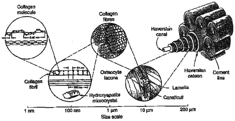
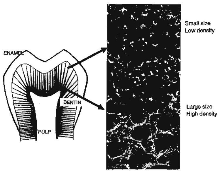
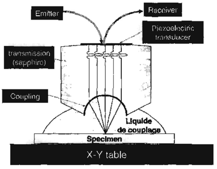
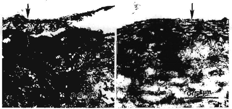
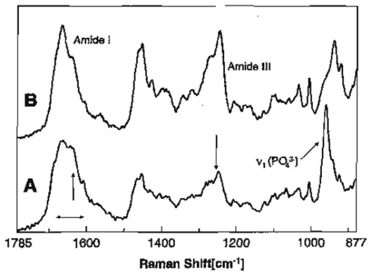
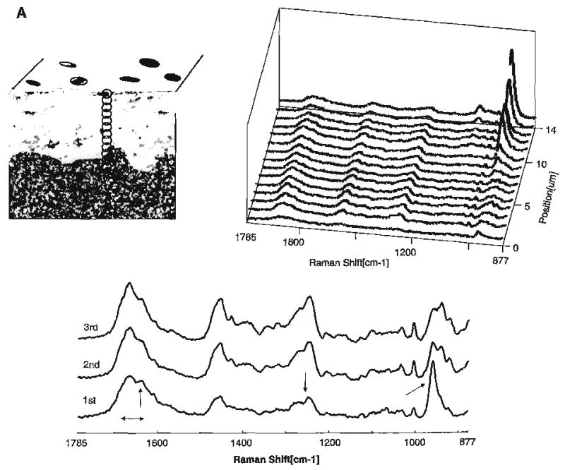
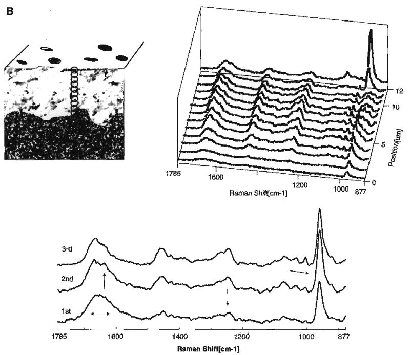
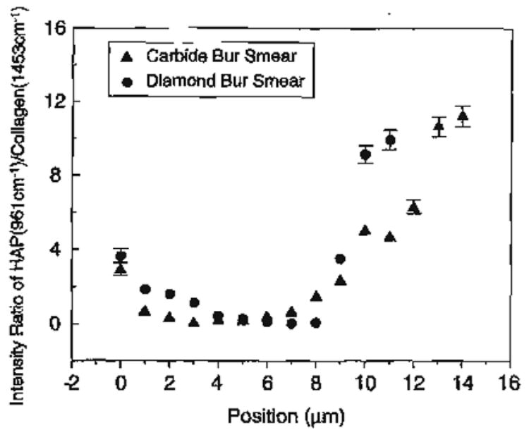
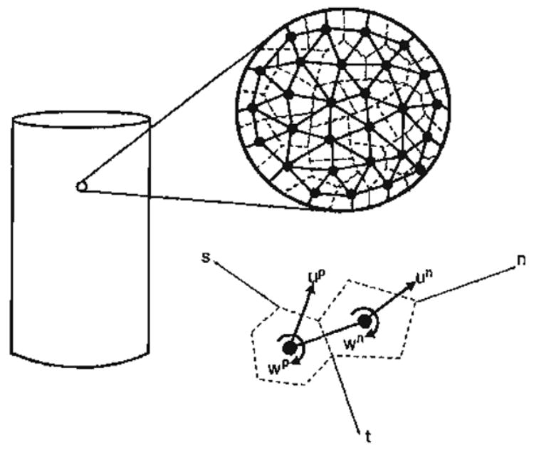
This paper presents a review plus new data that describes the role hierarchical nanostructural properties play in developing an understanding of the effect of scale on the material properties (chemical, elastic and electrical) of calcified tissues as well as the interfaces that form between such tissues and biomaterials. Both nanostructural and microstructural properties will be considered starting with the size and shape of the apatitic mineralites in both young and mature bovine bone. Microstructural properties for human dentin and cortical and trabecular bone will be considered. These separate sets of data will be combined mathematically to advance the effects of scale on the modeling of these tissues and the tissue/biomaterial interfaces as hierarchical material/structural composites. Interfacial structure and properties to be considered in greatest detail will be that of the dentin/adhesive (d/a) interface, which presents a clear example of examining all three material properties, (chemical, elastic and electrical). In this case, finite element modeling (FEA) was based on the actual measured values of the structure and elastic properties of the materials comprising the d/a interface; this combination provides insight into factors and mechanisms that contribute to premature failure of dental composite fillings. At present, there are more elastic property data obtained by microstructural measurements, especially high frequency ultrasonic wave propagation (UWP) and scanning acoustic microscopy (SAM) techniques. However, atomic force microscopy (AFM) and nanoindentation (NI) of cortical and trabecular bone and the dentin-enamel junction (DEJ) among others have become available allowing correlation of the nanostructural level measurements with those made on the microstructural level.


Figures

Fig. 1
Fig. 1
Hierarchical structure of human bone (R. Lakes, Viscoelastio Solids, 1998, with permission of CRC Press, Boca Raton, FL [160]).
Fig. 2
Fig. 2
Hierarchical structure of human tooth, inserts representing tubule characteristics as a function of position in reference to dentin-enamel junction (DEJ). In close proximity to the DEJ, low density distribution of tubules and small size. Tubules of large size and high density are noted in close proximity to the pulp and high-resolution image of intertubular dentin from this region treated with EDTA to reveal collagen network.
Fig. 3
Fig. 3
Diagram of the lens and specimen configuration for scanning acoustic microscopy (SAM).
Fig. 4
Fig. 4
(A) Transmission electron micrograph of carbide bur smear layer. (B) Transmission electron micrograph of diamond bur smear layer (Spencer et al. with permission of IADR/AADR, Alexandria, VA [70]).
Fig. 5
Fig. 5
Raman spectrum of acid-etched carbide bur-created smear layer (A); Raman spectrum of completely demineralized dentin (B).
Fig. 6
Fig. 6
(A) Raman map of dentin specimen fractured to provide cross-sectional view of the acid etched carbide bur-created smear layer/demineralized dentin/undisturbed dentin. Spectra acquired at 1-μm intervals beginning at the acid etched carbide bur-created smear layer, extending into the demineralized dentin and, finally, into the undisturbed dentin. (B) Raman map of the molecular structure of the acid etched diamond bur-created smear layer and subjacent demineralized/undisturbed dentin. Spectra acquired at 1-μm intervals beginning at the acid etched diamond bur-created smear layer and continuing into the undisturbed dentin (Spencer et al. [70] with permission of IADR/AADR).
Fig. 6
Fig. 6
(A) Raman map of dentin specimen fractured to provide cross-sectional view of the acid etched carbide bur-created smear layer/demineralized dentin/undisturbed dentin. Spectra acquired at 1-μm intervals beginning at the acid etched carbide bur-created smear layer, extending into the demineralized dentin and, finally, into the undisturbed dentin. (B) Raman map of the molecular structure of the acid etched diamond bur-created smear layer and subjacent demineralized/undisturbed dentin. Spectra acquired at 1-μm intervals beginning at the acid etched diamond bur-created smear layer and continuing into the undisturbed dentin (Spencer et al. [70] with permission of IADR/AADR).
Fig. 7
Fig. 7
Effect of smear layer type on degree of demineralization produced by 35% H3PO4 etching for 15 s.
Fig. 8
Fig. 8
From a microscopic viewpoint, the material contains a large number of molecular bonds whose average behavior in a given direction may be represented by nanoscale grains interacting through pseudo-bonds in that direction.
Fig. 9
Fig. 9
Likely forms of pseudo-bond force–displacement relationship in normal, Fn, and tangential, Fw, directions under compression and tension.

References

    1. LeGeros RZ. In: Monographs in Oral Science. Meyers HM, editor. Karger: Basel; 1991. p. 121. - PubMed
    1. Eppell SJ, Tong WL, Katz JL, Spearing WL, Glimcher MJ. Journal of Orthopaedic Research. 2001;19:1027. - PubMed
    1. Cowin S. Bone Mechanics. CRC Press; Boca Raton: 2001.
    1. Katz JL. Advances in Bioengineering. ASME; New York, NY: 1976.
    1. Katz JL. Nature. 1980;283:106. - PubMed

LinkOut - more resources