. 2008 Feb 13;3(2):e1605.
doi: 10.1371/journal.pone.0001605.

Prevention of hypovolemic circulatory collapse by IL-6 activated Stat3

Affiliations

Prevention of hypovolemic circulatory collapse by IL-6 activated Stat3

Jeffrey A Alten et al. PLoS One. .

Erratum in

Abstract

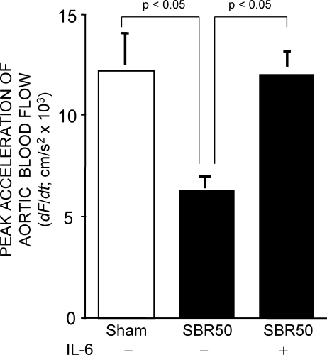
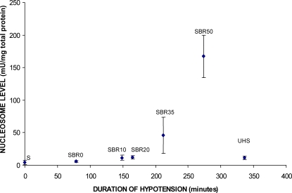
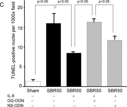
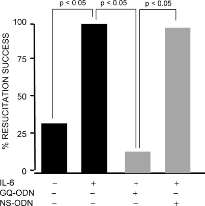
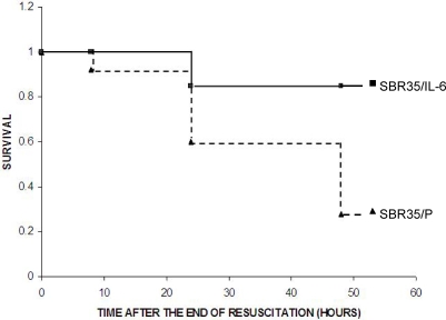
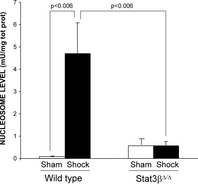
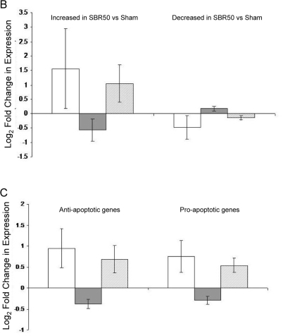
Half of trauma deaths are attributable to hypovolemic circulatory collapse (HCC). We established a model of HCC in rats involving minor trauma plus severe hemorrhagic shock (HS). HCC in this model was accompanied by a 50% reduction in peak acceleration of aortic blood flow and cardiomyocyte apoptosis. HCC and apoptosis increased with increasing duration of hypotension. Apoptosis required resuscitation, which provided an opportunity to intervene therapeutically. Administration of IL-6 completely reversed HCC, prevented cardiac dysfunction and cardiomyocyte apoptosis, reduced mortality 5-fold and activated intracardiac signal transducer and activator of transcription (STAT) 3. Pre-treatment of rats with a selective inhibitor of Stat3, T40214, reduced the IL-6-mediated increase in cardiac Stat3 activity, blocked successful resuscitation by IL-6 and reversed IL-6-mediated protection from cardiac apoptosis. The hearts of mice deficient in the naturally occurring dominant negative isoform of Stat3, Stat3beta, were completely resistant to HS-induced apoptosis. Microarray analysis of hearts focusing on apoptosis related genes revealed that expression of 29% of apoptosis related genes was altered in HS vs. sham rats. IL-6 treatment normalized the expression of these genes, while T40214 pretreatment prevented IL-6-mediated normalization. Thus, cardiac dysfunction, cardiomyocyte apoptosis and induction of apoptosis pathway genes are important components of HCC; IL-6 administration prevented HCC by blocking cardiomyocyte apoptosis and induction of apoptosis pathway genes via Stat3 and warrants further study as a resuscitation adjuvant for prevention of HCC and death in trauma patients.


Conflict of interest statement

Competing Interests: The authors have declared that no competing interests exist.

Figures

Figure 1
Figure 1. Effect of trauma/HS without or with IL-6 on peak acceleration of aortic blood flow.
Rats were subjected to the sham or the SBR50 protocol; SBR50 rats were randomly assigned to receive either placebo or IL-6 at the start of resuscitation (n≥3 in each group). Sixty minutes after start of resuscitation or at the equivalent time point for shams, rats underwent Doppler studies of the ascending aorta. Data presented is the mean±SEM of peak acceleration of aortic blood flow; significant differences are indicated (one-way ANOVA followed by Student-Newman-Kuels test).
Figure 2
Figure 2. Effect of duration of hypotension and resuscitation on cardiac nucleosome levels.
Rats (n≥3 in each group) were subjected to sham protocol (S) or the trauma/HS protocol with increasing severity of shock (SBR0, SBR10, SBR20, SBR35, and SBR50) as indicated followed by resuscitation. Hearts were harvested 60 minutes after the start of resuscitation. One group of rats (UHS) was subjected to the SBR50 protocol, but not resuscitated, rather, kept at the target MAP (35 mmHg) for an additional 60 minutes. Hearts were immediately harvested and snap frozen in liquid nitrogen. Nucleosome levels were measured in protein extracts of frozen sections of the heart and the results corrected for total protein and plotted as a function of the duration of the hypotensive period. Nucleosome levels increased exponentially with duration of hypotension (Pearson correlation coefficient = 0.764, p<0.0001, UHS group not included in the analysis).
Figure 3
Figure 3. Effect of trauma/HS without or with IL-6 on cardiac apoptosis; impact of Stat3 inhibition on the IL-6 effect.
Rats (n≥3 in each group) were randomly assigned to the sham or SBR50 protocol; SBR50 rats were randomized 24 hr prior to trauma/HS not to receive pretreatment or to receive pretreatment with GQ-ODN or non-specific (NS)-ODN by tail vein injection as indicated. At the start of resuscitation, rats in the first two SBR50 groups were randomized in a blinded fashion to receive either placebo or IL-6 at the start of resuscitation; each rat in the last two SBR50 groups received IL-6. In panel A, extracts of cryotome sections of flash-frozen ventricles obtained 60 minutes after the start of resuscitation were used to measure nucleosome levels by ELISA. Data presented are the mean±SEM of nucleosome levels (Units/mg protein); significant differences are indicated (one-way ANOVA followed by Student-Newman-Kuels test). In panel B, sections of fixed ventricles were stained using the TUNEL protocol; representative slides showing TUNEL-positive cells (arrows) are shown. In panel C, the number of TUNEL-positive nuclei was counted in twenty randomly chosen fields of each slide by an experienced microscopist blinded to the treatment the rats received. Data presented are mean number of TUNEL-positive nuclei per field±SEM for each group; significant differences are indicated (one-way ANOVA followed by Student-Newman-Kuels test).
Figure 4
Figure 4. Resuscitation success following trauma/HS without or with IL-6 treatment; impact of Stat3 inhibition on the IL-6 effect.
Rats were subjected to the 50% shed blood return protocol (SBR50) and randomized in a blinded fashion to receive either placebo (n = 6) or IL-6 (n = 7) at the start of resuscitation as indicated. Two additional groups of rats were randomly assigned 24 hr prior to SBR50 and IL-6 treatment to receive pretreatment with GQ-ODN or non-specific (NS)-ODN by tail vein injection as indicated (n≥7). Data presented is the percent resuscitation success for each group defined as achieving a MAP ≥mean MAP minus 2 SD of starting BP. Significant differences are indicated (Fisher's exact test).
Figure 5
Figure 5. Effect of IL-6 on mortality following HS.
Rats were subjected to the SBR35 protocol and randomized in a blinded fashion to receive either placebo (0.1 ml PBS; n = 25; SBR35/P; filled triangles) or IL-6 (10 µg/kg in 0.1 ml PBS; n = 26; SBR35/IL-6; filled squares) at the start of resuscitation. At the end of the resuscitation period, catheters were removed and anesthesia was reversed. Mortality was monitored and cumulative survival was plotted per Kaplan-Meier analysis (p<0.005).
Figure 6
Figure 6. Effect of IL-6 on cardiac Stat3 activity.
Rats were subjected to the SBR50 protocol and randomized in a blinded fashion to receive either placebo (0.1 ml PBS; −) or IL-6 (10 µg/kg in 0.1 ml PBS; +). Extracts (20 µg) of cryotome sections of flash-frozen ventricles were analyzed by EMSA; radiolabeled probe without cell extract (Neg) or an extract of cells previously shown to have Stat3 activity (Pos) were included as negative and positive controls, respectively. After separation, the gel was dried and autoradiographed (panel A) or subjected to phosphoimaging analysis (panel B). Data presented in panel B represents the mean±SD of the densitometry readings; significant differences are indicated (Student's t-test).
Figure 7
Figure 7. Effect of pretreatment with a Stat3 inhibitor on IL-6-induced Stat3 activity within the heart.
Rats were pre-treated with GQ-ODN or NS-ODN and subjected to the SBR50 protocol and resuscitated with IL-6 (10 µg/kg in 0.1 ml PBS). Extracts of cryotome sections of flash-frozen ventricles were analyzed by EMSA; radiolabeled probe without cell extract (Neg) or an extract of cells previously shown to have Stat3 activity (Pos) were included as negative and positive controls, respectively. After separation, the gel was dried and autoradiographed (panel A) or subjected to phosphoimaging analysis (panel B). Data presented in panel B represents the mean±SD of the densitometry readings; significant differences are indicated (Student's t-test).
Figure 8
Figure 8. Effect of Stat3β ablation on trauma/HS-induced cardiac apoptosis.
Stat3β homozygous-deficient (Stat3βΔ/Δ) mice and their littermate control wild type mice were subjected to the murine trauma/HS protocol or sham protocol and their hearts harvested 1 hr after the start of resuscitation. Nucleosome levels were measured in protein extracts of frozen sections of the heart and the results corrected for total protein. Data presented are the means±SEM of each group (n≥3). Significant differences are indicated (Student's t-test)
Figure 9
Figure 9. Effect of trauma/HS without or with IL-6 treatment on cardiac apoptosis-related gene expression; impact of Stat3 inhibition on the IL-6 effect.
A. Heat map of apoptosis pathway genes whose expression is altered by trauma/HS. Rows in the heat map represent genes as listed in Table 1. Columns represent samples from the 3 groups examined in Experiment 1 (left panel) as indicated (S, Sham; P, placebo-treated SBR50; and I, SBR50/IL-6) and the 2 groups examined in Experiment 2 (right panel) as indicated (N, SBR50/IL-6/NS and G, SBR50/IL-6/GQ-ODN). Red indicates a level of expression above the mean expression of a gene within the experiment. White indicates a level of expression at the mean within the experiment while blue indicates a level of expression below the mean within the experiment. B and C. Changes in expression levels of apoptosis-related genes are shown comparing SBR50 vs. sham (open bars), SBR50/IL-6 vs. SBR50 (gray bars) and SBR50/IL-6/G vs. SBR50/IL-6/N (stippled bars). In panel B, the 196 apoptosis-related genes whose expression levels were changed in SBR50 vs. sham in the first microarray experiment were separated into those genes whose transcript levels were increased in SBR50 vs. sham (135 genes; left side of panel) and those whose transcript levels were decreased in SBR50 vs. sham (61 genes; right panel). In panel C, the 196 apoptosis-related genes whose expression levels were changed in SBR50 vs. sham in the first microarray experiment were separated into anti-apoptotic genes whose transcript levels were altered in SBR50 vs. sham (115 genes; left side of panel) and pro-apoptotic genes whose transcript levels were altered in SBR50 vs. sham (81 genes; right panel).

References

    1. Minino AM, Anderson RN, Fingerhut LA, Boudreault MA, Warner M. Deaths: injuries, 2002. Natl Vital Stat Rep. 2006;54:1–124. - PubMed
    1. Peden M, MK, Krug E. Injury: A leading cause of burden of disease. 2002
    1. Heckbert SR, Vedder NB, Hoffman W, Winn RK, Hudson LD, et al. Outcome after hemorrhagic shock in trauma patients. J Trauma. 1998;45:545–549. - PubMed
    1. Wiggers C. Myocardial depression in shock. A survey of cardiodynamic studies. American Heart Journal. 1947;33:633–650. - PubMed
    1. Peitzman AB, Billiar TR, Harbrecht BG, Kelly E, Udekwu AO, Simmons RL. Hemorrhagic shock. [Review]. Current Problems in Surgery. 1995;32:925–1002. - PubMed

Publication types