. 2008 Mar 15;315(2):404-17.
doi: 10.1016/j.ydbio.2007.12.041. Epub 2008 Jan 8.

Target-dependent inhibition of sympathetic neuron growth via modulation of a BMP signaling pathway

Affiliations

Target-dependent inhibition of sympathetic neuron growth via modulation of a BMP signaling pathway

Jung-Il Moon et al. Dev Biol. .

Abstract

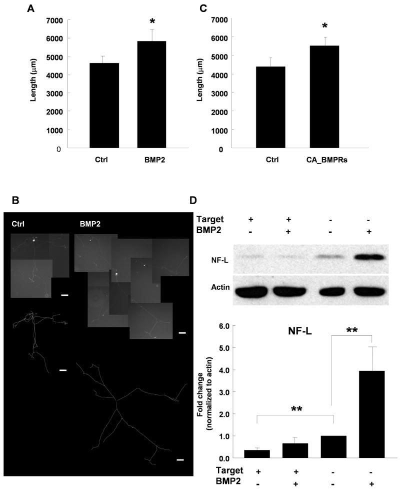
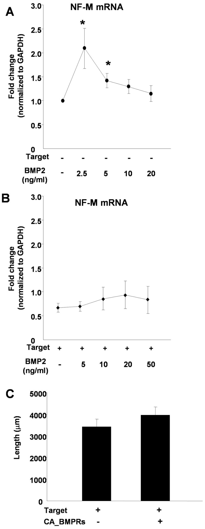
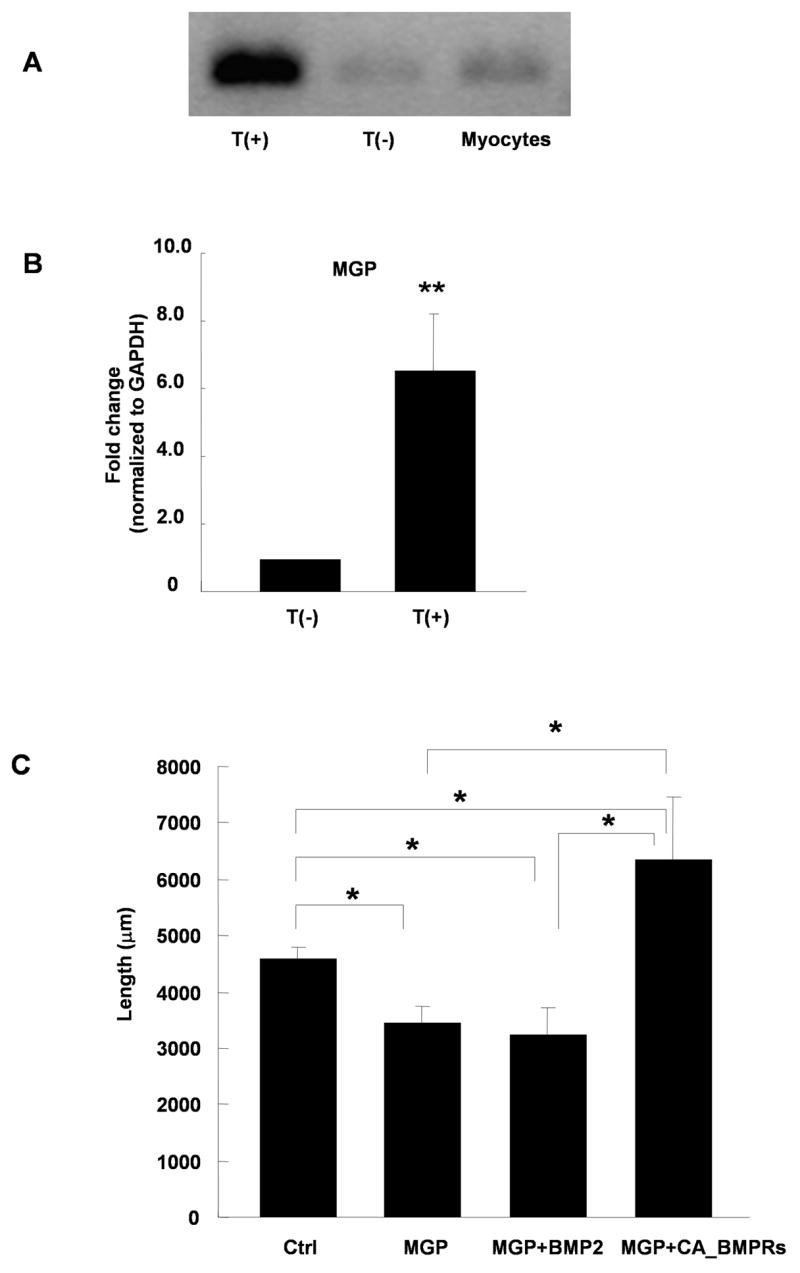
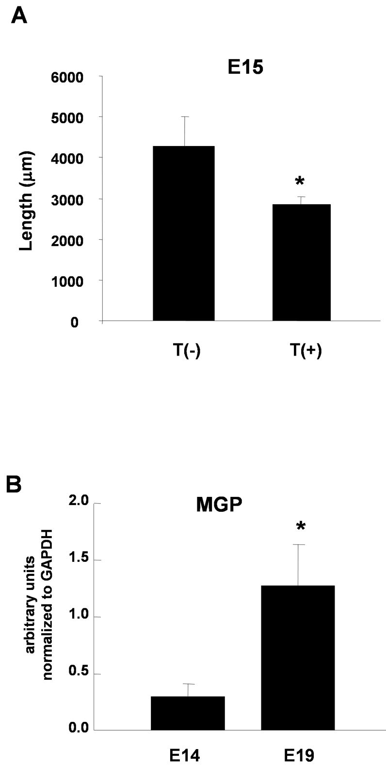
Target-derived factors modulate many aspects of peripheral neuron development including neuronal growth, survival, and maturation. Less is known about how initial target contact regulates changes in gene expression associated with these developmental processes. One early consequence of contact between growing sympathetic neurons and their cardiac myocyte targets is the inhibition of neuronal outgrowth. Analysis of neuronal gene expression following this contact revealed coordinate regulation of a bone morphogenetic protein (BMP)-dependent growth pathway in which basic helix-loop-helix transcription factors and downstream neurofilament expression contribute to the growth dynamics of developing sympathetic neurons. BMP2 had dose-dependent growth-promoting effects on sympathetic neurons cultured in the absence, but not the presence, of myocyte targets, suggesting that target contact alters neuronal responses to BMP signaling. Target contact also induced the expression of matrix Gla protein (MGP), a regulator of BMP function in the vascular system. Increased MGP expression inhibited BMP-dependent neuronal growth and MGP expression increased in sympathetic neurons during the period of target contact in vivo. These experiments establish MGP as a novel regulator of BMP function in the nervous system, and define developmental transitions in BMP responses during sympathetic development.


Figures

Figure 1
Figure 1
Sympathetic neuron growth is inhibited by contact with co-cultured cardiac myocytes. Neonatal rat sympathetic neurons were cultured alone, with cardiac myocytes or with CHO cells for 2 days. (A) Shows a neuron-myocyte co-culture stained with antibodies against peripherin (green) to stain neuronal fibers and alpha-actinin (red) to stain the cardiac myocyte. Scale bar = 50 μm. Image provided by Jeanine Hinterneder. (B) The neurons were transfected with EGFP-CD8 using a Gene Gun after the first day in culture to label the complete neuritic arbor of a few neurons on the dish and the total process length per neuron was analyzed. An image of EGFP-CD8 transfected neuron is shown with a traced image of the neuronal arbor shown below. Scale bars = 100 μm. (C) A plot of the average neurite length per neuron shows a decrease in total process length for sympathetic neurons cultured with myocytes (Co-cultures) compared to neurons cultured alone or with CHO cells. Data shown is the mean ± s.e.m. of a minimum of 27 transfected cells, p < 0.05.
Figure 2
Figure 2
Contact with cardiac myocytes decreases neurofilament expression in sympathetic neurons. Sympathetic neurons were cultured alone, T(−), or were co-cultured with cardiac myocytes, T(+). (A), (B) The mRNA expression levels of NF-L (A) and NF-M (B) were measured by real-time PCR and normalized to GAPDH. Data shown is the mean ± s.e.m. of a minimum of four independent mRNA preparations, p < 0.05. (C) Expression of NF-M protein in sympathetic neurons cultured alone, T(−), or with myocytes, T(+), was analyzed by immunocytochemistry. Cultures were grown for 2 days, fixed and stained with a NF-M antibody. The top panel shows example of images used for quantification, with averaged mean pixel intensity ± s.e.m. of a minimum of 12 images shown below.
Figure 3
Figure 3
Expression of basic helix-loop-helix (bHLH) transcription factors is regulated by target contact and influence sympathetic neurite extension. (A), (B) Sympathetic neurons were grown in the absence, T(−), or presence, T(+), of myocytes and RNA was isolated after 2 days. The expression of HAND2 (A) and HAND1 (B) mRNA was measured by real-time PCR and normalized to the GAPDH level. Data is shown as the mean ± s.e.m. of a minimum of 8 independent mRNA preparation, * p < 0.05; ** p < 0.01. (C), (D), (E) Sympathetic cultures were co-transfected with EGFP-CD8 and HAND2, HAND1, or E12. In control dishes neurons were transfected with EGRP-CD8 alone (Ctrl). The average neurite length was analyzed for control EGFP-labeled neurons and for neurons co-expressing HAND2, HAND1 or E12. Data is shown as the mean ± s.e.m of a minimum of 13 transfected neurons, *p < 0.05; ** p < 0.01. (F) Images of a neuron expressing EGFP-CD8 (top panel) and a neuron co-transfected with EGFP-CD8 and E12 (bottom panel). Scale bar = 100 μm. Traced arbors are shown to the right of the composite photographs.
Figure 4
Figure 4
BMP2 regulates the growth of sympathetic neurons in the absence of target. (A) The average neurite length per sympathetic neuron increased for EGFP-labeled neurons grown in absence (Ctrl) or presence of 10 ng/ml BMP2. (B) Images of EGFP-CD8 transfected neurons grown control medium (left panel) and grown with 10 ng/ml BMP2 in medium (right panel). The traced arbors are shown under the composite photographs. Scale bars = 100 μm. (C) Co-expression of constitutively active BMP receptor 1a & 1b with the EGFP label increased total neurite length per neuron. Data is shown as the mean ± s.e.m. of a minimum of 25 transfected neurons, *p < 0.05. (D) Expression of NF-L protein in sympathetic neurons is induced by BMP2 when the neurons were grown in the absence, but not the presence of myocytes. NF-L levels were analyzed by Western blot analysis and normalized to actin. The top panel shows an example of a Western blot, with the averaged data of 4 independent experiments shown below. Data is the mean ± s.e.m., **p < 0.01.
Figure 5
Figure 5
Target contact alters neuronal BMP responses. Sympathetic neurons were cultured alone (A) or with myocytes (B) in different concentrations of BMP2 (0–50 ng/ml). Real-time PCR analysis was used to measure NF-M expression and values were normalized to GAPDH expression levels. (A) BMP2 induced a dose-dependent increase in NF-M mRNA in neurons cultured in the absence of cardiac myocytes. (B) In the presence of co-cultured myocytes, BMP2 does not promote NF-M expression at any concentration tested. Data is shown as the mean of at least 4 experiments ± s.e.m., *p < 0.05. (C) Expression of constitutively active BMP receptors in sympathetic neurons does not increase neurite length for neurons cultured in the presence of myocyte targets. EGFP-CD8 was transfected alone or co-transfected with constitutively active BMP receptor 1a & 1b and the average neurite length of GFP-labeled sympathetic neurons was measured. Data is shown as the mean ± s.e.m. of a minimum of 30 transfected neurons.
Figure 6
Figure 6
Matrix Gla protein (MGP) is induced by target contact and modulates BMP2 growth responses. (A) Expression of MGP mRNA is increased in sympathetic neurons co-cultured with myocytes T(+) compared to neurons cultured alone T(−) or myocytes cultured alone. (B) Quantification of MGP expression as measured by real-time PCR and normalized to GAPDH levels for neurons grown alone T(−), or in the presence of myocytes T(+). Data shows the mean of 6 experiments ± s.e.m., **p < 0.01. (C) The average neurite length of sympathetic neurons expressing EGFP-CD8 alone (Ctrl) or co-expressing human MGP in the absence (MGP) or presence (MGP+BMP2) of 10 ng/ml BMP2, or co-expressing EGFP, MGP, and constitutively active BMP receptor 1a & 1b (MGP+CA_BMPRs). Neurite growth was inhibited by MGP expression, and this decrease was not rescued by the addition BMP2. Co-expression of CA-BMPRs with MGP resulted in increased neurite growth. Data is shown as the mean ± s.e.m. of a minimum of 17 transfected neurons in each condition, *p < 0.05.
Figure 7
Figure 7
Regulation of growth pathways in embryonic neurons and developmental regulation of MGP expression. A. E15 developing sympathetic neurons were cultured alone T(−) or with neonatal cardiac myocytes T(+) for 3 days. Cultures were stained with a NCAM antibody and the total process length per neuron was analyzed. Data shown is the mean ± s.e.m. of a minimum of 14 labeled neurons, p < 0.05. B. MGP mRNA expression increased in immuno-purified developing embryonic rat sympathetic neurons between E14 and E19, a period in which sympathetic neurons are establishing target contacts. MGP mRNA expression was measured by real-time PCR and was normalized to GAPDH expression. Data is shown as the mean of 4 experiments ± s.e.m., *p < 0.01.

Similar articles

Cited by

References

    1. Baird DH, Hatten ME, Mason CA. Cerebellar target neurons provide a stop signal for afferent neurite extension in vitro. J Neurosci. 1992;12:619–34. - PMC - PubMed
    1. Baird DH, Trenkner E, Mason CA. Arrest of afferent axon extension by target neurons in vitro is regulated by the NMDA receptor. J Neurosci. 1996;16:2642–8. - PMC - PubMed
    1. Beck HN, Drahushuk K, Jacoby DB, Higgins D, Lein PJ. Bone morphogenetic protein-5 (BMP-5) promotes dendritic growth in cultured sympathetic neurons. BMC Neurosci. 2001;2:12. - PMC - PubMed
    1. Bharmal S, Slonimsky JD, Mead JN, Sampson CP, Tolkovsky AM, Yang B, Bargman R, Birren SJ. Target cells promote the development and functional maturation of neurons derived from a sympathetic precursor cell line. Dev Neurosci. 2001;23:153–64. - PubMed
    1. Bostrom K, Tsao D, Shen S, Wang Y, Demer LL. Matrix GLA protein modulates differentiation induced by bone morphogenetic protein-2 in C3H10T1/2 cells. J Biol Chem. 2001;276:14044–52. - PubMed

Publication types

MeSH terms