. 2008 Feb 19;105(7):2415-20.
doi: 10.1073/pnas.0712168105. Epub 2008 Feb 12.

A chemical method for fast and sensitive detection of DNA synthesis in vivo

Affiliations

A chemical method for fast and sensitive detection of DNA synthesis in vivo

Adrian Salic et al. Proc Natl Acad Sci U S A. .

Abstract

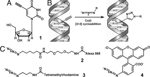
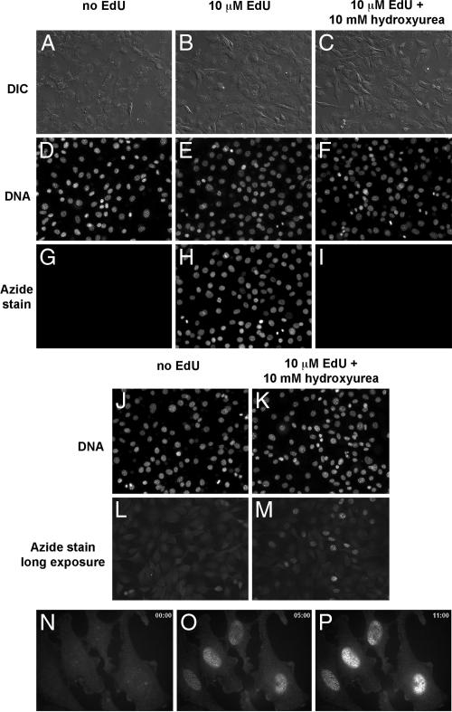
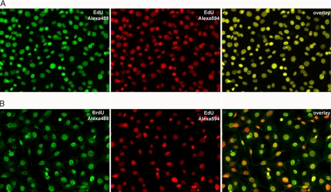
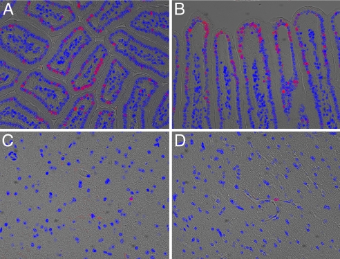
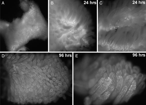
We have developed a method to detect DNA synthesis in proliferating cells, based on the incorporation of 5-ethynyl-2'-deoxyuridine (EdU) and its subsequent detection by a fluorescent azide through a Cu(I)-catalyzed [3 + 2] cycloaddition reaction ("click" chemistry). Detection of the EdU label is highly sensitive and can be accomplished in minutes. The small size of the fluorescent azides used for detection results in a high degree of specimen penetration, allowing the staining of whole-mount preparations of large tissue and organ explants. In contrast to BrdU, the method does not require sample fixation or DNA denaturation and permits good structural preservation. We demonstrate the use of the method in cultured cells and in the intestine and brain of whole animals.


Conflict of interest statement

The authors declare no conflict of interest.

Figures

Fig. 1.
Fig. 1.
Use of 5-ethynyl-2′-deoxyuridine (EdU) to label DNA in cells. (A) Structure of 5-ethynyl-2′-deoxyuridine, a thymidine analogue that carries a terminal alkyne group instead of a methyl in the 5 position of the pyrimidine ring. (B) Schematic of the click reaction for detecting EdU incorporated into cellular DNA. The terminal alkyne group, exposed in the major groove of the DNA helix readily reacts with an organic azide (R can be any fluorophore, hapten, electron-dense particle, quantum dot, etc.) in the presence of catalytic amounts of Cu(I). (C) Structures of the fluorescent azides used in the present study. Unlike azide 2, azide 3 is cell-permeable, allowing cells to be stained while alive, without fixation and/or permeabilization. Fluorescein azide 4 can be easily prepared in large amounts by using inexpensive starting materials. See text and Materials and Methods for details.
Fig. 2.
Fig. 2.
Detection of EdU incorporated into the DNA of cultured NIH 3T3 cells (A–M) and HeLa cells (N–P) by fluorescence microscopy. NIH 3T3 cells were incubated in media without EdU (A, D, G, J, and L), media supplemented with10 μM EdU (B, E, and H) or media with 10 μM EdU and 10 mM hydroxyurea to block DNA synthesis (C, F, I, K, and M). In A–M, the cells were fixed and then reacted with 10 μM Alexa568-azide (Fig. 1C, compound 2) for 10 min. The cells were then counterstained with Hoechst to reveal cellular DNA, washed, and imaged by fluorescence microscopy and differential interference contrast (DIC). Note the strong specific and low nonspecific azide stain in the presence (H) and absence (G) of EdU, respectively. Not all nuclei in H are labeled after overnight incubation with EdU, suggesting that only cells that went through S phase became labeled. Blocking DNA replication with hydroxyurea abolishes EdU incorporation almost completely (I). If cells labeled with EdU in the presence of hydroxyurea are photographed with longer exposure times (3-s exposure time in L and M, compared with 40-ms exposure time for G–I under otherwise identical illumination and camera settings), low levels of EdU incorporation can be seen in a small fraction of the hydroxyurea-treated cells. (N–P) Still images from a time-lapse recording of EdU staining of live cells by using the cell-permeable TMR-azide (Fig. 1C, compound 3). Live HeLa cells labeled with 10 μM EdU were stained with 500 nM TMR-azide in the presence of Cu(I) in PBS. Time is shown in minutes in the upper right corners of M–P. Note that Cu(I) is cytotoxic and the cells do not survive the staining reaction. See text and Materials and Methods for details.
Fig. 3.
Fig. 3.
Reproducibility of EdU labeling (A) and comparison with BrdU (B). (A) NIH 3T3 cells labeled by incubation overnight with 2 μM EdU were fixed and reacted successively with 10 μM Alexa488-azide and 10 μM Alexa594-azide, respectively, as described in Materials and Methods. The cells were imaged by fluorescence microscopy. (Left) Alexa488-azide stain. (Center) Alexa594-azide stain. (Right) Overlay of the Alexa488 and Alexa594 images. The complete colocalization of the two colors indicates that the two successive azide stains detect ethynyl groups that have the same distribution within the cell nucleus. (B) NIH 3T3 cells labeled by incubation overnight with 2 μM EdU and 2 μM BrdU were fixed and reacted with 10 μM Alexa594-azide, followed by standard BrdU immunodetection by using an anti-BrdU monoclonal antibody and an Alexa488-conjugated secondary antibody. (Left) BrdU stain. (Center) EdU stain. (Right) Overlay of the BrdU and EdU images. Note that each cell that incorporated BrdU also incorporated EdU. The BrdU micrograph was taken by using a five-times longer exposure than the exposure used for EdU (500 ms compared with 100 ms) under identical digital camera settings.
Fig. 4.
Fig. 4.
Labeling DNA in vivo by using EdU. An adult mouse was injected i.p. with 100 μg of EdU in PBS. Tissues were harvested, fixed, and sectioned 96 h later. Tissue sections on slides were then reacted for 10 min with 10 μM Alexa568-azide. The images shown are overlays of a DIC image of the sectioned tissue, a fluorescent image of cellular DNA (Hoechst stain; blue), and a fluorescent image of EdU-labeled DNA revealed by reaction with Alexa568 azide (red). (A and B) Mouse small intestine. Villi are seen in transverse section in A and in longitudinal section in B. The cells with red nuclei are descended from cells that had been in S phase during the EdU pulse and thus incorporated EdU into their DNA. (C and D) Mouse brain. The vast majority of nuclei on brain sections were not labeled with EdU, confirming that the label does not detectably incorporate into the DNA of nondividing cells. Each of the two images shows a field of cells containing an EdU-labeled nucleus, belonging to a cell of undetermined type.
Fig. 5.
Fig. 5.
Exploring cell proliferation and tissue dynamics in animals using EdU. Whole-mount fluorescent images of mouse small intestine, stained to detect EdU incorporation. Two adult mice were each injected i.p. with 100 μg of EdU in PBS and their small intestines were removed after 24 and 96 h, respectively. A freshly harvested 2-cm-long segment of the small intestine (A) was opened with a longitudinal cut, rinsed, and immediately stained for 10 min with 100 μM TMR-azide in the presence of Cu(I), without fixation or permeabilization. The intestine piece was then fixed, washed to remove unreacted TMR-azide, and imaged at low magnification on a dissecting microscope equipped with fluorescence. (B and C) After 24 h, the EdU shows strong incorporation in cells located at the base of each villus. (D and E) Ninety-six hours after the EdU pulse, the labeled cells have moved away from the base, near the tips of the villi. See text and Materials and Methods for details.

References

    1. Bick MD, Davidson RL. Total substitution of bromodeoxyuridine for thymidine in the DNA of a bromodeoxyuridine-dependent cell line. Proc Natl Acad Sci USA. 1974;71:2082–2086. - PMC - PubMed
    1. Gratzner HG. Monoclonal antibody to 5-bromo- and 5-iododeoxyuridine: A new reagent for detection of DNA replication. Science. 1982;218:474–475. - PubMed
    1. Waldman FM, et al. A comparison between bromodeoxyuridine and 3H thymidine labeling in human breast tumors. Mod Pathol. 1991;4:718–722. - PubMed
    1. Rakic P. Neurogenesis in adult primate neocortex: an evaluation of the evidence. Nat Rev Neurosci. 2002;3:65–71. - PubMed
    1. Tornoe CW, et al. Peptidotriazoles on solid phase: [1,2,3]-Triazoles by regiospecific copper(i) -catalyzed 1,3-dipolar cycloadditions of terminal alkynes to azides. J Org Chem. 2002;67:3057–3064. - PubMed

Publication types