

Case Reports
doi: 10.3174/ajnr.A0974.
Epub 2008 Feb 13.
Carcinoma ex pleomorphic adenoma of the parotid gland: radiologic-pathologic correlation with MR imaging including diffusion-weighted imaging
Affiliations
- PMID: 18272554
- PMCID: PMC8128585
- DOI: 10.3174/ajnr.A0974
Item in Clipboard
Case Reports
Carcinoma ex pleomorphic adenoma of the parotid gland: radiologic-pathologic correlation with MR imaging including diffusion-weighted imaging
AJNR Am J Neuroradiol.
2008 May.
Abstract
We present 4 cases of carcinoma ex pleomorphic adenoma of the parotid gland. In 3 of the 4 cases, diffusion-weighted and apparent diffusion coefficient (ADC) mapping images clearly revealed carcinoma as a hypercellular area with low ADC values and pleomorphic adenoma as a hypocellular area with high ADC values. Diffusion-weighted images demonstrated well complex tissue components in carcinoma ex pleomorphic adenoma, which may be useful for the diagnosis of this disease.
Figures
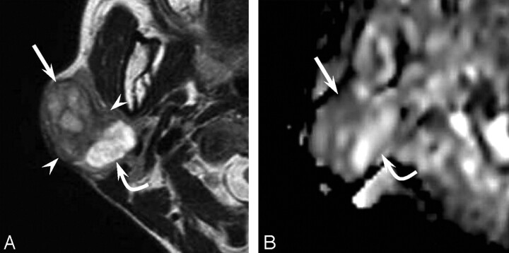
A 48-year-old woman with a carcinoma ex pleomorphic adenoma (case 1). A, Axial T2-weighted image (fast spin-echo: TR/TE, 4102/90) shows a well-demarcated mass with heterogeneous intensity of the right parotid gland. The lateral component shows mild high intensity (arrow), the intermediate component shows low intensity (arrowhead), and the medial component shows extremely high intensity (curved arrow). B, ADC mapping image shows the lateral component of hypercellularity with carcinoma (arrow) and the intermediate and medial component of hypocellularity with pleomorphic adenoma (curved arrow).
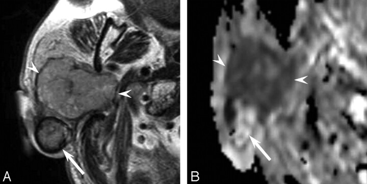
A 76-year-old man with a carcinoma ex pleomorphic adenoma (case 2). A, Axial T2-weighted image (fast spin-echo: TR/TE, 4102/90) shows a mainly well-demarcated mass with heterogeneous intensity of the right parotid gland. The anterior component shows mild high intensity (arrowheads), and the margin of the posterior component shows low intensity (arrow). B, ADC mapping image shows the anterior component of hypercellularity with carcinoma (arrowheads) and the posterior component of hypocellularity with pleomorphic adenoma (arrow).
References
-
- Seifert G. Histopathology of malignant salivary gland tumours. Eur J Cancer B Oral Oncol 1992;28:49–56 - PubMed
-
- Olsen KD, Lewis JE. Carcinoma ex pleomorphic adenoma: a clinicopathologic review. Head Neck 2001;23:705–12 - PubMed
-
- Joe VQ, Westesson PL. Tumors of the parotid gland: MR imaging characteristics of various histologic types. AJR Am J Roentgenol 1994;163:433–38 - PubMed
-
- Freling NJ, Molenaar WM, Vermey A, et al. Malignant parotid tumors: clinical use of MR imaging and histologic correlation. Radiology 1992;185:691–96 - PubMed
-
- Motoori K, Yamamoto S, Ueda T, et al. Inter- and intratumoral variability in magnetic resonance imaging of pleomorphic adenoma: an attempt to interpret the variable magnetic resonance findings. J Comput Assist Tomogr 2004;28:233–46 - PubMed
Publication types
MeSH terms
LinkOut - more resources
Full Text Sources
Medical