. 2008 Apr;82(8):3903-11.
doi: 10.1128/JVI.02227-07. Epub 2008 Feb 13.

Standardized and highly efficient expansion of Epstein-Barr virus-specific CD4+ T cells by using virus-like particles

Affiliations

Standardized and highly efficient expansion of Epstein-Barr virus-specific CD4+ T cells by using virus-like particles

Dinesh Adhikary et al. J Virol. 2008 Apr.

Abstract

Epstein-Barr virus (EBV)-specific T-cell lines generated by repeated stimulation with EBV-immortalized lymphoblastoid B-cell lines (LCL) have been successfully used to treat EBV-associated posttransplant lymphoproliferative disease (PTLD) in hematopoietic stem cell transplant recipients. However, PTLD in solid-organ transplant recipients and other EBV-associated malignancies respond less efficiently to this adoptive T-cell therapy. LCL-stimulated T-cell preparations are polyclonal and contain CD4(+) and CD8(+) T cells, but the composition varies greatly between lines. Because T-cell lines with higher CD4(+) T-cell proportions show improved clinical efficacy, we assessed which factors might compromise the expansion of this T-cell population. Here we show that spontaneous virus production by LCL and, hence, the presentation of viral antigens varies intra- and interindividually and is further impaired by acyclovir treatment of LCL. Moreover, the stimulation of T cells with LCL grown in medium supplemented with fetal calf serum (FCS) caused the expansion of FCS-reactive CD4(+) T cells, whereas human serum from EBV-seropositive donors diminished viral antigen presentation. To overcome these limitations, we used peripheral blood mononuclear cells pulsed with nontransforming virus-like particles as antigen-presenting cells. This strategy facilitated the specific and rapid expansion of EBV-specific CD4(+) T cells and, thus, might contribute to the development of standardized protocols for the generation of T-cell lines with improved clinical efficacy.


Figures

FIG. 1.
FIG. 1.
Virus production by LCL varies inter- and intraindividually. (A) The number of viral particles in the supernatants of LCL from five healthy donors (TG, MS, SM, CH, and IE) and from a patient with acute EBV-associated infectious mononucleosis (IM3) and from the supernatant of the EBV-producer cell line B95.8 was determined by quantitative PCR using BALF5-specific primers. Viral titers are given as EBV genome equivalents (geq)/milliliter. (B) The same EBV-positive target cells were tested for recognition by the BLLF1-specific CD4+ T-cell clone 1D6. Because none of the target cells expresses the restricting MHC-II molecule, the T cells recognized the target cells only after being cocultured with mini-LCL from donor JM (mini-LCL JM), which express the restricting MHC allele but are incapable of producing viral particles. T-cell recognition of the cell mixtures was target cell dependent but correlated with the amount of EBV genome equivalents detected in the culture supernatant as quantified by PCR. (C) Mini-LCL JM were cocultured for 24 h with MHC-mismatched LCL from donor GB (LCL GB) that had been cultured for different periods of time in vitro (T1 and T2). Subsequently, the cell mixtures were probed for recognition by the BLLF1-specific CD4+ T cells from donor JM (clone 1D6). As a control, the T-cell recognition of autologous LCL JM, autologous mini-LCL JM, and allogeneic LCL GB is shown. IFN-γ, gamma interferon.
FIG. 2.
FIG. 2.
Presentation of structural antigens of EBV is impaired after acyclovir treatment of LCL. Autologous LCL and mini-LCL from donor DA (mini-LCL DA), the MHC-mismatched LCL from donor JM (LCL JM), and LCL from donor TG (LCL TG), which had been left untreated or had been treated with 200 μM acyclovir (ACV) for 2 weeks, were tested for recognition by CD4+ T cells specific for the virion antigen BNRF1. In addition, the allogeneic lines were cocultured for 24 h with autologous mini-LCL DA and then tested for T-cell recognition. Except for LCL DA, none of the LCL was recognized by the T cells directly. However, the cell mix of autologous mini-LCL DA and allogeneic LCL was recognized, but the acyclovir treatment of the LCL abolished recognition. For a specificity control, the T cells also were tested for the recognition of autologous mini-LCL pulsed with the relevant EBV protein (BNRF1) or an irrelevant EBV protein (EBNA3C). IFN-γ, gamma interferon.
FIG. 3.
FIG. 3.
In LCL-stimulated CD4+ T-cell preparations, responses to FCS dominate virus-specific responses. CD4+ T-cell lines were established from EBV-positive donors by repeated stimulation with autologous LCL cultured in medium supplemented with either FCS or HS and tested against both types of stimulator cells in cytokine secretion assays. CD4+ T-cell lines that had been stimulated with LCL-FCS failed to recognize LCL-HS, suggesting that the lines predominantly recognized antigens derived from FCS. In contrast, CD4+ T-cell lines that had been stimulated with LCL-HS recognized both types of target cells, indicating that this line recognized viral or cellular antigen(s). GM-CSF, granulocyte-macrophage colony-stimulating factor.
FIG. 4.
FIG. 4.
Presentation of exogenous viral structure antigens is impaired by human serum components. (A) LCL grown in medium supplemented with either FCS or HS were tested for recognition by BLLF1-specific CD4+ T cells (clone 1D6). T-cell recognition was reduced when LCL were cultured in human serum. (B) Mini-LCL grown in FCS-containing media were pulsed with increasing amounts of virus supernatant that had been left untreated or had been incubated at a final concentration of 10% human serum from an EBV+ or EBV donor for 2 h. The recognition of the cells by BLLF1-specific CD4+ T cells (clone 1D6) was assayed 24 h later. IFN-γ, gamma interferon.
FIG. 5.
FIG. 5.
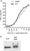
Mini-LCL pulsed with either VLP or WT EBV are recognized by virion antigen-specific CD4+ T cells with similar efficiencies. (A) Mini-LCL from donor JM (mini-LCL JM) were pulsed with supernatants from 293/TR− and 293/2089 cells containing 1 × 10⁷/ml VLP or WT EBV particles, respectively, and then were tested for recognition by autologous BLLF1-specific CD4+ T cells. Across the entire concentration range analyzed, WT EBV and VLP-pulsed mini-LCL were recognized by the T cells to the same extent. (B) The concentration of VLP and WT EBV in the supernatants used for the experiments shown in panel A was determined by Western blot analysis using an antibody against the tegument protein BNRF1. IFN-γ, gamma interferon.
FIG. 6.
FIG. 6.
EBV structural antigen-specific CD4+ T cells are efficiently expanded from peripheral blood of healthy EBV carriers by repeated stimulation with VLP-pulsed PBMC. (A) Autologous PBMC pulsed with VLP for 24 h were used for the repeated stimulation of peripheral CD4+ T cells of donors DA, JM, and GB. After five stimulations, all CD4+ T-cell lines recognized VLP-pulsed, but not barely unpulsed, PBMC, suggesting that these T cells recognized virion antigens. (B) When tested against autologous LCL and mini-LCL, these CD4+ T-cell lines showed much stronger responses against LCL, indicating that the VLP-stimulated T cells recognized antigens that also were presented by LCL. (C) After four to six rounds of VLP stimulation, CD4+ T cells became increasingly EBV specific, as indicated by increasing reactivity against VLP-pulsed autologous PBMC and autologous LCL and decreasing reactivity against unpulsed autologous PBMC. Results for donor JM are shown as an example. (D) To define the antigens recognized by the VLP-stimulated CD4+ T-cell lines, mini-LCL were pulsed separately with three structural proteins of EBV (BALF4, BNRF1, and BLLF1) and a nonstructural lytic cycle protein of EBV (BALF2). While none of the lines responded to BALF2, each line responded to at least one structural protein. IFN-γ, gamma interferon.
FIG. 7.
FIG. 7.
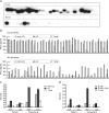
Clonality and specificity of VLP- and LCL-stimulated CD4+ T-cell lines. (A) Peripheral blood CD4+ T cells from donor GB were stimulated six times with either autologous LCL-HS or VLP-pulsed autologous PBMC and analyzed for T-cell receptor Vβ expression by PCR, and then the PCR products were subjected to Southern blot hybridization. T-cell lines stimulated with VLP-pulsed PBMC were oligoclonal at this passage, while LCL-stimulated T-cell lines were still polyclonal. (B) CD4+ T-cell clones were generated by limiting dilution from T-cell lines of donor GB at passage six after stimulation with either VLP-pulsed PBMC or LCL-HS. The specificity of the clones was determined by assessing their reactivity against autologous LCL and mini-LCL. After stimulation with VLP-pulsed PBMC, the majority of clones were specific for EBV lytic cycle proteins, as indicated by their exclusive reactivity against LCL. In contrast, most of the T-cell clones obtained from the LCL-stimulated T-cell line recognized both types of target cells and, thus, were not specific for EBV lytic cycle antigens. CD4+ T-cell lines from donor JM were stimulated eight times with either LCL-HS (LCL) or VLP-pulsed PBMC (VLP) and subsequently tested for recognition of autologous LCL and mini-LCL (C) or primary B cells incubated with T EBV (D). Both T-cell populations displayed a similar cytolytic phenotype and secreted perforin and granzyme B upon antigen recognition. Importantly, B cells newly infected with EBV were recognized to a much larger extent by VLP than by LCL-stimulated T cells, suggesting that T-cell lines generated by stimulation with VLP-pulsed PBMC contain a higher proportion of virion antigen-specific T cells. These experiments were performed twice in two different donors with similar results. IFN-γ, gamma interferon; OD(450), optical density at 450 nm.

References

    1. Adhikary, D., U. Behrends, H. Boerschmann, A. Pfunder, S. Burdach, A. Moosmann, K. Witter, G. W. Bornkamm, and J. Mautner. 2007. Immunodominance of lytic cycle antigens in Epstein-Barr virus-specific CD4+ T cell preparations for therapy. PLoS ONE 2e583. - PMC - PubMed
    1. Adhikary, D., U. Behrends, A. Moosmann, K. Witter, G. W. Bornkamm, and J. Mautner. 2006. Control of Epstein-Barr virus infection in vitro by T helper cells specific for virion glycoproteins. J. Exp. Med. 203995-1006. - PMC - PubMed
    1. Bevan, M. J. 2006. Cross-priming. Nat. Immunol. 7363-365. - PubMed
    1. Bollard, C. M., I. Kuehnle, A. Leen, C. M. Rooney, and H. E. Heslop. 2004. Adoptive immunotherapy for posttransplantation viral infections. Biol. Blood Marrow Transplant. 10143-155. - PubMed
    1. Chackerian, B. 2007. Virus-like particles: flexible platforms for vaccine development. Expert Rev. Vaccines 6381-390. - PubMed

Publication types

MeSH terms