. 2008 Mar;135(6):1169-78.
doi: 10.1242/dev.014142. Epub 2008 Feb 13.

Alternative promoter use in eye development: the complex role and regulation of the transcription factor MITF

Affiliations

Alternative promoter use in eye development: the complex role and regulation of the transcription factor MITF

Kapil Bharti et al. Development. 2008 Mar.

Abstract

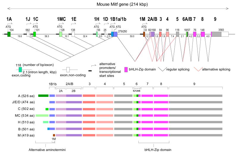
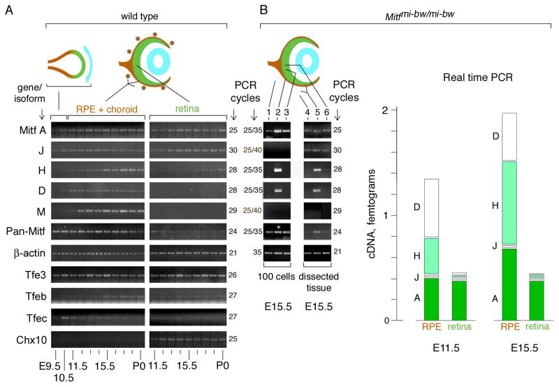
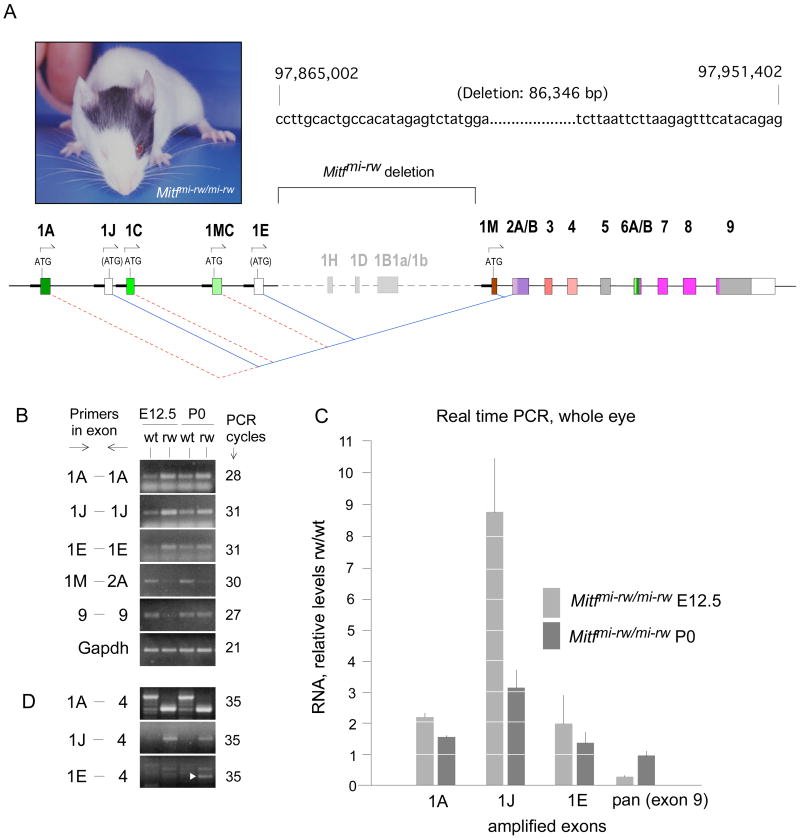
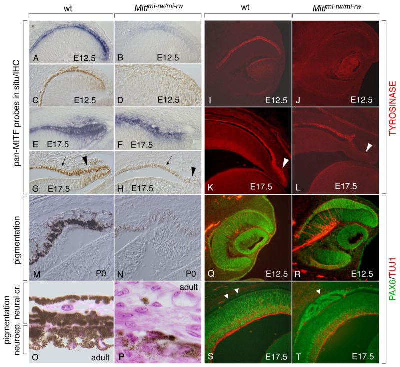
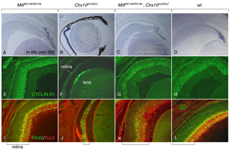
During vertebrate eye development, the transcription factor MITF plays central roles in neuroepithelial domain specification and differentiation of the retinal pigment epithelium. MITF is not a single protein but represents a family of isoforms generated from a common gene by alternative promoter/exon use. To address the question of the role and regulation of these isoforms, we first determined their expression patterns in developing mouse eyes and analyzed the role of some of them in genetic models. We found that two isoforms, A- and J-Mitf, are present throughout development in both retina and pigment epithelium, whereas H-Mitf is detected preferentially and D-Mitf exclusively in the pigment epithelium. We further found that a genomic deletion encompassing the promoter/exon regions of H-, D- and B-Mitf leads to novel mRNA isoforms and proteins translated from internal start sites. These novel proteins lack the normal, isoform-specific N-terminal sequences and are unable to support the development of the pigment epithelium, but are capable of inducing pigmentation in the ciliary margin and the iris. Moreover, in mutants of the retinal Mitf regulator Chx10 (Vsx2), reduced cell proliferation and abnormal pigmentation of the retina are associated with a preferential upregulation of H- and D-Mitf. This retinal phenotype is corrected when H- and D-Mitf are missing in double Mitf/Chx10 mutants. The results suggest that Mitf regulation in the developing eye is isoform-selective, both temporally and spatially, and that some isoforms, including H- and D-Mitf, are more crucial than others in effecting normal retina and pigment epithelium development.


Figures

Fig. 1
Fig. 1
Top: Schematic diagram of the mouse Mitf gene with its multiple promoters and regular and alternative splice choices. Bottom: Schematic diagram of the seven protein isoforms differing in their aminoterminal sequences due to different promoter choice.
Fig. 2
Fig. 2
Expression of Mitf RNAs and proteins containing exon 1B1b. (A) In situ hybridization with an exon 1B1b-specific probe on frontal sections of the developing eye at the indicated time points. Arrowheads at E9.5 indicate expression throughout the optic vesicle, and arrowhead at E10.5 indicates reduced expression in the distal optic vesicle. In a control section of an Mitfmi-rw/mi-rw embryo, which lacks exon 1B1b, there is no labeling. (B) Antibody characterization and Mitf expression in RPE/choroid and retina. A-, H-, D-, and M-Mitf cDNAs were expressed in NIH3T3 cells and extracts blotted with polyclonal anti-1B1b MITF antibodies (top panel) or monoclonal anti-MITF carboxyl terminal-domain antibodies (anti-CTD, middle panel). The bottom left panel shows a combined immunoprecipitation (IP)/immunoblot (IB) of NIH-3T3 cells transfected with A-Mitf. The bottom right panel shows a similar IP/IB of extracts from RPE/choroidal and retinal fractions from 100 wild type eye primordia (E12.5). Arrowheads point to MITF isoforms with distinct electrophoretic mobilities. (C) Immunohistochemistry of frontal cryosections of embryos of the indicated genotypes and ages using anti-1B1b MITF and anti-pan MITF antibodies. Arrows in panel labeled wt-E15.5 point to M-MITF expressing neural crest-derived melanocytes that are only seen with pan-MITF-specific antibodies. Note that “wt” refers to Tyrc (albino) embryos carrying a wild type Mitf allele. RPE, retinal pigment epithelium; ret: retina; CMZ: ciliary margin zone.
Fig. 3
Fig. 3
Differential expression of Mitf isoforms during mouse eye development. (A) RT-PCR analysis on RNA isolated from wild type whole optic vesicles (E9.5–10.5) or separate RPE+choroidal and retinal fractions (E11.5-P0). For primer choice and number of PCR cycles, see text and Supplemental Table S2. (B) RT-PCR and real-time PCR for RNA isolated from Mitfmi-bw/mi-bw eyes which lack neural crest-derived melanocytes. Left panel: E15.5 eyes were dissected as in (A) and 100 individual cells were microscopically selected as described in materials and methods. Lane 1, mesenchymal cells; Lane 2, RPE cells; Lane 3, retinal cells. The asterisk in lane 2 indicates that the corresponding cDNA was diluted 1:10 for the reaction with pan-specific primers. Middle panel: Pooled fractions from dissected E15.5 eyes as indicated. Lane 4, mesenchymal fraction; Lane 5, RPE/mesenchymal fraction; Lane 6, retinal fraction. Right panel: Real time PCR. RNA was prepared from separately pooled RPE/mesenchymal and retinal fractions from E11.5 and E15.5 eyes. Results are expressed as mean of absolute amounts and standard deviations (vertical lines in each column) of the respective cDNAs and are calculated taking into account that RPE cells represent 7% of the cells in the RPE/mesenchymal fraction (for details, see Supplemental Materials). Significance of the difference between RPE and retina (p values, Student’s t test, pools of 20 RPE/mesenchymal and retinal fractions each for E11.5; and 12 RPE/mesenchymal and retinal fractions each for E15.5, four separate assays in triplicates per pool): E11.5: A-Mitf, >0.1; J-Mitf, <0.1; H-Mitf, <0.001; D-Mitf, <0.00001. E15.5: A-Mitf, <0.01; J-Mitf, <0.01; H-Mitf, <0.0001; D-Mitf, <0.001.
Fig. 4
Fig. 4
Mitfmi-rw/mi-rw mutant mice carry a genomic deletion in Mitf encompassing the exons 1H, 1D and 1B. (A) Phenotype of an Mitfmi-rw/mi-rw mutant mouse. Generally, coat pigment patches and eye size are not correlated, as eye size is determined by the development of the RPE and coat patches by neural crest-derived melanocytes. The sequence on the right marks the base pairs flanking the genomic deletion, with the numbers of the first and last base of the depicted sequence referring to the positions on chromosome 6 according to assembly NCBIM36. The schematic diagram below shows the extent of the deletion and highlights the novel splice junctions that are generated between the upstream exons 1A, 1J, 1C, 1MC and 1E and the downstream exon 2A. For details, see text. (B) RT-PCR using RNA from E12.5 wild type and Mitfmi-rw (rw) mutant embryos and the corresponding P0 newborns. Note increased band intensity in rw with exons 1A, 1J and 1E but decreased band intensity at E12.5 with pan-specific primers (exon 9). (C) Real time PCR from RNA of whole eyes harvested at the indicated ages, using primers as in (B). The results are expressed as RNA levels in rw mutants relative to those in corresponding wild type embryos (groups of 14 eyes each, three measurements each in triplicates). The increases in rw over wild type in 1A and 1J as well as the decrease in exon 9 at E12.5 are statistically significant (p<0.01, Student’s t test). P values for the increase in 1E are <0.02 at E12.5 and =0.13 at P0. No significant difference was found for exon 9 at P0 (p=0.27). (D) RT-PCR using primers spanning four exons. Note different-size products in wt and rw for isoform 1A. For isoform 1J, the expected larger product in wt is not seen because its relative level is low (see Fig. 2), but the smaller-size product in rw is visible. In 1E-4, white arrowhead points to the correct E-Mitf band.
Fig. 5
Fig. 5
The Mitfmi-rw allele allows for the expression of Mitf and its target gene tyrosinase in the anterior RPE as well as for residual pigmentation in the iris but leads to an abnormal RPE dorsally. Sections of wild type and Mitfmi-rw/mi-rw mutant eyes of the indicated ages were stained with a pan-Mitf in situ probe (A,B,E,F), a pan-MITF antiserum (C,D,G,H), antibodies against TYROSINASE (I–L), or against PAX6 (green) and TUJ1 (red) (Q–T). Note that the mutant eye expresses Mitf RNA (B,F) and low levels of MITF protein, along with TYROSINASE, particularly at later stages (compare G and H, arrows; and K and L). Also note that in the mutant at E17.5, the distal ciliary margin, though positive for Mitf RNA (F), is relatively free of MITF and TYROSINASE (arrowhead in H and L, compare with arrowhead in G and K). (M,N) Strong pigmentation at P0 in wild type (M) and weak pigmentation in rw (N). (O,P) Pigmentation in adult wild type iris (O) and rw iris (P). (Q,R) Dorsal thickening of the E12.5 RPE in rw (R) compared to wild type (Q). (S,T) RPE abnormalities in rw at E17.5. Arrowheads in (S) mark the location of the wt RPE which now is free of PAX6 staining. In contrast, mutant RPE retains PAX6 staining and in this eye showed epihelial folds rather than homogeneous thickening (arrowhead in T).
Fig. 6
Fig. 6
Lack of downregulation of Mitf in Chx10orJ/orJ retinas predominantly affects H- and D-Mitf. (A) Genetic pathway showing the downregulation of Mitf in the future retina by FGFs emanating from the surface ectoderm (light blue) and Chx10 operating in the distal optic neurepithelium. In Chx10 mutant animals, Mitf is not downregulated in the future retina and the retina hypoproliferates. (B–E) Cryostat sections from E13.5 wild type eyes (B,D) and Chx10orJ/orJ eyes (C,E) labeled for phosphohistone H3 (B,C) or by in situ hybridization using an Mitf exon 1B1b probe (D,E). Note upregulation of exon-1B1b containing RNA in mutant retina (E) compared to wild type retina (D). Also note that the section in (E) comes from an embryo with a pigmented RPE (brown stain) while the one in (D) comes from an albino embryo. (F) Limited cycle RT-PCR analysis performed on RNA isolated from wild type and Chx10orJ/orJ whole eyes at E13.5. Primers for A-, J-, H-, D- and M-Mitf are as those used for Fig. 3A. For pan-specific amplification, primers in exon 5 and 7 were used. Note that for A- and J-Mitf, at the lower number of PCR cycles, the intensities of the bands are slightly increased in mutant compared to wild type eyes, but at the higher number of cycles, the difference is no longer visible. No differences are seen for M-Mitf. H- and D-Mitf, however, show a clear difference between wild type and mutant at both 29 and 30 cycles of amplification. The use of primers specific for CyclinD1 indicates a reduction in mutant, consistent with the corresponding retinal hypoproliferation.
Fig. 7
Fig. 7
The Mitfmi-rw allele partially rescues the Chx10orJ mutant phenotype. Eyes from newborns of the indicated genotypes were sectioned and processed for in situ hybridization with a pan-Mitf probe (A–D), CYCLIND1 immunofluorescence (E–H), or PAX6/TUJ1 double immunofluorescence (I–L). Compared with wild type, Mitfmi-rw/mi-rw retinas appear normal both in thickness and in staining. In contrast, Chx10orJ/orJ retinas retain Mitf expression and are severly hypoplastic, with a pigmented monolayer replacing the retina particularly in the distal part (B). Moreover, they show fewer CYCLIND1-positive and PAX6-positive cells (F,J). Eyes from Mitfmi-rw/mi-rw;Chx10orJ/orJ double mutants, however, have retinas of relatively normal appearance and thickness even though their PAX6 staining and lamination is still abnormal (C,G,K). Brackets at the bottom mark the thickness of the retina.

References

    1. Amae S, Fuse N, Yasumoto K, Sato S, Yajima I, Yamamoto H, Udono T, Durlu YK, Tamai M, Takahashi K, et al. Identification of a novel isoform of microphthalmia-associated transcription factor that is enriched in retinal pigment epithelium. Biochem Biophys Res Commun. 1998;247:710–715. - PubMed
    1. Arnheiter H, Hou L, Nguyen MTT, Bismuth K, Csermely T, Murakami H, Skuntz S, Liu W, Bharti K. Mitf—A matter of life and death for the developing melanocyte. In: Hearing V, Leong SPL, editors. From Melanocytes to Melanoma: The progression to malignancy. Totowa, NJ: Humana Press; 2006.
    1. Bäumer N, Marquardt T, Stoykova A, Spieler D, Treichel D, Ashery-Padan R, Gruss P. Retinal pigmented epithelium determination requires the redundant activities of Pax2 and Pax6. Development. 2003;130:2903–2915. - PubMed
    1. Bejar J, Hong Y, Schartl M. Mitf expression is sufficient to direct differentiation of medaka blastula derived stem cells to melanocytes. Development. 2003;130:6545–6553. - PubMed
    1. Bharti K, Nguyen MT, Skuntz S, Bertuzzi S, Arnheiter H. The other pigment cell: specification and development of the pigmented epithelium of the vertebrate eye. Pigment Cell Research. 2006;19:380–394. - PMC - PubMed

Publication types

MeSH terms