. 2008 Feb 13;28(7):1640-8.
doi: 10.1523/JNEUROSCI.3677-07.2008.

Functional characterization of Rab7 mutant proteins associated with Charcot-Marie-Tooth type 2B disease

Affiliations

Functional characterization of Rab7 mutant proteins associated with Charcot-Marie-Tooth type 2B disease

Maria Rita Spinosa et al. J Neurosci. .

Abstract

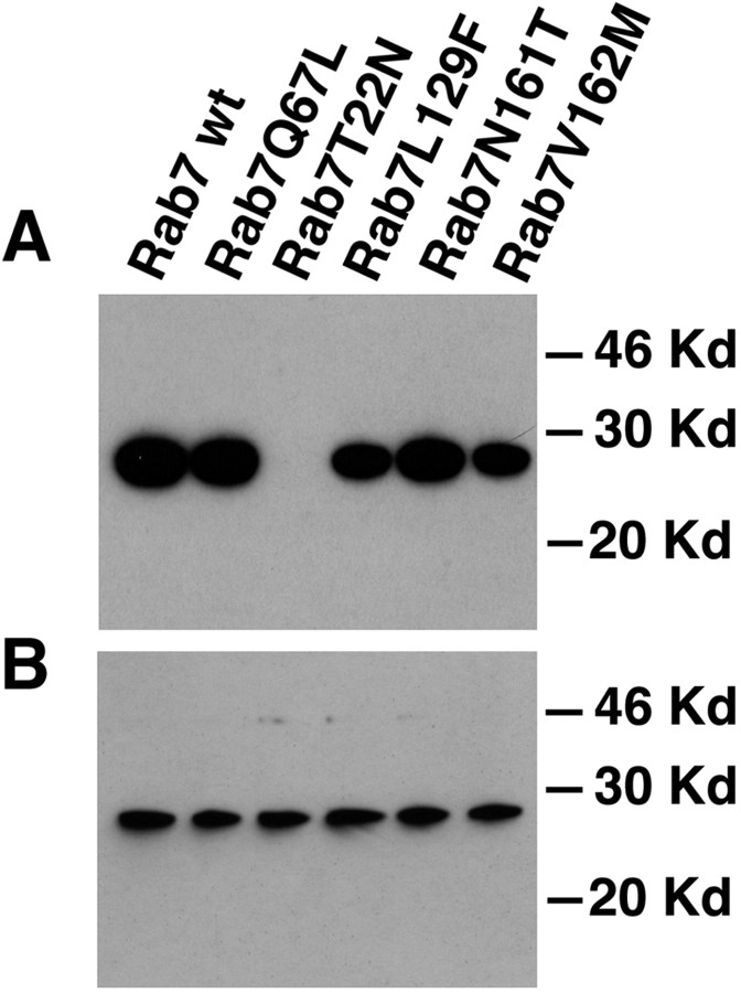
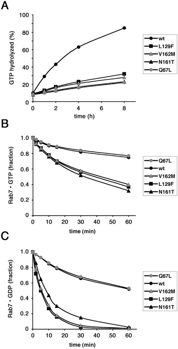
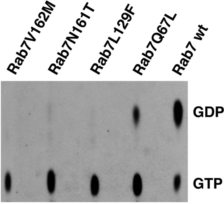
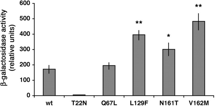
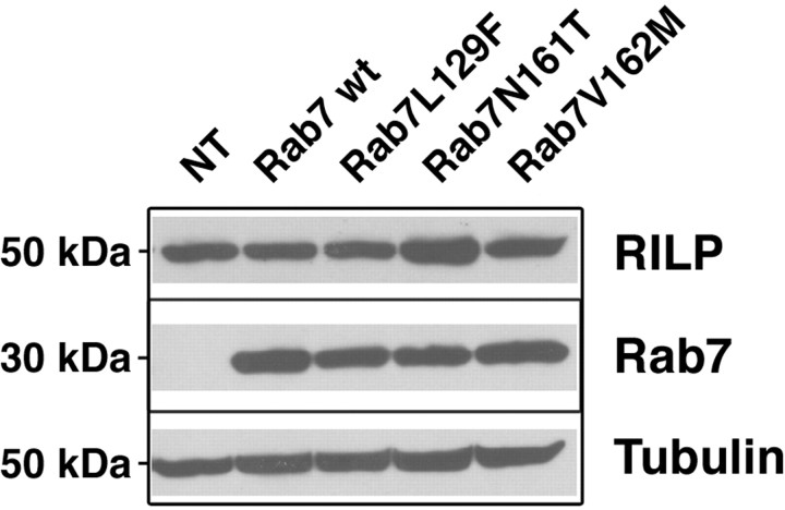
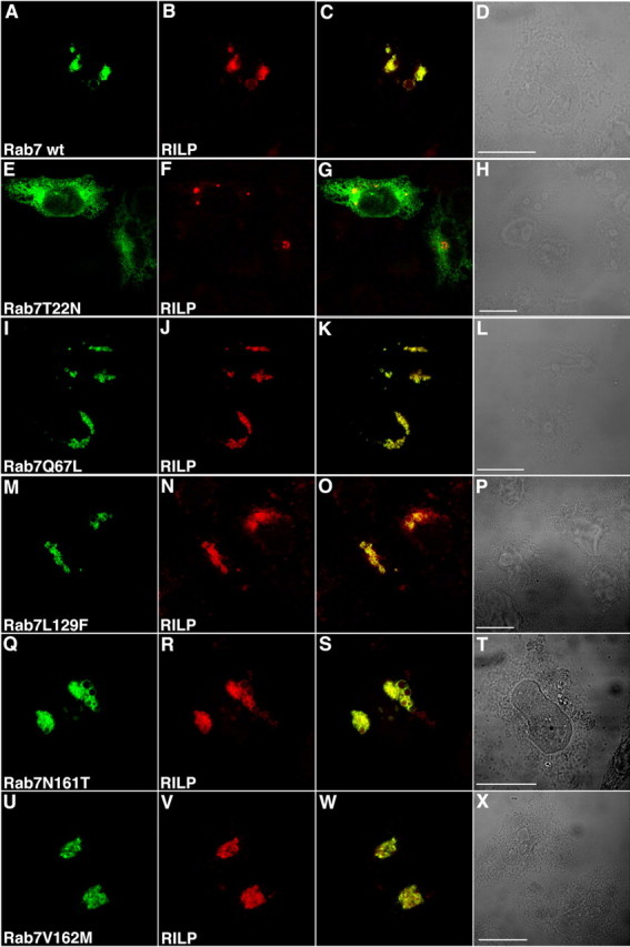
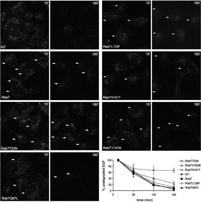
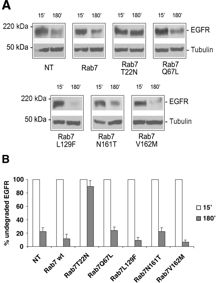
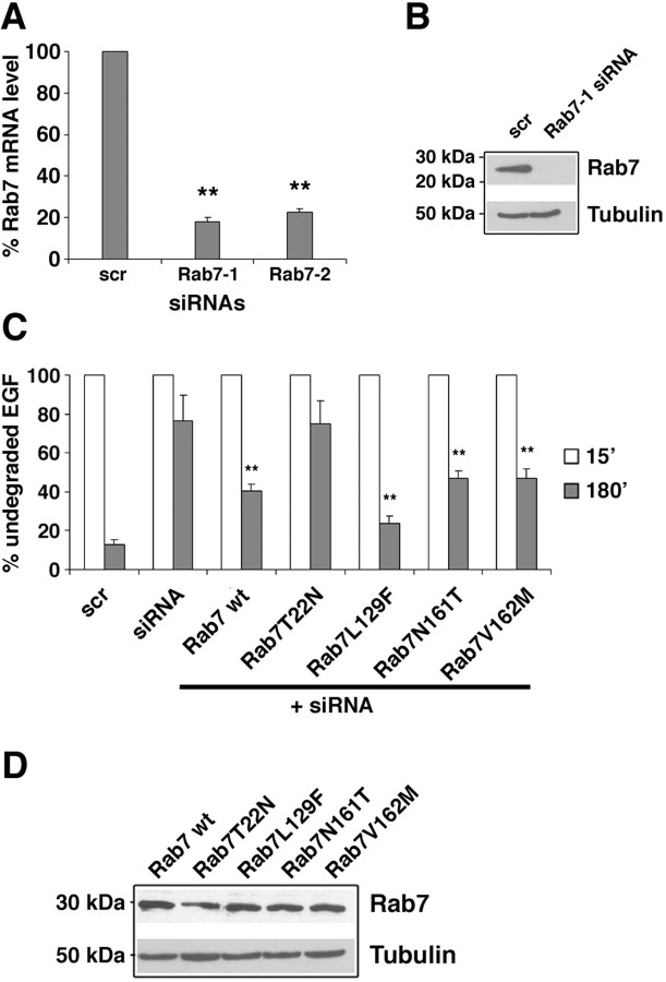
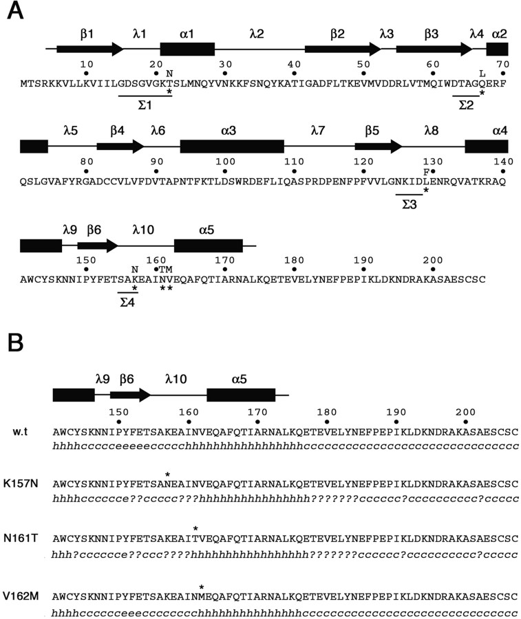
Charcot-Marie-Tooth (CMT) type 2 neuropathies are a group of autosomal-dominant axonal disorders genetically and clinically heterogeneous. In particular, CMT type 2B (CMT2B) neuropathies are characterized by severe sensory loss, often complicated by infections, arthropathy, and amputations. Recently, four missense mutations in the small GTPase Rab7 associated with the Charcot-Marie Tooth type 2B phenotype have been identified. These mutations target highly conserved amino acid residues. However, nothing is known about whether and how these mutations affect Rab7 function. We investigated the biochemical and functional properties of three of the mutant proteins. Interestingly, all three proteins exhibited higher nucleotide exchange rates and hydrolyzed GTP slower than the wild-type protein. In addition, whereas 23% of overexpressed wild-type Rab7 was GTP bound in HeLa cells, the large majority of the mutant proteins (82-89%) were in the GTP-bound form, consistent with the data on GTP hydrolysis and exchange rates. The CMT2B-associated Rab7 proteins were also able to bind the Rab7 effector RILP (Rab-interacting lysosomal protein) and to rescue Rab7 function after silencing. Altogether, these data demonstrate that all tested CMT2B-associated Rab7 mutations are mechanistically similar, suggesting that activated forms of the Rab7 are responsible for CMT2B disease.


Conflict of interest statement

The authors declare no competing financial interests.

Figures

Figure 1.
Figure 1.
GTP overlay and Western blot analysis on HeLa cells expressing Myc-tagged Rab7 wt and mutant proteins. Cells were transfected with the indicated Myc-tagged Rab7 wt and mutant proteins and lysed, and the extracts were analyzed by SDS-PAGE. A, GTP overlay was performed as indicated in Materials and Methods. B, Western blot analysis with the same lysates was performed using monoclonal anti-Myc antibody as detailed in Materials and Methods.
Figure 2.
Figure 2.
Biochemical characterization of CMT2B-associated Rab7 mutant proteins. A, Measurement of the intrinsic GTPase activity of His-tagged Rab7 wt, Rab7 Q67L, Rab7 L129F, Rab7 N161T, and Rab7 V162M, as indicated. Proteins were loaded with labeled GTP, and then 100-fold molar excess of GTP or GDP was added. GTP hydrolysis was monitored during 8 h. Differences between the values of all Rab7 mutated proteins and those of Rab7 wt are statistically significant (p value <0.01) at 1, 2, 4, and 8 h. B, C, Dissociation of guanine nucleotides from Rab7 wt, Rab7 Q67L, Rab7 L129F, Rab7 N161T, and Rab7 V162M. Purified proteins were incubated with [3H]GTP (B) or [3H]GDP (C). In the experiment shown, prebinding was allowed to proceed to 30% saturation. One hundred-fold molar excess of cold competitor (GDP or GTP) was then added, and dissociation of nucleotide was monitored for 1 h. In B, differences between the values of all Rab7 CMT2B mutated proteins and those of Rab7 wt are statistically significant (p value <0.01) at 15, 30, and 60 min. In C, differences between the values of all Rab7 CMT2B mutated proteins and those of Rab7 wt are statistically significant (p value <0.01) at 5, 10, 15, 30, and 60 min.
Figure 3.
Figure 3.
GTP:GDP ratio of Rab 7 wt and mutant proteins in transfected HeLa cells. Cells were transfected with the indicated HA-tagged constructs and lysed, and immunoprecipitation was performed using an anti-HA antibody. The amounts of nucleotides bound to the immunoprecipitated proteins were determined by TLC, as described in Materials and Methods. The positions of GTP and GDP are indicated.
Figure 4.
Figure 4.
Interaction of Rab7 wt and mutant proteins with RILP. L40 yeast cells were cotransformed with pGADGH-RILPC33 and the indicated pLexA-Rab7 wt or mutant constructs. The β-galactosidase activity of double transformants was measured using o-nitrophenyl-β-d-galactoside as substrate as detailed in Materials and Methods. Activities are measured as arbitrary relative units and represent mean ± SEM values of six independent transformants. Differences between the values of Rab7 CMT2B mutated proteins and that of Rab7 wt are statistically significant at p values <0.05 (*) or <0.01 (**).
Figure 5.
Figure 5.
Transient expression of CMT2B-associated Rab7 mutant proteins does not alter RILP expression. HeLa cells were nontransfected (NT) or transfected with HA-tagged Rab7 wt, Rab7 L129F, Rab7 N161T, or Rab7 V162M for 20 h and then lysed. Lysates were analyzed by Western blot using anti-RILP antibody. Anti-tubulin was used to verify equal loadings, and anti-HA antibody was used to check expression of the different Rab7 constructs.
Figure 6.
Figure 6.
Colocalization of CMT2B-associated Rab7 wt and mutant proteins with RILP. HeLa cells were cotransfected with RILP and GFP-tagged Rab7 wt and mutants as indicated. A, E, I, M, Q, U, GFP signal. B, F, J, N, R, V, TRITC signal of secondary anti-rabbit antibody used after a primary anti-RILP antibody. C, G, K, O, S, W, Overlays of the two colors. D, H, L, P, T, X, Phase-contrast images. Scale bars, 10 μm.
Figure 7.
Figure 7.
Effect of CMT2B-associated Rab7 mutations on EGF degradation. HeLa cells were transfected with GFP-tagged Rab7 wt, Rab7 T22N, Rab7 Q67L, Rab7 L129F, Rab7 N161T, Rab7 V162M, or nontransfected (NT) as indicated. Then cells were incubated 1 h at 4°C with rhodamine-labeled EGF, washed, and incubated at 37°C for 15 min or 3 h. Finally, they were processed for confocal fluorescent microscopy. Transfected cells were selected, and the total red signal in the entire cell (representing undegraded EGF) was quantified. Staining of EGF at 15 and 180 min is shown. Arrows indicate transfected cells. Intensities of EGF were quantified and plotted as percentage of the respective intensities after 15 min incubation at 37°C (bottom right). Values are calculated on 30 cells for each construct and represent means ± SEM. Differences between the values of Rab7 CMT2B mutated proteins and that of Rab7 T22N are statistically significant (p value <0.01) at 180 min. Differences between the values of Rab7 V162M and those of Rab7 wt or the other Rab7 CMT2B mutated proteins are not statistically significant.
Figure 8.
Figure 8.
Effect of CMT2B-associated Rab7 mutations on EGFR degradation. HeLa cells were transfected for 20 h with HA-tagged Rab7 wt, Rab7 T22N, Rab7 Q67L, Rab7 L129F, Rab7 N161T, Rab7 V162M, or nontransfected (NT) as indicated. Cells were then treated with cycloheximide for 1 h and subsequently with EGF for 15 or 180 min. A, Lysates were subjected to Western blot analysis using an anti-EGFR antibody. Western blot with anti-tubulin antibody on the same membrane was used to verify equal loadings. B, The intensities of EGFR staining were quantified and plotted as a percentage of the respective intensities after 15 min of EGF stimulation. Values at 15 min were set to 100%. Values are means of four independent experiments ± SEM (error bars). Differences between the values of cells expressing Rab7 T22N and the other samples are statistically significant (p value <0.01); differences between the values of cells expressing Rab7 wt and cells expressing CMT2B associated Rab7 mutants are not statistically significant.
Figure 9.
Figure 9.
Expression of CMT2B-associated Rab7 mutant proteins restores Rab7 function. A, HeLa cells were treated with control RNA (scr) and two different siRNAs against Rab7 (Rab7-1 or Rab7-2). Rab7 mRNA levels were determined by real-time PCR as indicated in Materials and Methods. Decreases in Rab7 mRNA levels in siRNA-treated samples are statistically significant at p value <0.01 (**). B, HeLa cells were treated with control siRNA (scr) or siRNA against Rab7 (Rab7-1), lysed, and subjected to Western blot analysis using anti-Rab7 antibody. Anti-tubulin antibody was used to verify equal loadings. C, HeLa cells were treated with control RNA (scr) or with Rab7-1 siRNA. Cells silenced for Rab7 were then transfected with the different Rab7 wt or mutant constructs as indicated. All cells were incubated for 1 h at 4°C with rhodamine-labeled EGF, washed, and incubated at 37°C for 15 min or 3 h. Transfected cells were selected, and the red signal (representing undegraded EGF) was quantified by confocal microscopy. The intensities of EGF labeling were quantified and plotted as percentage of the respective intensities after 15 min of incubation at 37°C. Values at 15 min were set to 100%. Values were calculated on 30 cells for each construct and represent means ± SEM. Differences between the values of cells expressing Rab7 CMT2B mutated proteins and that of siRNA-treated control cells are statistically significant at p value <0.01 (**). Differences between the values of cells transfected with the different CMT2B mutated proteins are not statistically significant. D, The amount of expression of Rab7 wt and mutant proteins was checked by Western blot analysis using anti-Rab7 antibody. Anti-tubulin antibody was used to verify equal loadings.
Figure 10.
Figure 10.
Amino acid sequence and secondary structure of wild-type and CMT2B-associated Rab7 proteins. A, Amino acid sequence of the human wild-type Rab7 protein. Asterisks mark the amino acid substitutions in the mutated proteins Rab7 T22N, Rab7 Q67L, Rab7 L129F, Rab7 K157N, N161T, and V162M. Amino acids in the four sequence motifs (Σ1–Σ4) conserved in all GTP-binding proteins are underlined. Secondary structure above the amino acid sequence has been predicted on the basis of the available structural data for Rab proteins (Merithew et al., 2001). α1–α5, α helices; β1–β6, β sheets; λ1–λ10, loops. B, Secondary structure prediction in the C-terminal region of wild-type and CMT2B-associated Rab7 proteins. The software Secondary Structure Prediction at the Pôle Bioinformatique Lyonnais (http://pbil.univ-lyon1.fr/) has been used with the methods DSC, MLRC, and PHD (with default parameters) that generated the secondary consensus shown below each amino acid sequence. h, α helix; e, extended strand; c, random coil; ?, ambiguous state.

References

    1. Auer-Grumbach M, Mauko B, Auer-Grumbach P, Pieber TR. Molecular genetics of hereditary sensory neuropathies. Neuromolecular Med. 2006;8:147–158. - PubMed
    1. Bartel P, Chien CT, Sternglanz R, Fields S. Elimination of false positives that arise in using the two-hybrid system. Biotechniques. 1993;14:920–924. - PubMed
    1. Bronfman FC, Escudero CA, Weis J, Kruttgen A. Endosomal transport of neurotrophins: roles in signaling and neurodegenerative diseases. Dev Neurobiol. 2007;67:1183–1203. - PubMed
    1. Bucci C, Chiariello M. Signal transduction gRABs attention. Cell Signal. 2006;18:1–8. - PubMed
    1. Bucci C, Parton RG, Mather IH, Stunnenberg H, Simons K, Hoflack B, Zerial M. The small GTPase rab5 functions as a regulatory factor in the early endocytic pathway. Cell. 1992;70:715–728. - PubMed

Publication types

MeSH terms