. 2008 Apr;19(4):1637-45.
doi: 10.1091/mbc.e07-11-1114. Epub 2008 Feb 13.

Na/H exchange regulatory factor 1, a novel AKT-associating protein, regulates extracellular signal-regulated kinase signaling through a B-Raf-mediated pathway

Affiliations

Na/H exchange regulatory factor 1, a novel AKT-associating protein, regulates extracellular signal-regulated kinase signaling through a B-Raf-mediated pathway

Bin Wang et al. Mol Biol Cell. 2008 Apr.

Abstract

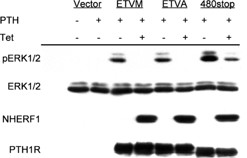
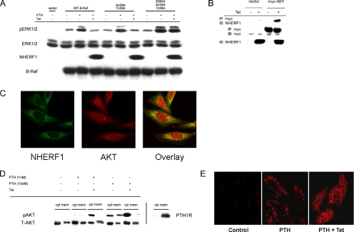
Na/H exchange regulatory factor 1 (NHERF1) is a scaffolding protein that regulates signaling and trafficking of several G protein-coupled receptors (GPCRs), including the parathyroid hormone receptor (PTH1R). GPCRs activate extracellular signal-regulated kinase (ERK)1/2 through different mechanisms. Here, we characterized NHERF1 regulation of PTH1R-stimulated ERK1/2. Parathyroid hormone (PTH) stimulated ERK1/2 phosphorylation by a protein kinase A (PKA)-dependent, but protein kinase C-, cyclic adenosine 5'-monophosphate-, and Rap1-independent pathway in Chinese hamster ovary cells stably transfected with the PTH1R and engineered to express NHERF1 under the control of tetracycline. NHERF1 blocked PTH-induced ERK1/2 phosphorylation downstream of PKA. This suggested that NHERF1 inhibitory effects on ERK1/2 occur at a postreceptor locus. Forskolin activated ERK1/2, and this effect was blocked by NHERF1. NHERF1 interacted with AKT and inhibited ERK1/2 activation by decreasing the stimulatory effect of 14-3-3 binding to B-Raf, while increasing the inhibitory influence of AKT negative regulation on ERK1/2 activation. This novel regulatory mechanism provides a new model by which cytoplasmic adapter proteins modulate ERK1/2 activation through a receptor-independent mechanism involving B-Raf.


Figures

Figure 1.
Figure 1.
PTH-stimulated ERK1/2 phosphorylation and inhibition by NHERF1. (A) Representative time course of PTH-stimulated ERK1/2 phosphorylation in CHO-N10-R3 cells (top). Cells were treated with 10 nM PTH(1-34) for the indicated time. Data from three independent time course and concentration dependence experiments were quantified, normalized to total ERK2, and expressed as the -fold change of basal pERK2. Data are summarized as mean ± SEM (bottom left). At the 10-min time point, PTH (10−11–10−6 M) induced concentration-dependent increases of ERK1/2 phosphorylation (bottom right). (B) NHERF1 inhibition of PTH-stimulated ERK1/2 at graded PTH concentrations. CHO-N10-R3 cells were pretreated where indicated with 50 ng/ml Tet for 48 h. The cells were then serum starved for 3–5 h and incubated at 37°C for 10-min with the indicated concentration of PTH(1-34). Levels of phosphorylated and total ERK1/2, NHERF1 and HA-PTH1R in whole-cell lysates were determined by immunoblotting as described in Materials and Methods. (C) Graded induction of NHERF1 expression causes progressive inhibition of PTH-stimulated ERK stimulation. CHO-N10-R3 cells were pretreated with the indicated concentration of Tet. Cells were then serum starved for 3–5 h, and then they were incubated at 37°C for 10 min with 10 nM PTH(1-34). The figure is representative of three or four independent experiments performed with similar results.
Figure 2.
Figure 2.
NHERF1 inhibition of ERK1/2 does not require the PTH1R PDZ recognition domain. CHO-N10 cells were pretreated with 50 ng/ml Tet as indicated, and then they were transiently transfected with 1.0 μg of DNA/well of empty vector (pcDNA3.1+), wild-type PTH1R(ETVM), mutant PTH1R (ETVA), or truncated PTH1R(480stop). After 48 h, the cells were serum starved for 3–5 h, and then they were treated for 10 min with 1 nM PTH. The extent of phosphorylated and total ERK1/2, NHERF1, and PTH1R was determined as described in Materials and Methods. The results represent one of three independent experiments performed with similar results.
Figure 3.
Figure 3.
NHERF1 inhibition of ERK1/2 activation is not mediated by transactivation of inhibition of Gi/o. CHO-N10-R3 cells were pretreated with 50 ng/ml Tet for 48 h or with 100 ng/ml PTX for 16 h. The cells were then serum-starved for 3–5 h and treated with the PDGF receptor inhibitor AG1295 (10 μM) for 40 min or EGF receptor inhibitor AG1478 (500 nM) for 15 min before addition of 1 nM PTH for 10 min. The extent of phosphorylated and total ERK1/2 and NHERF1 in whole-cell lysates was determined as described in Materials and Methods. The results represent one of three independent experiments performed with similar results.
Figure 4.
Figure 4.
NHERF1 inhibits ERK1/2 at a postreceptor locus. CHO-N10-R3 cells were pretreated with 50 ng/ml Tet for 48 h as indicated. The cells were then serum starved for 3–5 h and treated for 10 min with 20 μM H89 before addition of 10 μM forskolin for 10 min or 1 nM PTH for 10 min. (A) Phosphorylated and total MEK1/2 and ERK1/2, and NHERF1 in whole-cell lysates determined by immunoblotting. (B) ERK1/2 activity (pElk-1) was measured as described in Materials and Methods. The results are representative of three independent experiments performed with similar results.
Figure 5.
Figure 5.
NHERF1 inhibition of ERK1/2 activation is mediated by decreased of 14-3-3 binding to B-Raf. (A) NHERF1 disrupts 14-3-3 binding to B-Raf. Interaction of 14-3-3 with FLAG-tagged wild-type B-Raf in CHO-N10-R3 cells was determined by coimmunoprecipitation (IP) and immunoblotting (IB) as described in Materials and Methods. (B) B-Raf S728 is critical for activation of ERK1/2. CHO-N10-R3 cells were pretreated with 50 ng/ml Tet as indicated, and then they were transiently transfected with 1.0 μg DNA/well of empty vector, wild-type B-Raf, B-Raf(S364A), B-Raf(S728A), or B-Raf(S364A, S728A). After 48 h, the cells were serum starved for 3–5 h and incubated with 1 nM PTH for 10 min. The extent of phosphorylated and total ERK1/2, NHERF1, and B-Raf in whole-cell lysates was determined by immunoblotting. (C) NHERF1 blocks PTH-stimulated B-Raf S728 phosphorylation. CHO-N10-R3 cells were transfected with HA-tagged wild-type B-Raf or B-Raf(S728A). phospho-Ser728 expression was determined by coimmunoprecipitation with HA monoclonal affinity matrix. The blot was probed with an antibody specific for Raf-1 phospho-Ser621 Raf corresponding to Ser728 in the catalytic domain of B-Raf (Qiu et al., 2000). All figures represent one of three independent experiments performed with comparable results.
Figure 6.
Figure 6.
NHERF1 effects on B-Raf regulatory domain. (A) Mutation of the inhibitory B-Raf regulatory domain AKT phosphorylation sites enhances PTH-stimulated ERK1/2 and prevents inhibition by NHERF1. CHO-N10-R3 cells were pretreated with 50 ng/ml Tet as indicated, and then they were transiently transfected with 1.0 μg DNA/well of empty vector, wild-type B-Raf, B-Raf(S428A,T439A), or B-Raf(S364A, S428A, T439A). After 48 h, the cells were serum starved for 3–5 h, and then they were pretreated with 20 μM H89 for 10 min before addition of 1 nM PTH for 10 min. Phosphorylated and total ERK1/2, NHERF1 and B-Raf in whole-cell lysates were determined by immunoblotting (IB). The results are representative of three independent experiments. (B) AKT directly interacts with NHERF1. CHO-N10-R3 cells were treated with 50 ng/ml Tet as indicated, and then they were transfected with myc-AKT. After 48 h, the interaction with NHERF1 was determined by coimmunoprecipitation (IP) and immunoblotting. (C) Colocalization of AKT and NHERF1. CHO-N10-R3 cells were treated with 50 ng/ml Tet for 48 h, and then they were fixed and incubated with polyclonal anti-AKT antibody and monoclonal anti-NHERF1 antibody. Fluorescent staining was analyzed by confocal microscopy as described in Materials and Methods. (D) NHERF1 blocks PTH-induced AKT translocation from membrane to cytosol. The extent of phospho-AKT in cytoplasm (cyt) and membrane (mem) was determined by immunoblotting of the respective fractions that were prepared as described in Materials and Methods. PTH1R was similarly analyzed. (E) Phospho-AKT localization. In control condition, pAKT is barely detectable. PTH (10 nM) for 10 min significantly increased pAKT in membrane punctae. Induction of NHERF1 restricts pAKT to cytoplasm upon PTH application.
Figure 7.
Figure 7.
Model of NHERF1 inhibition of PTH-stimulated ERK1/2 activity. PTH binding PTH1R activates adenylyl cyclase and PKA. B-Raf phosphorylated by PKA binds to 14-3-3, thereby activating B-Raf. The activated B-Raf subsequently phosphorylates its substrate MEK1/2 and stimulates ERK1/2 activation. Akt phosphorylates B-Raf in its regulatory domain and decreases B-Raf activation. Higher concentration of PTH stimulates AKT activity, which negatively regulates ERK1/2 activity. The magnitude of PTH-stimulated ERK1/2 phosphorylation is determined by both PKA and AKT activation. In the presence of NHERF1, both B-Raf phosphorylation at position of Ser728 and the association of 14- 3-3 with B-Raf are inhibited. NHERF1 also increases the AKT activity and negatively regulates ERK1/2 activation, thereby inhibiting ERK1/2 phosphorylation.

Similar articles

Cited by

References

    1. Ahmed I., Gesty-Palmer D., Drezner M. K., Luttrell L. M. Transactivation of the epidermal growth factor receptor mediates parathyroid hormone and prostaglandin F2α-stimulated mitogen-activated protein kinase activation in cultured transgenic murine osteoblasts. Mol. Endocrinol. 2003;17:1607–1621. - PubMed
    1. Aitken A., Baxter H., Dubois T., Clokie S., Mackie S., Mitchell K., Peden A., Zemlickova E. Specificity of 14-3-3 isoform dimer interactions and phosphorylation. Biochem. Soc. Trans. 2002;30:351–360. - PubMed
    1. Bretscher A., Chambers D., Nguyen R., Reczek D. ERM-Merlin and EBP50 protein families in plasma membrane organization and function. Annu. Rev. Cell Dev. Biol. 2000;16:113–143. - PubMed
    1. Bretscher A., Edwards K., Fehon R. G. ERM proteins and merlin: integrators at the cell cortex. Nat. Rev. Mol. Cell Biol. 2002;3:586–599. - PubMed
    1. Calipel A., Mouriaux F., Glotin A. L., Malecaze F., Faussat A. M., Mascarelli F. Extracellular signal-regulated kinase-dependent proliferation is mediated through the protein kinase A/B-Raf pathway in human uveal melanoma cells. J. Biol. Chem. 2006;281:9238–9250. - PubMed

Publication types

MeSH terms