

Clonally related follicular lymphomas and histiocytic/dendritic cell sarcomas: evidence for transdifferentiation of the follicular lymphoma clone
- PMID: 18272816
- PMCID: PMC2424145
- DOI: 10.1182/blood-2007-11-124792
Clonally related follicular lymphomas and histiocytic/dendritic cell sarcomas: evidence for transdifferentiation of the follicular lymphoma clone
Abstract
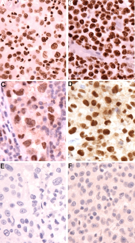
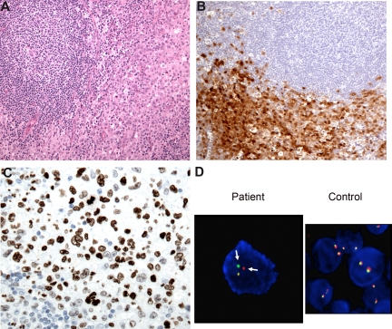
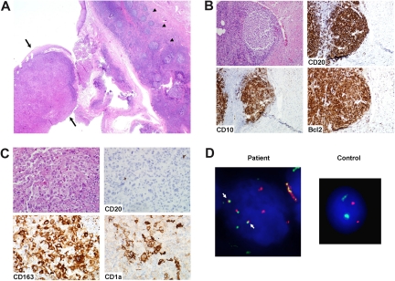
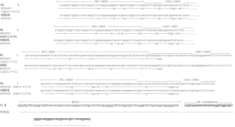
Rare cases of histiocytic and dendritic cell (H/DC) neoplasms have been reported in patients with follicular lymphoma (FL), but the biologic relationship between the 2 neoplasms is unknown. We studied 8 patients with both FL and H/DC neoplasms using immunohistochemistry, fluorescence in situ hybridization (FISH) for t(14;18), and polymerase chain reaction (PCR)/sequencing of BCL2 and IGH rearrangements. There were 5 men and 3 women (median age, 59 years). All cases of FL were positive for t(14;18). The H/DC tumors included 7 histiocytic sarcomas, 5 of which showed evidence of dendritic differentiation, and 1 interdigitating cell sarcoma. Five H/DC tumors were metachronous, following FL by 2 months to 12 years; tumors were synchronous in 3. All 8 H/DC tumors showed presence of the t(14;18) either by FISH, or in 2 cases by PCR with the major breakpoint region (MBR) probe. PCR and sequencing identified identical IGH gene rearrangements or BCL2 gene breakpoints in all patients tested. All H/DC tumors lacked PAX5, and up-regulation of CEBPbeta and PU.1 was seen in all cases tested. These results provide evidence for a common clonal origin of FL and H/DC neoplasms when occurring in the same patient, and suggest that lineage plasticity may occur in mature lymphoid neoplasms.
Figures
Comment in
-
Follicular lymphomas and histiocytic/dendritic neoplasms related?Blood. 2008 Jun 15;111(12):5418-9. doi: 10.1182/blood-2008-02-140962. Blood. 2008. PMID: 18544687 Free PMC article.
References
-
- Akashi K, Traver D, Miyamoto T, Weissman IL. A clonogenic common myeloid progenitor that gives rise to all myeloid lineages. Nature. 2000;404:193–197. - PubMed
-
- Akashi K, Reya T, Dalma-Weiszhausz D, Weissman IL. Lymphoid precursors. Curr Opin Immunol. 2000;12:144–150. - PubMed
-
- Jaffe ES, Harris NL, Stein H, Vardiman J. Lyon, France: IARC Press; 2001. Pathology and Genetics of Tumours of Haematopoietic and Lymphoid Tissues.
-
- Feldman AL, Berthold F, Arceci R, et al. Clonal relationship between precursor T-lymphoblastic leukaemia/lymphoma and Langerhans-cell histiocytosis. Lancet Oncol. 2005;6:435–437. - PubMed
-
- Feldman AL, Minniti C, Santi M, Downing JR, Raffeld M, Jaffe ES. Histiocytic sarcoma after acute lymphoblastic leukaemia: a common clonal origin. Lancet Oncol. 2004;5:248–250. - PubMed
Publication types
MeSH terms
LinkOut - more resources
Full Text Sources
Other Literature Sources
Research Materials
