. 2004 May;1(2):101-12.
doi: 10.1017/s1740925x04000225.

Lateral neuron--glia interactions steer the response of axons to the Robo code

Affiliations

Lateral neuron--glia interactions steer the response of axons to the Robo code

Edward F V Kinrade et al. Neuron Glia Biol. 2004 May.

Abstract

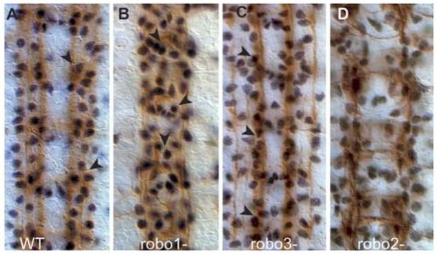
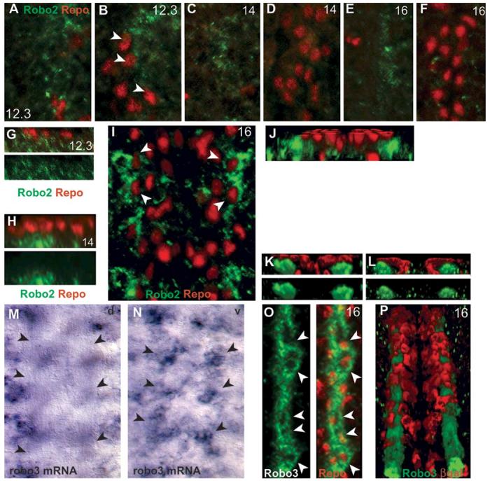
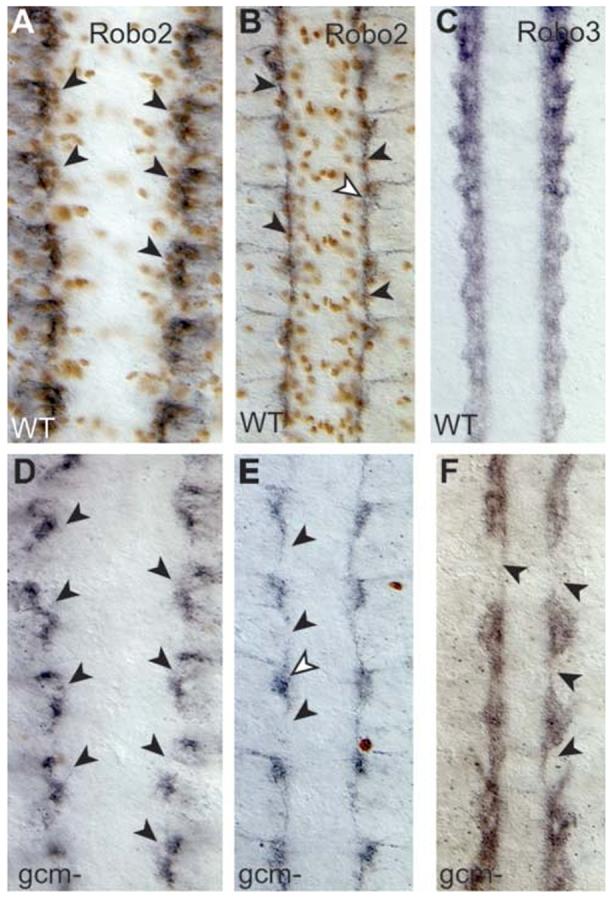
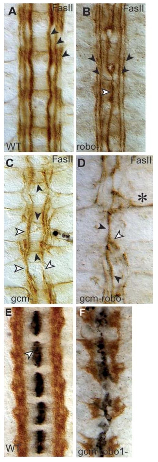
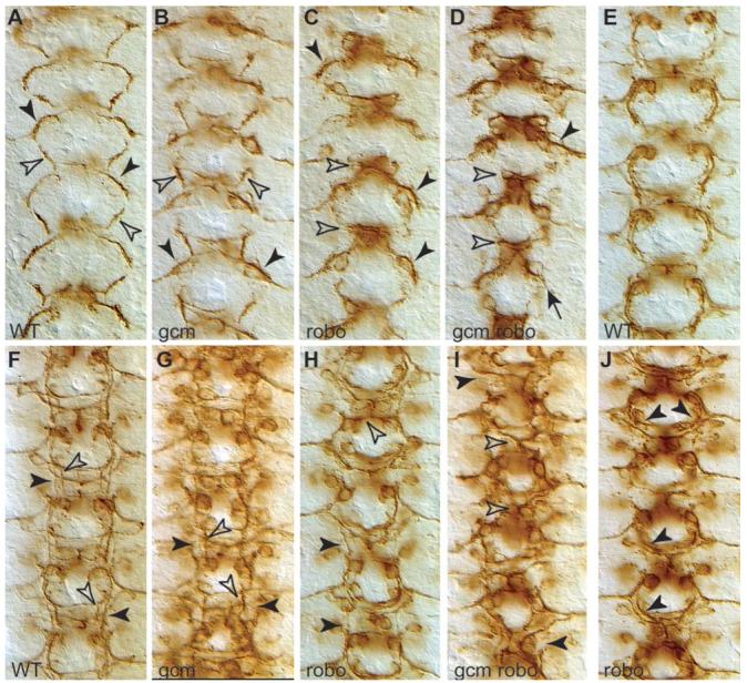
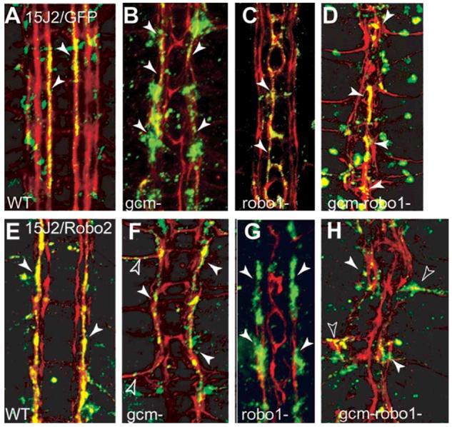
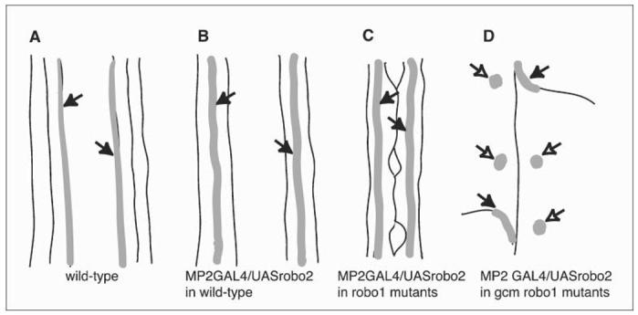
Glia are required for axon pathfinding along longitudinal trajectories, but it is unknown how this relates to the molecular paradigm of axon guidance across the midline. Most interneuron axons in bilateral organisms cross the midline only once. Preventing them from recrossing the midline requires the expression of Robo receptors on the axons. These sense the repulsive signal Slit, which is produced by the midline. The lateral positioning of longitudinal axons depends on the response to Slit by the combination of Robo receptors expressed by the axons, on selective fasciculation, and on longitudinal (lateral) glia. Here, we analyse how longitudinal glia influence reading of the 'Robo code' by axons. We show that whereas loss of robo1 alone only affects the most medial axons, loss of both glial cells missing (gcm) and robo1 causes a severe midline collapse of longitudinal axons, similar to that caused by the loss of multiple Robo receptors. Furthermore, whereas ectopic expression of robo2 is sufficient to displace the medial MP2 axons along a more lateral trajectory, this does not occur in gcm-robo1 double-mutant embryos, where axons either do not extend at all or they misroute exiting the CNS. Hence, lateral neuron-glia interactions steer the response of axons to the Robo code.

Keywords: Glia; axon guidance; gcm; neuron-glia interactions; robo.


Figures

Fig. 1
Fig. 1
LG are misplaced in Robo-mutant embryos. Embryos stained with the glial nuclear anti-Repo (black) and neuronal FasII (brown) antibodies at stage 16. (A) In wild-type embryos glia (arrowheads) are deployed in discrete rows over the three FasII fascicles. (B) In robo1-mutant embryos, some glia migrate over the midline (arrowheads) and some remain over the lateral axons. (C) In robo3-mutant embryos, many glia are displaced medially and they overlie the fascicle formed by the fused medial and central fascicle (arrowheads). (D) In robo2 mutants the lateral FasII fascicle and glial patterns are only slightly disorganised. Anterior is to the top.
Fig. 2
Fig. 2
Robo2 and Robo3 are not expressed in LG. LG nuclei are visualised in wild-type embryos with the glial nuclear marker anti-Repo (red) (A-K,O), and LG cytoplasm with anti-βgal on the LG-lacZ reporter (red) (L,P). (A-J) Temporal profile of Robo2 distribution (green) in the ventral nerve cord. Ventral (A) and dorsal (B) longitudinal and (G) sagittal sections of the same stage-12.3 embryo. Robo2 is first detected at stage 12.3 lateral to the LG and mostly in a ventral plane under the LG. Some LG nuclei appear partly surrounded by Robo2 signal (arrowheads in B), but in a transverse view (G) the glial nuclei overlie Robo2 signal. At stage 14, from a frontal view Robo2 signal is ventral (C) and physically separate from the dorsal, Repo-positive LG nuclei (D,H). (G,H) Single channel images of anti-Robo2 are also shown (green). (E,F,I) At stage 16, Robo2 is lateral to the LG but it appears to surround some Repo-positive LG nuclei (arrowheads in I), however these are in separate planes: ventral Robo2 (E); dorsal Repo glia (F); and a whole-stack projection (I). (J) A tilted, 3D-reconstruction section of the same embryonic stack as in (I), showing that Robo2 lies ventrally and laterally to the LG. (M,N) Distribution of robo3 transcripts by in situ hybridisation with DIG probes. (M) There is no signal in the dorsal plane of the neuropile, axons are seen clear of signal by Nomarski optics (arrows). (N) All DIG signal is in a ventral plane (arrows) below the neuropile. (K,L,O,P) Distribution of anti-Robo3 (green) and either Repo or anti-βgal (red) in the LG at stage 16. (O,P) From a frontal view, Robo3 appears to surround the nuclei of LG (arrowheads, whole-stack projections). (O) Single channel image shown in green next to merged image. (K) 3D transverse view of the embryo in (O) reveals that the glial nuclei are dorsal to and separated from the Robo3 axons (bottom, single channel view of Robo3, green; top, merge). (P) LG are visualised with anti-βgal (red) using the LG-lacZ reporter overlie the Robo3 axons (tilted 3D reconstruction, whole stack). (L) 3D transverse view of the same embryo as in (P) revealing that the βgal glial signal overlies the Robo3 axonal bundles, but it is not coincident with them. All images were processed with Volocity (Improvision). Anterior is up. A-F,G,H show one hemisegment; O just over one hemisegment; the midline is to the left. I shows one segment; M,N, 4 segments; P, 5 segments; G,H, lateral sections; J,K,L, cross-section views. A-F are projections of sections restricted to or excluding glial nuclei; I,O,P are projections of the whole stack of sections through the neuropile.
Fig. 3
Fig. 3
Robo2 and Robo3 in the absence of glia. Anti-Robo2 (black) and anti-Repo (brown) in wild-type embryos (A,B) and gcm mutant (D,E) embryos. Anti-Robo3 in wild-type (C) and gcm mutant (F) embryos. At stage 13 in wild-type embryos, Robo2 clusters lie under the LG (A). Robo2 is present but disarranged in gcm mutants (D). At stage 16, Robo2 longitudinal axons are frequently missing in gcm mutants (E, arrowheads), but Robo2 signal (white arrowhead) is present at the base of the commissures. (F) Robo3 is still present in gcm mutants, but the longitudinal projection of axons is severely compromised (arrowheads). Anterior is up.
Fig. 4
Fig. 4
Midline collapse of longitudinal axons in gcm-robo1 double-mutant embryos. Axons are visualised with anti-FasII antibodies (brown) at stage 16 (A-D). (A) Wild-type embryo. (B) robo1 mutant. Note that the medial fascicle runs along the midline (white arrowhead) whereas the outer two fascicles are well formed and run parallel to the midline (black arrowheads). (C) In gcm mutants, axons sporadically misroute across the midline (black arrowheads) and the lateral fascicles can be fused into one that runs parallel to the midline along the central trajectory (white arrowheads). This embryo has also been stained with anti-Repo (in black, there are a few glia present). (D) In gcm-robo1 double-mutant embryos, axons can fuse (white arrowhead) into one single fascicle running along the midline (black arrowhead) or exit the CNS (asterisk) and grow towards the muscle, but no axons run along the lateral trajectories. (E,F) Axons are stained with anti-Robo3 (brown) and midline glia with anti-Slit (black) at stage 16. (E) Robo3 axons never cross the midline in wild-type embryos, and anti-Slit stains the midline glia in each segment. (F) gcm-robo1 double mutant. Slit (black) is still produced by the midline glia, although the midline glia are disorganised. No longitudinal axons remain, instead axons cross the midline despite expressing robo3. Anterior is up.
Fig. 5
Fig. 5
Axonal defects occur from the earlier phases of pioneer axon extension. Pioneer axons visualised with 22c10 antibodies in wild-type (A,E,F), gcm-mutant embryo (B,G), robo1 mutant (C,H,J) and gcm-robo1 double mutant (D,I) at stages 12.1 (A,B,C,D); 13 (E,J) and 14 (F,G,H,I). (A) In wild-type embryos, the descending dMP2/MP1 axons (black arrowhead) meet the ascending vMP2/pCC axons (white arrowhead) at the end of stage 12. (B) In gcm mutants, the dMP2/MP1 fascicle misroutes to the muscle, so its descending trajectory is missing (black arrowheads), but the vMP2/pCC fascicle is unaffected (white arrowheads). (C) In robo mutants, the pCC/vMP2 fascicle does not grow but is trapped in the midline (white arrowheads), whereas the dMP2/MP1 fascicle begins its normal descending trajectory. This is also seen slightly later, at stage 13, (J) where the dMP2/MP1 fascicle extends normally all the way down (arrowheads) and only then curves towards the midline as it fails to encounter the ascending vMP2/pCC fascicle, which is missing (compare to E, wild-type). (D) In gcm-robo1 double-mutant embryos, the pCC/vMP2 fascicle is completely collapsed on the midline (white arrowheads) and the dMP2/vMP2 fascicle is either missing or misrouted to the muscle (black arrowheads). (F) At stage 14, the two fascicles, dMP2/MP1 (black arrowheads) and pCC/vMP2 (white arrowheads) are clearly distinguishable. (G) In gcm mutants, only the pCC/vMP2 fascicle is present (white arrowheads) and the dMP2/MP1 fascicle is missing (black arrowheads). (H) In robo1 mutants, the pCC/vMP2 fascicle collapses on the midline (white arrowhead) but the dMP2/MP1 fascicle (black arrowhead) is normal. (I) In gcm-robo1 double mutants, the pCC/vMP2 fascicle collapses on the midline, but the dMP2/MP1 fascicle is not discernible. Anterior is up.
Fig. 6
Fig. 6
Ectopic expression of robo2 does not displace the MP2 axons along lateral longitudinal tracts in the absence of glia. FasII axons are visualised in red at stage 16. The MP2 axons are visualised with anti-GFP (green) by the expression of 15J2/UASGAPGFP in multiple genotypes (A-D). Colocalisation of GFP and FasII is seen in yellow. Ectopic expression of robo2 with 15J2/UASrobo2-myc, visualised with anti-Myc (green) antibodies in embryos of different genotypes (E-H). Colocalisation of Myc and FasII is seen in yellow. (A) Wild-type. The MP2 axons run parallel to the midline along the medial FasII fascicle at this stage (arrowheads, 15J2/UASGAPGFP). (E) When expression of robo2 is driven ectopically in the MP2 axons, their trajectories are displaced to the central fascicle (arrowheads, 15J2/UASRobo2myc). (B) In gcm-mutant embryos, the MP2 axons still run along the medial fascicle (arrowheads, gcmΔP1/gcmΔP1; 15J2/UASGAPGFP). (F) When robo2 is expressed ectopically in the MP2 neurons in gcm mutants (gcmΔP1/gcmΔP1; 15J2/UASrobo2myc), the MP2 axons stay within the fused, single fascicle (arrowheads). This gcm-mutant embryo is phenotypically more severe than in B, where the fasicles are defasciculated but not fused. (C) In robo1 mutants, the MP2 axons run on the medial fascicle, collapsed along the midline (arrowheads, robo1/robo1; 15J2/UASGAPGFP). (G) Expression of robo2 in the MP2 axons in robo1 mutants (robo1/robo1; 15J2/UASrobo2myc) displaces the MP2 axons laterally onto the central fascicle (arrowheads). (D) gcm-robo1 double mutant with midline collapse of the longitudinal fascicles, in which the MP2 axons run along the midline (arrowheads, gcmΔP1 robo/gcmΔP1 robo; 15J2/UASGAPGFP). (H) gcm-robo1 double mutant embryo with midline collapse of the longitudinal fascicles, expressing robo2 in the MP2 axons (gcmΔP1 robo1/gcmΔP1 robo1; 15J2/UASrobo2myc): the MP2 axons leave the CNS and extend towards the periphery (empty arrowheads) or they do not grow at all (white arrowheads). There are no MP2 axons projecting longitudinally in these embryos. Anterior is up.
Fig. 7
Fig. 7
Schematic of the behaviour of MP2 longitudinal axons in different genetic backgrounds. In wild-type embryos, MP2 axons extend along the medial fascicle (A, arrows) and are displaced laterally on expression of robo2 (B, arrows). Lateral displacement still takes place in robo1 mutant embryos (C, arrows). However, if embryos are double mutant for robo1 and gcm and, therefore, lack functional glia, MP2 axons either do not grow (D, empty arrows) or they misroute out of the CNS towards the muscle (D, arrows).

References

    1. Ba-Charvet KTN, Brose K, Marillat V, Kidd T, Goodman CS, Tessier-Lavigne M, Sotelo C, Chedotal A. Slit2-mediated chemorepulsion and collapse of developing forebrain axons. Neuron. 1999;22:463–473. - PubMed
    1. Bagri A, Martin O, Plump AS, Mak J, Pleasure SJ, Rubenstein JLR, Tessier-Lavigne M. Slit proteins prevent midline crossing and determine the dorsoventral position of major axonal pathways in the mammalian forebrain. Neuron. 2002;33:233–248. - PubMed
    1. Bastiani MJ, du Lac S, Goodman CS. Guidance of neuronal growth cones in the grasshopper embryo. I. Recognition of a scpecific pathway by the pCC neuron. Journal of Neuroscience. 1986;6:3518–3531. - PMC - PubMed
    1. Battye R, Stevens A, Jacobs R. Axon repulsion from the midline requires Slit function. Development. 1999;126:2475–2481. - PubMed
    1. Booth G, Kinrade E, Hidalgo A. Glia maintain follower neuron survival during Drosophila CNS development. Development. 2000;127:237–244. - PubMed

LinkOut - more resources